 |

|
 |
|
Case Series
| ||||||
| Ipsilateral fracture of the supracondylar humerus and forearm in children | ||||||
| Shahid Hussain | ||||||
|
Registar, Department of Orthopedics, Sheri Kashmir Institue of Medical Sciences and Hospital, Bemina, Srinagar, Jammu and Kashmir, India.
| ||||||
| ||||||
|
[HTML Abstract]
[PDF Full Text]
[Print This Article]
[Similar article in Pumed] [Similar article in Google Scholar] 
|


| How to cite this article: |
| Hussain S. Ipsilateral fracture of the supracondylar humerus and forearm in children. International Journal of Case Reports and Images 2014;5(3):189–194. |
|
Abstract
|
|
Introduction:
Simultaneous ipsilateral fracture of the elbow and forearm (floating elbow) is an uncommon injury and treatment recommendations are controversial. The aim of our study was to evaluate the incidence of ipsilateral fractures of the upper limb and to present our experience in dealing with such injuries and to review the literature relating to this topic. The following variables were used: age, gender, side, mechanism of injury, type of fracture, classification, treatment methods, complications and outcome.
Case Report: We prospectively followed five children who presented with displaced supracondylar fractures of the humerus associated with a forearm fracture of the same limb. All patients underwent emergency procedures in the form of closed reduction and K-wire fixation/cast fixation. At a minimum follow up of 24 months, all patients were assessed clinically and radiologically and the results evaluated according to a conventional scoring system. Four patients had excellent or good outcomes, and there was one poor result. Conclusion: The floating elbow is an indicator of a high energy injury. The incidence of open fractures, compartment syndrome and nerve injury and the need to perform an open reduction were higher than those recorded for isolated supracondylar or forearm fractures. The existing controversies in the management of such a complex injury and associated problems are outlined. A pertinent literature review is also included. | |
|
Keywords:
Children, Musculoskeletal injuries, Floating elbow, closed reduction and internal fixation
| |
|
Introduction
| ||||||
|
In children, isolated supracondylar fractures constitute 50–70% of elbow injury while the distal forearm bone fractures are equally common injury comprising 75% all forearm fracture. [1] The single bone fractures of radius or ulna or both constitute up to 10% of all pediatric fractures. But ipsilateral supracondylar and forearm bone fracture is an uncommon injury. [2] [3] Some of the textbooks of pediatric orthopedics do not make a mention of floating elbow injury. [4] The incidence of floating elbow injuries is between 3–13%. [5] It is a severe injury and some authors have reported a high incidence of compartment syndrome and in most cases they are associated with high-energy trauma. [6] [7] [8] Stanitski and Micheli were the first to use the term floating elbow to describe combination of supracondylar and both bone forearm fractures where in the elbow is effectively dissociated from the rest of the limb. [7] In a study on 3,472 patients, Buckley et al. stated that the second most frequent associations was between radial, ulnar and humeral fractures (nine cases) in concomitant fractures. [9] In his study of 1199 patients with 1722 injuries, Malheiros found combination of fractures of humerus and forearm bones to be most frequent (25 cases) among multiple injured patients. [10] Complications like open fractures, compartment syndrome and neurovascular injuries are more frequent in children with floating elbow injury than in isolated supracondylar fractures. [11] The recommended treatment for this combination of fractures remains controversial. Various combinations of treatment options has been suggested for this injury such as primary closed reduction of both fractures and long arm cast application or olecranon pin traction till swelling subsides and delayed long arm cast application or pinning supracondylar fracture and short arm cast application for forearm bone injury or closed reduction and percutaneous pinning for both injuries. [5] [8] Choosing best management options for floating elbow is challenging. Because of rarity of injury and limited availability of literature. Although conservative management has been described in literature, [12] [13] most authors recommend percutaneous fixation of humeral fractures with pins for better results and to reduce risk of neurovascular complications. [7] [14] [15] [16] Templeton and Graham recommended fixation of forearm fractures for neurovascular monitoring and better wound care in open injuries. [17] The aim of the present study was to evaluate the incidence and treatment modalities of ipsilateral supracondylar and forearm fractures in five children who attended our hospital between 2010 and 2012. A detailed review of literature is also included for better understanding of this uncommon injury. | ||||||
|
Case Series
| ||||||
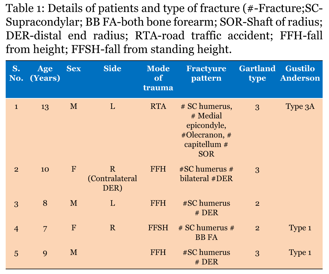
|
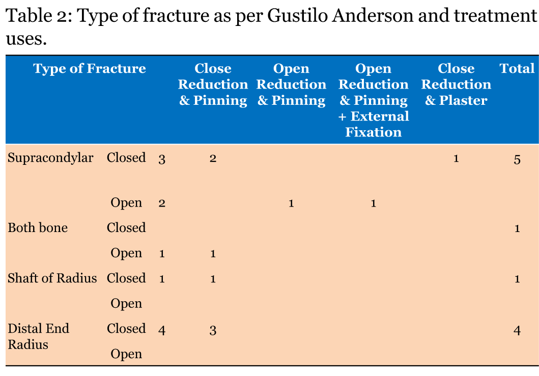
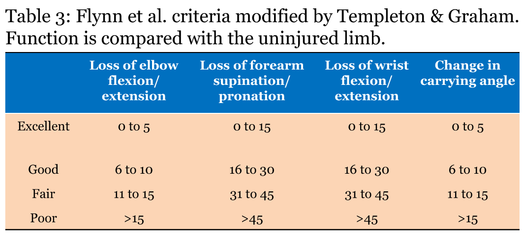
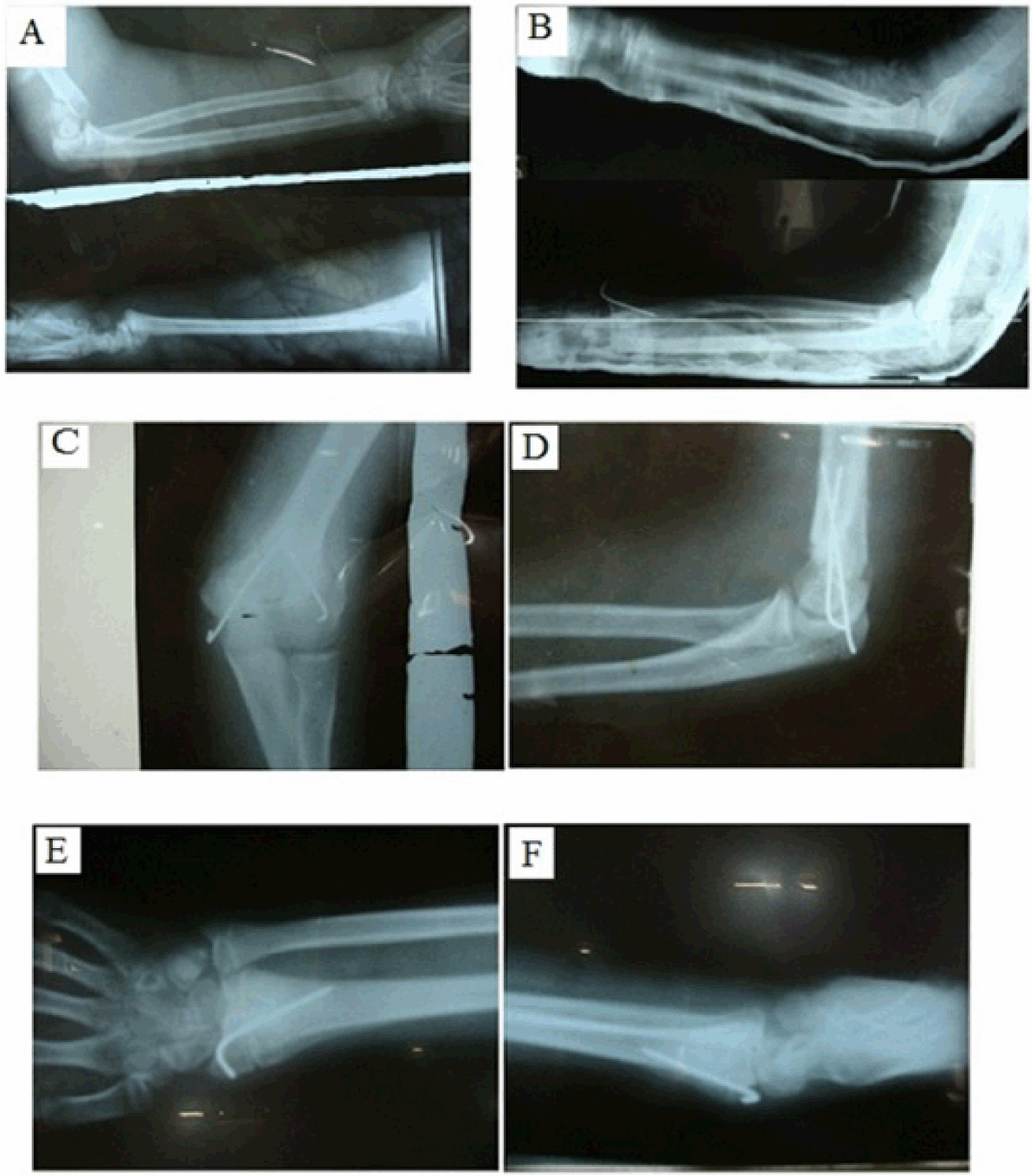
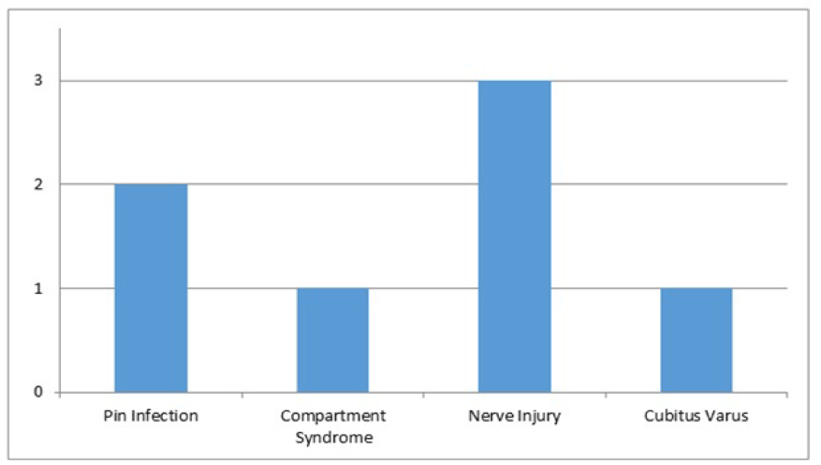
Over a two-year period from September 2010 to September 2012 we conducted prospective analysis of 122 pediatric patients at Department of Orthopedics, Sheri Kashmir Institute of Medical Sciences, Srinagar with different musculo-skeletal injuries and came across five (4.09%) children with ipsilateral supracondylar humerus and forearm fractures. The five children were treated prospectively by closed reduction and percutaneous pinning/external fixation/cast fixation depending upon the fracture anatomy. There were three (60%) boys and two (40%) girls with a mean age of 9.4 years (range 7–13 years). The fractures were right sided in two (40%) and left-sided in three (60%). The cause of injury was a fall from a height in three (60%) cases, road traffic accident in one (20%) and fall from standing height in one (20%). Details of the patients are given in Table 1. There were three Gartland type-III and 2 Gartland type-II supracondylar fractures of the humerus. In the type-III injuries the displacement was posteromedial in four cases. There were four fractures of distal end radius in three patients (with one patient having bilateral injury), both bones of the forearm in one and of the radius alone in one patient. One patient with RTA with compound type 3B elbow injury had, in addition ipsilateral fractures of medial epicondyle, capitellum and olecranon. One patient had ipsilateral fractures of olecranon. Fasciotomy was performed in one patient with ipsilateral both bone forearm fracture who presented with compartment syndrome the type of fracture and treatment used is given in Table 2. One supracondylar fracture was classified as Gustilo Anderson compound type 3B, one supracondylar and one both bone fractures were classified as Gustilo type-1 open fractures. [18] All four distal end radius fractures were Salter Harris type 2 injuries. There were two median nerve injuries. Preoperatively, the radial pulse was absent in two patients. In these patients, the pulses returned after reduction. The forearm both bone (n=1) and proximal radial shaft fractures (n=1) were reduced and stabilized with K-wires. Three distal radial epiphyseal injuries were fixed with K-wires while one fracture was stabilized in plaster cast. The supracondylar fractures were then reduced by longitudinal traction and manipulation and fixed with two crossed K-wires; one lateral and one medial entry pins (in four (80%) patients). One patient with compound elbow injury needed external fixator in addition to cross K-wire fixation. One patient (20%) was managed with plaster fixation. The lateral pin is inserted first and must traverse the lateral portion of ossified capitellum, cross the physis, proceed up the lateral column and engage the opposite medial cortex. The medial pin is placed through the medial epicondyle and should traverse the medial column and engage the opposite lateral cortex. Care must be taken to protect the ulnar nerve. The elbow is placed short of 90 degree flexion. The nerve may be palpated if possible or a small 1.5 cm incision made over the medial epicondyle. Continuous fluoroscopy was used during reduction of the supracondylar fracture and pinning of both bone fracture. The pins were bent outside the skin to prevent migration and to make later removal easier and an above elbow slab was applied. One patient developed iatrogenic ulnar nerve palsy in immediate post-operative period which eventually recovered at three months follow-up. The two median nerve palsies also resolved with time. Postoperatively, an above-elbow long arm posterior plaster slab was retained for three weeks. The wires were removed from the elbow at 3–4 weeks and active exercises started. The wires were removed from the forearm six weeks after operation. Patients were recalled for a detailed clinical and radiographic evaluation at between 12 and 24 months from injury and were assessed for pain, stiffness and cosmesis. The range of elbow flexion-extension, forearm rotation, wrist flexion-extension and the carrying angles were measured by goniometers in both the injured and non-injured limbs. Radiographs were obtained of elbows, forearms and wrists. We performed a detailed neurovascular examination and compared the findings with those recorded on admission and after operation. The final outcome was graded according to a combination of Flynn’s criteria for isolated SC fractures and a grading of forearm rotation and wrist. [16] The mean follow-up time was 24 months (range 20–27). We found an incidence of combined fractures of 4.09%, which is similar to the incidence of 3–13% reported in other studies. The left upper extremity was more commonly affected than the right (3:2) and there was higher incidence of open fractures and nerve injuries; which is well documented in literature. Patients who suffered road traffic accidents had multiple fractures, some of which are open injuries. The patients were followed-up at second week, sixth week and third month by the operating surgeon. The final follow-up was at 24th months. Patients were examined for active/passive movements of the elbow and wrist (flexion-extension), measuring loss of motion, and the carrying angle loss (cosmetic factor). Following Flynn criteria, (Table 3) there were excellent results in three (60%) cases, good result in one (20%) case and poor results in one (20%) case. [16] One patient with open elbow injury and managed with external fixation developed stiffness of elbow and had less than satisfactory results. There were no cases of failure of fixation and no delayed union or nonunion. We had one case with cubitus varus in a patient managed with closed reduction and plaster. There were two superficial pin-track infections, one in supracondylar and one in forearm region which healed after removal of the wires. The nerve injuries were all temporary and recovered by final follow-up. There were one iatrogenic ulnar nerve injuries that resolved with pin removal. | ||||||
|
| ||||||
| ||||||
| ||||||
| ||||||
|
| ||||||
| ||||||
|
Discussion
| ||||||
|
The combination of a supracondylar fracture of the humerus and an ipsilateral fracture of the forearm is rare but a severe injury in the growing child. The reported incidence varies between 3–13%. [15] [16] The upper segment injury may include supracondylar, intercondylar, lateral condyle, medial epicondyle fractures. The lower segment fractures may be also at different locations: olecranon, radial neck, Monteggia lesions, and various levels of the bone shaft and distal forearm. [19] [20] [11] Palmer et al. in their analysis of 78 supracondylar fractures found four ipsilateral fractures of the radius and ulna, two ipsilateral fractures of the radius alone and one ipsilateral midshaft ulna fracture. [21] Stanitski coined the term floating elbow for such injury, considered a high energy fracture. [7] [20] The force is so much that single fracture could not dissipate all the energy of trauma. [11] In the true sense floating elbow should include fracture supracondylar humerus with fracture of both bones forearm wherein the elbow is effectively dissociated from rest of the upper limb. However, various reports have included association of single bone fracture also in floating elbow. [4] [7][17] The incidence of compartment syndrome is higher in these injuries [11] [22] so, the management is sometimes challenging. Blakemore et al. reported an incidence of compartment syndrome of 33% in ipsilateral displaced supracondylar humeral and forearm fractures. [6] In our patients all supracondylar fractures were of the extension type. The mechanism of this injury is usually a fall on the outstretched hand and arm with the wrist dorsiflexed and the elbow extended. The direction of the supracondylar fracture was posteromedial in 80% of our patients which is comparable to an incidence of 75% to 90% in isolated supracondylar fractures. [23] [24] In general, the supracondylar component shows complete (Type III) displacement and the forearm fracture is generally seen in the distal one third. This combination of fractures is usually caused by a fall from a height, three (60%) in our series and emphasizes the fact that this association of fractures occurs as a result of high-energy trauma in most cases. Fall from standing height and road traffic accidents accounted for one patient (20%) each. The incidence of associated nerve injuries (40%) and of open fractures (40%) was higher than expected because of the severity of the injury. [23] [25] Tabak AY recommended conservative treatment of the nerve injuries as spontaneous resolution may be expected. [26] While majority of isolated displaced supracondylar fractures of the humerus are managed with closed reduction and percutaneous fixation, the treatment of a supracondylar fracture in the presence of an ipsilateral forearm fracture is still controversial. [7] [12][14] [15] [25] Though good results have been reported after conservative treatment [15] [16] many authors consider pinning of the supracondylar fracture the best choice. [5] [13] [20] Others recommend percutaneous pinning of both proximal and distal fractures. [11] In 1960s, closed reduction and casting was recommended but an increased frequency of cubitus varus, of up to 25% of cases was observed. [5] [14] Reed in an early series (15 cases) treated all of his patients by conservative methods. [5] [19] Fowles (n=175) reported six cases of this injury, all of them were managed by pinning of the supracondylar fracture and closed reduction and cast immobilization of the forearm fracture. [27] Williamson and Cole managed the supracondylar fracture by traction or manipulative reduction and percutaneous pinning and the forearm fractures were managed by reduction and casting. [15] Reed et al. in a series of 15 patients treated conservatively reported good functional outcome. [14] Stanitski recommends early closed reduction and pinning of superior fracture and closed reduction of inferior fracture and casting. [7] Biyani A et al. reported primarily posterior slab for supracondylar facture and short arm cast for forearm fracture and olecranon pin traction or K- wire fixation for supracondylar fracture only when closed reduction failed [13]. Similarly, Reed et al. also used plaster slab for both injuries after fracture reduction. [14] Both of these studies reported cubitus varus deformity with incidence of 20% but did not find any compartment syndrome. Roposch reported three of his 18 patients with forearm fractures displaced in cast while none of the 29 cases pinned displaced. [5] Harrington et al. stated that they achieved good results from similar treatment for four of their 12 patients with floating elbow. [4] In a series of 21 cases of ipsilateral supracondylar and forearm fractures, Pierce and Hodorski in 1976 concluded that nerve injuries are a predictive factor for poor. [28] We agree with Shaw and Kasser who advocated stabilization of the elbow without exploration unless capillary refill is compromised. [29] Priority of reduction of stabilization of supracondylar fracture or forearm injury first varies among authors though no definitive study has been done in terms of outcome and complications. Templeton and Graham recommended reduction and stabilization of the supracondylar fracture first because they suggested that maintenance of reduction and access to the limb for neurovascular monitoring, dressings and the closure of open fractures may be difficult if the forearm fracture is treated first. [17] The forearm fractures were fixed first in a series by Tabak followed by closed reduction and percutaneous fixation of the supracondylar fracture. [26] This protocol was followed in the Suresh’s series. We too reduced and fixed the forearm fractures first because leaving the forearm dangling during reduction of supracondylar fracture can cause more soft tissue injury of forearm and can increase chance of compartment syndrome. [30] In addition, we found subsequent reduction of supracondylar fractures easier. If the forearm fracture is not reduced and fixed first, it will remain mobile during flexion of the elbow and rotational maneuvers. We managed 1 (20%) supracondylar fracture conservatively who, however, developed cubitus varus. The remaining 4 (80%) were managed with pinning. The distal end radius fractures were managed conservatively with closed reduction and casting (n =1) and percutaneous pinning (n=3). We stabilized the forearm and radial shaft fractures with pinning and did not see any loss of reduction. The rate of remanipulation of the forearm fractures after closed reduction and immobilization in a cast is reported to be between 7% and 15%. [31] [32] Various techniques of K- wire fixation such as crossed K-wires, lateral two K-wires have been described in literature for better biomechanical stability and to decrease chance of potential iatrogenic nerve injury. [26] There was one case of ulnar nerve injury associated with introduction of the medial wire, the nerve function returned after removal of the K-wires. Although fixation with crossed wires is more stable, there is greater risk of injuring the ulnar nerve if the medial wire is passed through the bone blindly. [33] We had no complications such as loss of reduction or neurovascular injury in forearm fractures after K-wire fixation. We therefore recommend stabilization of displaced both bone forearm fractures which are associated with supracondylar fractures. We achieved good results with closed reduction and application of plaster for distal radial epiphyseal injury. The overall results were excellent or good in four (80%) patients but poor in one (20%) patients. | ||||||
|
Conclusion
| ||||||
|
Ipsilateral supracondylar and forearm fractures area result of high energy trauma. The injury is uncommon and treatment recommendations are controversial. However, these injuries need prompt management and close observation for early signs and symptoms of development of compartment syndrome. In this series the good results obtained after surgical treatment allow us to state that floating elbow injuries are best managed aggressively with surgical stabilization. However, given the scarce sources in a third world setting as ours, we suggest conservative management in selected cases only. | ||||||
|
Acknowledgements
| ||||||
|
I must thank my tender child patients for allowing me to spend time with them to compile this paper. I must also thank Dr Shabir Dhar, consultant orthopedician, who is a teacher and a senior colleague and helped me in compiling this paper in its final stages. | ||||||
|
| ||||||
|
References
| ||||||
| ||||||

|
[HTML Abstract]
[PDF Full Text]
|
|
Author Contributions:
Shahid Hussain – Substantial contributions to conception and design, Acquisition of data, Analysis and interpretation of data, Drafting the article, Revising it critically for important intellectual content, Final approval of the version to be published |
|
Guarantor of submission
The corresponding author is the guarantor of submission. |
|
Source of support
None |
|
Conflict of interest
Authors declare no conflict of interest. |
|
Copyright
© Shahid Hussain et al. 2014; This article is distributed the terms of Creative Commons Attribution License which permits unrestricted use, distribution and reproduction in any means provided the original authors and original publisher are properly credited. (Please see Copyright Policy for more information.) |
|
|