  |

|
 |
|
Case Report
| ||||||
| Diagnosis of a case of papillary-cystic variant of acinic-cell carcinoma on fine needle aspiration cytology: Myriad of cytomorphological features | ||||||
| Suma Mysore Narayan1, Jeyachandran Padmini2, Raja Parthiban3, Jena Madhusmita3, Gandhi Natarajan2, Parappa Sangappa Revadi1 | ||||||
|
1MBBS, MD, DNB, Assistant Professor, Department of Pathology, MVJ MC & RH, Bangalore, Karnataka, India.
2MBBS, MD, Professor, Department of Pathology, MVJ MC & RH, Bangalore, Karnataka, India. 3MBBS, MD, Associate Professor, Department of Pathology, MVJ MC & RH, Bangalore, Karnataka, India. | ||||||
| ||||||
|
[HTML Abstract]
[PDF Full Text]
[Print This Article]
[Similar article in Pumed] [Similar article in Google Scholar] 
|


| How to cite this article |
| Narayan SM, Padmini J, Parthiban R, Madhusmita J, Natarajan G, Revadi PS. Diagnosis of a case of papillary-cystic variant of acinic-cell carcinoma on fine needle aspiration cytology: Myriad of cytomorphological features. International Journal of Case Reports and Images 2014;5(1):18–22. |
|
Abstract
|
|
Introduction:
The diagnosis of papillary-cystic variant of acinic-cell carcinoma (ACC-PCV) is easy on histology. However, the diagnosis on cytology usually poses a problem because of the cytoarchitecture which is different from classic type.
Case Report: A 39-year-old male presented with left parotid swelling for last four months. Fine needle aspiration cytological smears revealed papillary fragments and tumor cells with varied morphological features. Cell block prepared from the fluid aspirates confirmed the diagnosis of papillary-cystic variant of acinic- cell carcinoma. Histological study of the excised specimen confirmed the diagnosis of acinic cell carcinoma, papillary cystic variant. Conclusion: This case describes papillary-cystic variant of acinic-cell carcinoma and discusses the myriad of cytological features exhibited by it. | |
|
Keywords:
Salivary gland neoplasm, Papillary-cystic variant of acinic-cell carcinoma (ACC-PCV), Fine needle aspiration cytology (FNAC)
| |
|
Introduction
| ||||||
|
Acinic cell carcinoma is an uncommon low grade tumor of the salivary glands that constitutes 2.5–4% of parotid gland tumors. Papillary-cystic variant of acinic-cell carcinoma (ACC-PCV) is histologically composed of tumor with papillary and cystic growth patterns, with varying proportions of one or more cell types. An ACC-PCV has been mostly reported to occur in younger patients (16–40 years), [1] [2] [3] [4] as compared to the classic type that characteristically presents in the fifth decade of life. The diagnosis of ACC-PCV is easy on histology. However, the diagnosis on cytology usually poses a problem because of the cytoarchitecture which is different from classic type as it shows prominent papillary architecture with more cohesive cells and change in morphology of tumor cells due to suspension in cystic fluid. This report describes the detailed cytological features on fine needle aspiration cytology (FNAC) and the use of cell block preparation from the cyst fluid aiding in the diagnosis of this special variant. | ||||||
|
Case Report
| ||||||
|
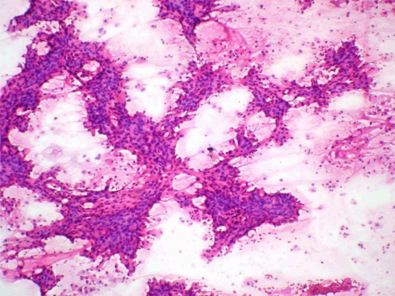
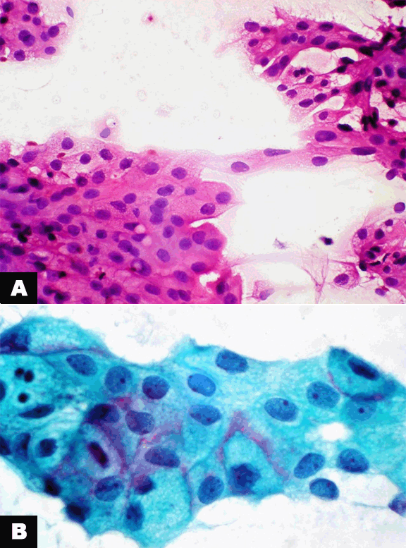
A 39-year-old male presented to the surgical outpatient department for a firm, painless, freely movable, swelling measuring 5x6 cm, extending from the lower left ear lobule to the angle of the mandible and enlarging for the last four months. The skin over the swelling was stretched and was not pinchable. Fluctuation and transillumination were negative. Ultrasonography revealed a cystic lesion in the left parotid gland probably of inflammatory etiology. Further contrast–enhanced computed tomography (CECT) scan was done and was reported as a complex thick walled, peripherally enhancing cystic mass lesion involving the left parotid region suggestive of neoplastic etiology. Fine-needle aspiration cytology was done and 10 mL of turbid brownish fluid was aspirated. After evacuating the cyst, FNAC was repeated on the nodular areas felt from the collapsed site of the cyst. Smears were stained with Giemsa, hematoxylin-eosin (H&E), and Papanicolaou. The cystic fluid was centrifuged and the pellet was made into cell block. Cytological examination revealed moderate cellularity with cells in cohesive clusters forming papillary pattern. (Figure 1) Cells had well defined cell borders and showed varied features from finely vacuolated to oncocytic cytoplasm. (Figure 2A) Densely granulated and ductal types of cells were seen. The nuclei in all these myriad of cells revealed bland morphology with a single nucleolus. (Figure 2B) No frequent mitosis or pleomorphism was seen. Background showed cystic degenerated material with a few macrophages engulfing hemosiderin and cholesterol crystals. Occasional individual neoplastic cells were seen. Cell block stained with H&E revealed microcystic and papillary cystic pattern of acinic-cell carcinoma. (Figure 3A) A diagnosis of ACC-PCV was given on FNAC and advised excision. Under general anaesthesia, superficial parotidectomy was done. The facial nerve trunk was identified and traced anteriorly. Superficial parotidectomy with the mass was excised and sent to histopathology. The gross specimen measured 6x5x4 cm. It was well circumscribed with adjacent normal parotid salivary gland. The tumor showed cystic degeneration with yellowish nodular areas embedded in part of the cyst wall. Histopathological examination revealed predominantly papillary cystic pattern along with solid and micro cystic pattern. Acinic, oncocytic, granular and vascular cells were seen. No atypical mitoses or necrosis was seen. Nuclei were placed eccentrically with normal nuclear-cytoplasmic ratio (N:C ratio) and bland nuclear chromatin with single nucleolus. Lymphoid stroma was seen surrounding the tumor. There was no vascular, extracapsular or perineural invasion seen. A diagnosis of papillary cystic variant of ACC was confirmed on histology. (Figure 3B) | ||||||
| ||||||
|
| ||||||
|
| ||||||
|
Discussion
| ||||||
|
A variety of appearances of ACC of salivary gland is described. [5] The architectural patterns which may be seen in ACC are: solid, solid-lobular, acinar-microcystic, papillary cystic, tubuloductal, follicular and macrocystic, and dedifferentiated. The solid and microcystic are the most common subtypes and the papillary cystic variant accounts for one-fourth of ACC and because of these variants, ACC-PCV is a diagnostic challenge in FNAC. [6] Fine needle aspiration cytological features of classic ACC are well known. [6] [7] Smears show loosely clustered and acinar groups of cells that are generally bland looking, resembling non-neoplastic salivary acinar cells. [1] Cell dissociation and presence of some bare tumor cell nuclei are some of the features that distinguish well differentiated ACC from normal salivary gland acini. [7] In ACC-PCV, cell dissociation is sparse and acinar structures and bare tumor cell nuclei are missing. On the other hand, numerous small and large papillary groups and monolayered sheets of moderately pleomorphic tumor cells have been described. [1] [2] [8] The papillary cystic variant is believed to have two forms of genesis: one a retrogressive phenomenon and the other solely neoplastic. The former is a histopathologic reflection of a self destructive quality of some acinic cell carcinomas, the end stage of which is a solitary loculated cyst with attenuated and hydropically altered neoplastic cells in company with neoplastic papillary excrescences in various stages of degeneration. [6] The mean age of occurrence is in the fifth decade, but the papillary cystic variant is reported to occur in younger patients compared to the classic type. Females are involved more commonly unlike in our case who was a male patient. The reasons for difficulty in cytodiagnosis is firstly cystic fluid dilutes the overall cellularity leading to a benign diagnosis. Hence, it is a fair proposition to subject the fluid for cell block preparation. Secondly, cytoarchitecture is different from classic type with more cohesive cells and papillary fragments. Thirdly, change in morphology of tumor cells due to suspension in cystic fluid and presence of metaplastic oncocytic cells. [6] Although a single pattern usually dominates, a mixture of histologic patterns may be observed. Papillary-cystic variant typically involves transition of usual dense cellularity into papillary folds interspersed with cystic spaces. It may vary from small cysts with scanty papillary projections to large cystic spaces into which extend delicate papillary growths supported by thin, vascular stalks. [9] | ||||||
|
Acknowledgements
| ||||||
|
Amrit Kaur Kaler, MBBS, MD Asst Professor of Pathology Department of Pathology, MVJ MC & RH | ||||||
|
Conclusion
| ||||||
|
Papillary-cystic variant of acinic-cell carcinoma is a rare variant of salivary gland neoplasms. Fine needle aspiration cytology remains the primary modality for the diagnosis of salivary gland swellings. This case highlights the importance of detailed examination of cytological features on fine needle aspiration cytology and using ancillary technique like cell block preparation aiding in preoperative diagnosis of papillary cystic variant of acinic cell carcinoma. | ||||||
|
References
| ||||||
| ||||||


|
[HTML Abstract]
[PDF Full Text]
|
|
Author Contributions
Suma Mysore Narayan – Substantial contributions to conception and design, Acquisition of data, Analysis and interpretation of data, Drafting the article, Revising it critically for important intellectual content, Final approval of the version to be published Jeyachandran Padmini – Substantial contributions to conception and design, Analysis and interpretation of data, Drafting the article, Revising it critically for important intellectual content, Final approval of the version to be published Raja Parthiban – Substantial contributions to conception and design, Analysis and interpretation of data, Drafting the article, Revising it critically for important intellectual content, Final approval of the version to be published Jena Madhusmita – ubstantial contributions to conception and design, Analysis and interpretation of data, Drafting the article, Revising it critically for important intellectual content, Final approval of the version to be published Gandhi Natarajan – Substantial contributions to conception and design, Analysis and interpretation of data, Drafting the article, Revising it critically for important intellectual content, Final approval of the version to be published Parappa Sangappa Revadi – Substantial contributions to conception and design, Analysis and interpretation of data, Drafting the article, Revising it critically for important intellectual content, Final approval of the version to be published |
|
Guarantor of submission
The corresponding author is the guarantor of submission. |
|
Source of support
None |
|
Conflict of interest
Authors declare no conflict of interest. |
|
Copyright
© Suma Mysore Narayan et al. 2014; This article is distributed the terms of Creative Commons Attribution License which permits unrestricted use, distribution and reproduction in any means provided the original authors and original publisher are properly credited. (Please see Copyright Policy for more information.) |
|
|
|
About The Authors
| |||
| |||
| |||
| |||
| |||
| |||
| |||


