  |

|
 |
|
Case Report
| ||||||
| Reversible cerebral vasoconstriction syndrome in HELLP syndrome | ||||||
| Shinji Katsuragi1, Masato Osaki2, Rieko Suzuki3, Tomoaki Ikeda4, Kazunori Toyoda5, Jun Yoshimatsu6 | ||||||
|
1MD, Chirman, Obstetric Cardiology, Sakakibara Heart Institute, Fuchu, Tokyo, Japan.
2MD, Resident, Cerebrovascular Medicine, National Cerebral and Cardiovascular Center, Suita, Osaka, Japan. 3MD, Staff, Cerebrovascular Medicine, National Cerebral and Cardiovascular Center, Suita, Osaka, Japan. 4MD, Professor, Obstetrics and Gynecology, Mie University, Tsu, Mie, Japan. 5MD, Chirman Cerebrovascular Medicine, National Cereral and Cardiovascular Center, Suita, Osaka, Japan. 6MD, Chairman, Perinatology and Gynecology, National Cerebral and Cardiovascular Center, Suita, Osaka, Japan. | ||||||
| ||||||
|
[HTML Abstract]
[PDF Full Text]
[Print This Article]
[Similar article in Pumed] [Similar article in Google Scholar] 
|


| How to cite this article |
| Katsuragi S, Osaki M, Suzuki R, Ikeda T, Toyoda K, Yoshimatsu J. Reversible cerebral vasoconstriction syndrome in HELLP syndrome. International Journal of Case Reports and Images 2013;4(12):735–738. |
|
Abstract
|
|
Introduction:
Reversible cerebral vaso-constriction syndrome (RCVS) is characterized by severe and hyperacute headaches with or without additional focal neurological deficits, and evidence of vasoconstriction of cerebral arteries which resolves spontaneously within 1–3 months. However, the precise mechanism of RCVS is not fully understood.
Case Report: We report a 33-year-old postpartum woman with RCVS with HELLP syndrome and eclampsia. Magnetic resonance angiography (MRA) of head revealed transient segmental stenosis and dilatation. Especially, most part of the middle cerebral artery (MCA) was irregularly thinner at the acute phase than have ever previously reported. Transcranial color flow imaging (TCCFI) showed the highest peak systolic velocity of middle cerebral artery that has ever reported in RCVS cases (330 cm/s). Histologically, the large areas that showed cytotoxic edema on magnetic resonance imaging at acute phase, which indicated possible infarction at the chronic stage, have eventually become normal in chronic stage. Conclusion: This RCVS case occurred to a postpartum woman in HELLP syndrome after eclampsia. The peak velocity of MCA measured by TCCFI was the highest so far ever reported in RCVS cases, and vasoconstriction on MRA was severe. However, the severe vasoconstriction did not last long enough to cause infarction in brain tissue in this case. TCCFI also shows the steep decline of the peak velocity of MCA after acute phase and through this case we reconfirm the usefulness of TCCFI as this bed-side test reflects quite well the changes of vasoconstriction in severe RCVS case. | |
|
Keywords:
RCVS (Reversible Cerebral Vasoconstriction Syndrome), Pregnancy, TCCFI (Transcranial color flow imaging), peak systolic velocity
| |
|
Introduction
|
|
Reversible cerebral vasoconstriction syndrome (RCVS) is characterized by severe headaches, often thunderclap headaches, with or without focal deficits and seizures, and a multifocal constriction of cerebral arteries, which resolve spontaneously within three months. This syndrome has a female preponderance and a mean age of onset of approximately 42 years. [1] [2] The pathophysiology of RCVS is not fully understood. Here, we report a case of RCVS in a patient with HELLP syndrome who developed an eclamptic attack and had caudate hemorrhage. The time-course of reversible vasoconstriction was documented by magnetic resonance angiography (MRA), and transcranial color flow imaging (TCCFI). |
|
Case Report
|
|
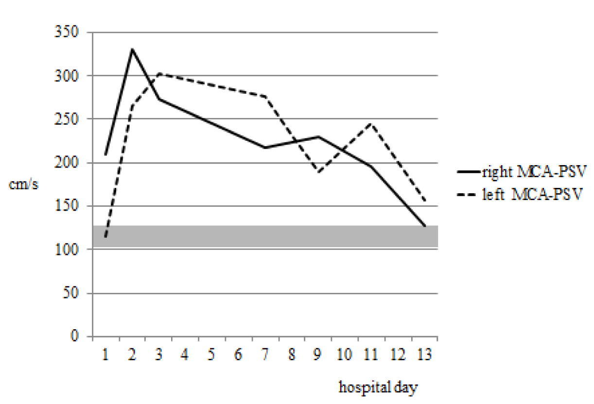
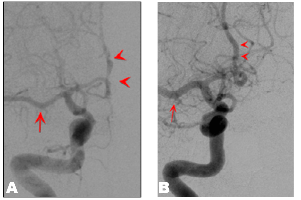
A 33-year-old primipara women was referred to our hospital because of severe headache on third day postpartum following vaginal delivery at 37th week. Her delivery course at a previous hospital was eventful because she developed an eclamptic attack just before delivery and was diagnosed with HELLP syndrome based on findings of hemolysis, elevation of liver enzymes (AST/ALT: 1455/663 U/L), and a low platelet count (40×103/µL). Her blood pressure on tonic-clonic seizure was 192/108 mmHg. On admission to our hospital, the patient was alert and conscious, but had severe headache over the temples with a positive Kernig's sign. Computed tomography of head showed hemorrhage in the left caudate nucleus and magnetic resonance imaging (MRI) of the corpus callosum on both sides showed fluid attenuated inversion recovery (FLAIR) and diffusion-weighted image (DWI) hyperintensity, and apparent diffusion coefficient (ADC) map hypointensity. The anterior cerebral arteries (ACA) and middle cerebral arteries (MCA) were barely detectable by brain MRA. (Figure 1A) Therapy for brain edema and continuous intravenous infusion of nicardipine were performed as conservative management. On hospital day 2, TCCFI indicated MCA peak systolic velocities (PSV) of 330 (right side) and 266 (left side) cm/s. (Figure 2) On hospital day 3, nicardipine was discontinued and replaced by oral administration of a calcium blocking agent, which maintained blood pressure at approximately 130/80 mmHg. Headache was also relieved from hospital day 7. TCCFI on hospital day 13 showed that the PSV of both MCAs had improved to approximately 150 cm/s. MRA on hospital day 14 and at 43rd day postpartum revealed progressive improvement of the lesions in the ACAs and MCAs. (Figure 1B-C) On hospital day 17, cerebral angiography showed improvement of segmental changes of the ACA and a larger diameter of the MCA compared to that in previous tests. (Figure 3) MRI scan of head at 43rd day postpartum showed decreased caudate hemorrhage and disappearance of the lesions in the corpus callosum. |
|
|
|
|
|
|
|
Discussion
|
|
The pathophysiology of eclampsia is thought to involve vasoconstriction. (Figure 3) The patient in this case developed juvenile cerebral hemorrhage during puerperium, and the observation of multiple reversible stenoses and the absence of findings suggesting vasculitis indicated the presence of reversible cerebral vasoconstriction syndrome (RCVS). Calabrese et al. suggested that the important factors for diagnosis of RCVS are as follows: (Figure 1)
Reversible cytotoxic edema and cerebral hemorrhage were observed in our patient. In MRI, the corpus callosum showed FLAIR and DWI, and hypointensity on an ADC map in DWI, which indicated development of cytotoxic edema that was probably due to RCVS, since segmental stenosis and dilatation were found in the ACA, which supplies the corpus callosum. Of further interest, the lesions in the corpus callosum eventually almost disappeared, although they were relatively widely spread, which indicated possible infarction at the chronic stage. This unusual phenomenon may be explained as follows. A sudden increase of blood pressure in eclamptic seizure during delivery may have increased the cerebral blood flow volume (Figure 3) and then administration of ergometrine and packed red blood cell transfusion triggered vasoconstriction. (Figure 1) (Figure 2) Under these circumstances, blood perfusion to the ACA and MCA areas may have suddenly increased and then decreased. This vasoconstriction occurred suddenly and intensively enough to cause cytotoxic edema, but did not persist for long enough to result in irreversible tissue damage. The measurement of MCA blood flow velocity in patients with RCVS can be achieved with TCCFI, which is a combination of transcranial color Doppler imaging and transcranial Doppler sonography. (Figure 5) (Figure 6) Recent developments in diagnostic equipment have led to significant upgrades of TCCFI, which is now widely used in clinical settings. (Figure 7) (Figure 8) (Figure 9) The mean blood flow velocity of the right and left MCAs in our patient were quite rapid, at 275 and 249 cm/s, respectively, and much higher than that found in a prospective study in 32 RCVS patients in Taiwan, (Figure 10) in which no subjects had a blood flow velocity >200 cm/s. This is well supported by the severely constricted MCA on MRA at admission. Improvement of the MCA-PSV was coincident with the improvement of blood flow in the MCA observed by angiography and MRA. This indicates that TCCFI can be used to monitor the therapeutic effect of drugs used for treatment of RCVS. This case shows that the cerebral circulation changes dramatically after an eclamptic seizure attack. It is of particular interest in this case that cytotoxic edema disappeared with time. Posterior reversible encephalopathy syndrome (PRES) is another possible cause of eclampsia (Figure 3) that is mainly confined to the occipital lobe, but does not, necessarily, involve a severe headache such as that in RCVS. Based on our observations in this case, we hypothesize that certain regions of cytotoxic edema related to eclampsia may also be reversible encephalopathy, similarly to PRES, even though the etiology of PRES is vasogenic edema and completely differs from RCVS. This hypothesis requires testing by physical examinations, blood pressure measurements, and longitudinal investigations using MRI, MRA, angiography and TCCFI in future cases. The results of these tests will provide further important clues to understanding of the mechanism of eclampsia. |
|
Conclusion
|
|
The peak velocity of middle cerebral artery by transcranial color flow imaging was the highest in reversible cerebral vasoconstriction syndrome cases that have ever reported, and large areas of cytotoxic edema disappeared in the chronic stage. These facts and magnetic resonance angiography/angiography reflects the vasoconstriction was most severe but rapidly normalized which was clearly shown by transcranial color flow imaging. These results further support the idea that transcranial color flow imaging demonstrates the rapid change of vasoconstriction even in the most acute type of reversible cerebral vasoconstriction syndrome. |
|
References
|
|


|
[HTML Abstract]
[PDF Full Text]
|
|
Author Contributions
Shinji Katsuragi – Substantial contributions to conception and design, Acquisition of data, Analysis and interpretation of data, Drafting the article, Revising it critically for important intellectual content, Final approval of the version to be published Masato Osaki – Analysis and interpretation of data, Drafting the article, Revising it critically for important intellectual content, Final approval of the version to be published Rieko Suzuki – Analysis and interpretation of data, Drafting the article, Revising it critically for important intellectual content, Final approval of the version to be published Tomoaki Ikeda – Analysis and interpretation of data, Drafting the article, Revising it critically for important intellectual content, Final approval of the version to be published Kazunori Toyoda – Analysis and interpretation of data, Drafting the article, Revising it critically for important intellectual content, Final approval of the version to be published Jun Yoshimatsu – Analysis and interpretation of data, Drafting the article, Revising it critically for important intellectual content, Final approval of the version to be published |
|
Guarantor of submission
The corresponding author is the guarantor of submission. |
|
Source of support
None |
|
Conflict of interest
Authors declare no conflict of interest. |
|
Copyright
© Shinji Katsuragi et al. 2013; This article is distributed the terms of Creative Commons attribution 3.0 License which permits unrestricted use, distribution and reproduction in any means provided the original authors and original publisher are properly credited. (Please see Copyright Policy for more information.) |
|
|