| |
|
|
|
Case Report
| ||||||
| Chest X-Ray appearance of total opacification of the hemithorax following central venous line insertion: A cautionary tale | ||||||
| Victor Kong1, Leah Naidoo2, Damon Jeetoo3, George Oosthuizen4, Grant Laing5, Damian Clarke5 | ||||||
|
1MBChB, Surgical Registrar, Pietermaritzburg Metropolitan Trauma Service, Department of Surgery, Edendale Hospital, Pietermaritzburg, KwaZulu Natal, South Africa.
2MBChB, Surgical House Officer, Pietermaritzburg Metropolitan Trauma Service, Department of Surgery, Edendale Hospital, Pietermaritzburg, KwaZulu Natal, South Africa. 3MBChB, Radiology Registrar, Department of Radiology, Edendale Hospital, Pietermaritzburg, KwaZulu Natal, South Africa. 4FCS(SA), Senior Consultant Trauma Surgeon, Pietermaritzburg Metropolitan Trauma Service, Department of Surgery, Edendale Hospital, Pietermaritzburg, KwaZulu Natal, South Africa. 5FCS(SA), Consultant Trauma Surgeon, Pietermaritzburg Metropolitan Trauma Service, Department of Surgery, Edendale Hospital, Pietermaritzburg, KwaZulu Natal, South Africa | ||||||
| ||||||
|
[HTML Abstract]
[PDF Full Text]
[Print This Article]
[Similar article in Pumed] [Similar article in Google Scholar] 
|
| How to cite this article: |
| Kong V, Naidoo L, Jeetoo D, Oosthuizen G, Laing G, Clarke D. Chest X-Ray appearance of total opacification of the hemithorax following central venous line insertion: A cautionary tale. International Journal of Case Reports and Images 2013;4(12):686–690. |
|
Abstract
|
|
Introduction:
Central venous line insertion is associated with significant complications. Procedural complications relating to lung injury is relatively common, and these may require tube thoracostomy for management. Although a post-procedure chest X-ray is routinely undertaken in many centers, erroneous interpretation can lead to potentially incorrect and unnecessary further intervention.
Case Report: We report a case of a 25-year-old male who had a central venous line inserted, with the Chest X-ray appearance of a massive opacification of the hemithorax misdiagnosed as a hemothorax and planned tube thoracostomy. It was subsequently confirmed as a complete pulmonary collapse caused by a large mucus plug with obstruction of the main bronchus. This completely resolved after an awake flexible bronchoscopic clearance, without the need for tube thoracostomy. Conclusion: Whilst Chest X-ray allows identification of most mechanical complications of central venous line insertion, accurate interpretation and correct clinical correlation are absolutely critical in order to avoid unnecessary interventions. | |
|
Keywords:
Central venous line, Hemothorax
| |
|
Introduction
| ||||||
|
Central venous line insertion is associated with significant complications which are often underappreciated. [1] Mechanical complications, especially pneumothorax, are common, and injuries to major structures (including the major vessels) are reported to be as high as 20%. [2] A chest X-ray post-insertion of a central venous line is commonly performed. [3] Technical complications need to be addressed in a timely fashion, but requires correct interpretation of radiological findings. We report a case of a 25-year-old male who had misinterpreted post insertion chest radiography and a tube thoracostomy was subsequently planned inappropriately by the junior surgical resident. | ||||||
|
Case Report
| ||||||
|
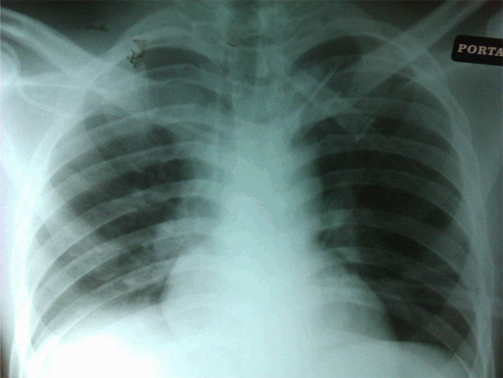
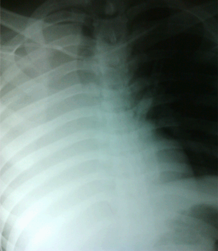
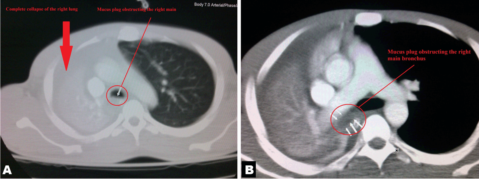
A 25-year-old male was presented who had a laparotomy for a gastric perforation following a stab injury. Due to poor peripheral venous access, a right subclavian central venous line was inserted by the anaesthetic resident intra-operatively. However, several attempts were undertaken due to difficult positioning of the patient. Ultrasound guidance was not utilised because the machine was not available due to maintenance. The central line remained patent and functional, and the patient remained well throughout the operation. The surgery was uneventful and he was discharged to the general ward. A routine post procedural chest X-ray (Figure 1) was done immediately, but this was not reviewed by the surgical resident until 12 hours later. The chest X-ray appeared to show that the central venous line entered the right subclavian vein, and then traversed the brachiocephalic vein before re-entering the left subclavian vein. There was no pneumothorax or hemothorax noted. The patient had no complains and was not in distress. Therefore, it was decided that the central line should be removed as soon as the patient could tolerate orally, likely to be the following day. However, he began complaining of abdominal pain and increasing dull pain of the right chest throughout the night. He had poor respiratory effort, refused chest physiotherapy and was unable to mobilize out of the bed due to ongoing pain. On the following morning, he was noted to be in mild respiratory distress, with a respiratory rate of 24/min, shallow pattern, SpO2: 90% (FiO2: 100% via a Hudson mask with reservoir bag) with marked dullness and reduce air entry on the right chest. His heart rate was 90/min and blood pressure 105/70 mmHg. Laboratory results were: hemoglobin 11.5 g/dL, white blood cell 15x103/µL. A repeat chest X-ray (Figure 2) showed a complete opacification of the right hemithorax. The junior resident who reviewed the chest X-ray felt it was due to a delayed presentation of iatrogenic hemothorax related to the insertion of the central line, and a tube thoracostomy was planned. However, prior to tube thoracostomy, the patient was eventually reviewed by the duty attending trauma surgeon who advised on an emergency computed tomography (CT) scan of the thorax prior to proceeding. The CT scan (Figure 3) revealed a complete collapse of the right lung, with a large mucus plug obstructing the right main bronchus. An emergency awake flexible bronchoscopy was performed (under conscious sedation), and a large amount of mucus was removed using suction. Clinical condition of the patinent improved immediately. A subsequent chest X-ray, six hours after the procedure, showed a complete expansion of the right lung. Following this, his analgesia regime was further escalated, and he was treated with aggressive chest physiotherapy, which was well tolerated. He had an uneventful recovery, did not eventually require a tube thoracostomy, and subsequently discharged on the sixth day. | ||||||
| ||||||
|
| ||||||
|
| ||||||
|
Discussion
| ||||||
|
Central venous line insertion is a common procedure which is frequently performed by hospital doctors, but is associated with significant complications. Overall complication rate varies, but it has been reported to be as high as 20%. [1] Complications are generally divided into mechanical, thromboembolic and infectious. Of the mechanical complications, vascular and lung injuries (hemothorax, pneumothorax) are the most common. [4] The choice of insertion site, however, is often dictated by the operator, usually dependent on the individual level of skills and experience. [4] Malpositioning has been reported in up to 15%, even when inserted by experienced clinicians. [3] Current evidence appears to suggest that the subclavian route is more likely to be associated with malposition in comparison with the internal jugular route. [1] However, there is no evidence to suggest any difference in the incidence of hemothorax, pneumothorax and vessel occlusion between the two different routes. [1] It is also important to stress that although free venous back flow must be present after insertion, it does not absolutely exclude malpositioning or even subsequent migration. [5] Whilst insertion under ultrasound guidance has become increasingly common, the traditional anatomical landmark technique continues to be used frequently, [6] especially in developing countries where the resources and facilities may be limited. Death following significant subclavian artery injury has been reported. [7] The artery is particularly vulnerable due to its close proximity to the subclavian vein. Furthermore, this complication appears to be directly proportional to the number of attempts undertaken. More than three attempts are associated with a complication rate of approximately 50%. [8] Several important issues are highlighted in this unusual case. Firstly, the initial placement position was clearly incorrect, but this was only recognised 12 hours after the insertion. Also, because the line appeared functional, this initially gave a false sense of security. Although some literatures suggest that chest radiography may not be absolutely necessary in all cases, especially when inserted by experienced operators, most clinicians continue to do so, especially when difficulties are encountered or when multiple attempts have been made. [3] In the context of central line insertion, the appearance of pneumothorax or hemothorax on the chest X-ray is usually obvious, but erroneous interpretation (as in this case) almost leads to unnecessary further procedures (such as tube thoracostomy) being carried out. Whilst the appearance of total opacification of the hemithorax could be caused by hemothorax (secondary to vascular injury), another important diagnosis to consider is complete pulmonary collapse, which has a number of causes. In this case, the patient remained hemodynamically stable for more than 24 hours after the original procedure (with hemoglobin of 11.5 g/dL), with predominantly respiratory symptoms, makes the diagnosis unlikely. The importance of accurate interpretation of radiograph and the integration of these findings within the clinical context cannot be over-emphasized. Erroneous interpretation and failure to recognize alternative diagnoses could have potentially led to a disastrous situation. It is important to re-emphasize that one has to remain opened to alternative diagnoses and constantly question oneself if the findings do not add up to the clinical picture. Secondly, tube thoracostomy is usually reserved for the treatment of significant symptomatic hemothorax and pneumothorax. Given the significance of the patient’s symptomatology, it was probably reasonable for the junior resident to consider this as an option. However, tube thoracostomy itself is invasive which is also associated with appreciable morbidity, and even mortality. [9] Therefore, it should only be performed if absolutely necessary. In our case, even if tube thoracostomy had been performed based on the initial suspicion of a hemothorax, it would have been highly unlikely to have resolved the situation appropriately. Finally, postoperative pulmonary complications are common and may occur in up to 20% of all emergency surgical patients. [10] Pulmonary collapse (atelectasis) is one of the most common complications following major abdominal surgery. [10] While numerous factors contribute the development of pulmonary collapse, it is generally agreed that the major precipitating factors are limited chest movement and bronchial obstruction caused by a thick mucus plug. [11] Suboptimal analgesia, poor pulmonary toileting, and immobility were the most likely contributory factors in this case. These were not recognised early and contributed to the progressive deterioration in this patient. This was subsequently recognized prior to embarking on tube thoracostomy an appropriate definitive intervention was performed, albeit somewhat delayed. Earlier recognition and preventative measures would probably have enabled the avoidance of a potentially dangerous situation. | ||||||
|
Conclusion
| ||||||
|
Central venous line insertion is associated with significant morbidity, and is often under-appreciated. Although vascular and lung injuries are relatively common, chest radiographic findings may be easily misinterpreted. Whilst clinicians must always remain vigilant of potential complications, it is equally important to remain open to alternative diagnoses, especially if the overall clinical picture does not seem to fit together. | ||||||
|
References
| ||||||
| ||||||
|
[HTML Abstract]
[PDF Full Text]
|
|
Author Contributions
Victor Kong – Substantial contributions to conception and design, Acquisition of data, Analysis and interpretation of data, Drafting the article, Revising it critically for important intellectual content, Final approval of the version to be published Leah Naidoo – Analysis and interpretation of data, Drafting the article, Revising it critically for important intellectual content, Final approval of the version to be published Damon Jeetoo – Analysis and interpretation of data, Drafting the article, Revising it critically for important intellectual content, Final approval of the version to be published George Oosthuizen – Analysis and interpretation of data, Drafting the article, Revising it critically for important intellectual content, Final approval of the version to be published Grant Laing – Analysis and interpretation of data, Drafting the article, Revising it critically for important intellectual content, Final approval of the version to be published Damian Clarke – Analysis and interpretation of data, Drafting the article, Revising it critically for important intellectual content, Final approval of the version to be published |
|
Guarantor of submission
The corresponding author is the guarantor of submission. |
|
Source of support
None |
|
Conflict of interest
Authors declare no conflict of interest. |
|
Copyright
© Victor Kong et al. 2013; This article is distributed the terms of Creative Commons Attribution License which permits unrestricted use, distribution and reproduction in any means provided the original authors and original publisher are properly credited. (Please see Copyright Policy for more information.) |
|
|