  |

|
 |
|
Case Report
| ||||||
| Solitary ileal bezoar: A rare cause of acute ileal obstruction in a teenager | ||||||
| Sefu Juma Uledi1, Fauzia Ayubu Masumai2 | ||||||
|
1MMed. Surgery, Head of Surgery Department, Mzuzu Central Hospital, Mzuzu, Malawi.
2MB.ChB, Senior Medical Officer, Department of Obstetric and Gynaecology, Mzuzu Central Hospital, Mzuzu, Malawi. | ||||||
| ||||||
|
[HTML Abstract]
[PDF Full Text]
[Print This Article]
[Similar article in Pumed] [Similar article in Google Scholar] 
|


| How to cite this article: |
| Uledi SJ, Masumai FA. Solitary ileal bezoar: A rare cause of acute ileal obstruction in a teenager. International Journal of Case Reports and Images 2013;4(11):645–649. |
|
Abstract
|
|
Introduction:
Bezoars may be described as conglomerates of partially digested or indigestible organic substances in the gastrointestinal tract. Bezoars are classified based on their composition. There are four main categories—phytobezoars, trichobezoars, pharmacobezoars, and lactobezoars. Typically, bezoars are ingested and primarily conglomerate in the stomach over time. Occasionally, they may migrate distally and cause obstruction of small bowel. Bezoar formation mainly occurs in patients with predisposing factors like altered gastrointestinal anatomy due previous surgery or impaired gastric motility. The presence of isolated bezoar in the small bowel without synchronous existence of primary gastric bezoar or any apparent predisposing factors for bezoar formation remains an exceedingly a rare presentation.
Case Report: We hereby, report a case of isolated ileal bezoar causing acute small bowel obstruction in rather a healthy 15-year-old girl. Conclusion: Solitary bezoar induced ileal obstruction in a healthy teenager is a rarity occurrence. This report is, therefore, aimed at highlighting this atypical cause of small bowel obstruction in this age group and cautions clinicians in our environment that bezoars form an essential part of the differential diagnosis when assessing patients with small bowel obstruction. | |
|
Keywords:
Isolated bezoar, Small intestinal obstruction, Enterotomy, Laparotomy
| |
|
Introduction
| ||||||
|
Bezoar refers to impacted concretions of partially digested or indigestible organic substances in the gastrointestinal tract. Various types of bezoars have been described. However, bezoars are classified based on their composition. There are four main categories. These are phytobezoars, trichobezoars, pharmacobezoars, and lactobezoars. [1] Phytobezoars, being the most common, consist of non-digestible fibers of fruits and vegetables and mainly occurs in patients with predisposing factors like altered gastrointestinal anatomy due previous surgery or impaired gastric motility. Trichobezoars, these consist of hair fibers and seen mostly in patients with behavioral or psychiatric illness. Pharmacobezoars are composed of medicines such as antacids, ferrous sulfate and cholestyramine. Less common category is lactobezoars which are composed of milk, curd and occasionally encountered in premature infants on formula feeds. [1] [2] [3] Typically, bezoars are ingested and primarily conglomerate in the stomach over time. Occasionally, they may migrate distally and cause obstruction of small bowel. Occurrence of isolated bezoar in the small bowel without synchronous existence of primary gastric bezoar or any apparent predisposing factors for bezoar formation has been barely reported and remains exceedingly a rare presentation. [3] [4] We hereby, report a case of isolated ileal bezoar causing acute small bowel obstruction in a healthy 15-year-old girl. This case report is, principally, aimed at documenting an extraordinary aetiology of small bowel obstruction in this age group and concurrently alerts clinicians that bezoars form an essential part of the differential diagnosis when evaluating patients with small bowel obstruction. | ||||||
|
Case Report
| ||||||
|
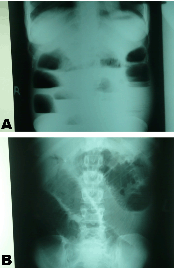
A 15-year-old girl presented to our hospital with a history of severe colicky abdominal pains, abdominal distension, and bilious vomiting for two days. Patient gave a history that she had been relatively well until about two days prior to admission when she started experiencing severe abdominal pains. Pains were, colicky in nature, non-radiating and more marked on the periumbilical region. Patient sited no aggravating or relieving factors for the pain. Associated with abdominal pains patient gave history of nausea and non-projectile bilious vomiting, she reported to have vomited five times prior to admission. Vomitous was foul smelling, and mainly consisted of watery feculent fluid which was heavily bile stained. There was no history of hematemesis or fever. In the same duration, patient gave a history of progressive abdominal distension and absolute constipation. Patient was a primary school girl who had been experiencing normal menses and had no history of previous surgery or any history suggestive mental illness. Also, she had no history of similar complaints in the past and generally gave no history suggestive of impaired mastication, abnormal deglutition or chronic diseases such as diabetes mellitus. On general examination we saw a young girl in good nutritional status, afebrile, not pale, and moderately dehydrated. She was well oriented but looked rather anxious and distressed. She had a pulse rate of 116 beats per minute, regular with good volume, her blood pressure was 110/60 mmHg, with obvious tachypnea. Per abdomen examination revealed moderately and uniformly distended abdomen which was moving with respiration. There was visible peristalsis on the epigastric region. The abdomen was rather tense with slightly generalized tenderness. There was no obvious palpable mass or organomegaly. Tympanic note was elicited on percussion. Bowel sounds were present with a high-pitched metallic tinkling tone. Digital rectal examination revealed an empty rectum. The rest of systemic examination was essentially normal. She had complete blood count (CBC), serum electrolytes, blood grouping and cross matching, abdominal ultrasonography as well as plain abdominal X-rays. Complete blood count revealed moderate leucocytosis where as serum electrolytes showed mild hypokalemia. Abdominal ultrasonography showed moderate amount of fluid in peritoneal cavity and dilated loops of small intestine. Erect view of the plain abdominal radiograph showed multiple air fluid levels in a step ladder pattern, there was no free gas under the diaphragm. Supine view revealed diffuse gaseous distension of small bowel loops. (Figure 1A–B) In view of the history, physical examination and investigations a diagnosis of small bowel obstruction was made, though at this juncture we could not precisely ascertain the exact underlying aetiology. After adequate resuscitation and obtaining informed consent from the patient’s parents, she was planned for emergency surgery whereby laparotomy was performed, through a midline incision which revealed significantly distended proximal ileal loops with an impacted palpable intraluminal hard mass which was well circumscribed and situated at about 55 cm from the Ileocecal junction, small bowel distal to the mass was collapsed. (Figure 2) Endeavors to carry out extramural fragmentation of this firm mass by using fingers or atraumatic babcock forceps were futile due to its large size and consistency. Therefore, we had to resort to enterotomy. Enterotomy was performed and a darkish unclean foul smelling bezoar was found. The ileal bezoar measured 6×4×4 cm, was completely delivered out with minimal difficulties. (Figure 3) (Figure 4) There were no synchronous existence of primary bezoars noted in the stomach, duodenum or proximal jejunum. The postoperative period was uneventful and the patient was well and had no complaints at last follow-up six months postsurgery. | ||||||
|
| ||||||
|
| ||||||
| ||||||
| ||||||
|
Discussion
| ||||||
|
Our reported patient had no obvious predisposing factors which lead to small bowel bezoar formation and subsequently bowel obstruction. Bezoar formation in rather healthy subjects has been previously reported though still remains an extremely infrequent clinical entity. [2] [3][4] Bezoars mostly stem from the stomach and occasionally drift into small bowel. Primary small bowel bezoar development may be foreseeable in patients with underlying small bowel disease such as stricture due to previous surgery, tuberculosis or Crohn’s disease. Other predisposing factors include poor mastication, bolus intakes of indigestible vegetables, diverticuli formation, and small bowel tumors. It is hypothesized that these conditions provide areas of sufficient stagnation within a dilated bowel segment for a bezoar to conglomerate over time. [2] [3] [4] Often patients exhibit diverse clinical presentation depending on the size, type and location of the bezoar. The most common clinical features comprises abdominal pains, poor appetite, vomiting, weight loss and anemia. Also patients may present with a wide range of gastrointestinal tract complications such as ulcer formation, bleeding, pressure necrosis, perforation and intestinal obstruction. [1] [2] [3] [4] The most frequent site of obstruction is at the level of the gastric outlet or duodenum. Obstruction of distal parts of the small bowel or large bowel remains a rarity occurrence. Features suggestive of psychiatric ailment and other chronic disease like diabetes mellitus or hyperthyroidism may as well be encountered. [1] [2] [3] [4] [5] The diagnosis of bezoar as an underlying aetiology of gastrointestinal obstruction has always been arduous. In most instances, a definitive diagnosis has been made during surgery. Usually apart from hematological and biochemical work up, a battery of radiological investigations have been recommended for detection of bezoars. These include the following plain abdominal X-ray, barium studies, ultrasonography, magnetic resonance imaging (MRI) scan, and computed tomography (CT) scan. [1] [2] [3] [4] [5] [6] A CT scan is very useful diagnostic tool in assessing patients with bezoars, it clearly delineate the site, size and nature of a bezoar. Characteristically, bezoar is revealed as a well-defined intraluminal mass with mottled gas on CT scan. When available, MRI is equally good in evaluating patients with suspected bezoar induced small bowel obstruction. [1] [2] [3] [6] Though not to the same magnitude such as CT scan or MRI, barium studies may as well be employed to confirm the presence of bezoar in the gastrointestinal tract and at the same time aid to detect other complications such as ulcers. [1] [2] [3] [4] Abdominal ultrasonography is another reliable modality in the diagnosis of gastrointestinal bezoars. Characteristically, sonographic visualization of an intraluminal mass with a hyper echoic arc-like surface and a marked acoustic shadow is greatly indicative of a bezoar. [6] Unlike the latter imaging modalities, plain abdominal X-ray mostly show features of bowel obstruction and occasionally depict an opaque intraluminal soft tissue air-containing mass which may be highly suggestive of bezoar. [1] [2] [3]Generally, plain abdominal X-ray lacks fine details and remains rather limited in evaluation of patients with bezoars. Where accessible, use of endoscopy is extremely crucial. Endoscopy could be utilized for both diagnostic and therapeutic purposes. Endoscopic examination allows visualization of both gastric and small bowel bezoars and where possible bezoar may be retrieved endoscopically. [1] [2] [3] [4] Often treatment of bezoars entails a wide range of options contingent upon the size, site and nature of the bezoar. However, retrieval of bezoar remains the core and primary treatment goal. In some cases surgical removal by gastrostomy or enterotomy is adequate but in others segmental bowel resection may be warranted due to bowel necrosis or denuded serosa. [1] [2] [3] [4] [5] The use of endoscopy and laparoscopic gadgets is becoming increasingly popular treatment modality for bezoar removal despite the fact that they may be of limited use when a bulky or rigid bezoar is involved. [2] [3] [7] Bezoars dissolution therapy has been mentioned in some series, however, by far there has been no tangible success with this treatment modality. [2] By and large, a timely intervention carries an excellent prognosis. | ||||||
|
Conclusion
| ||||||
|
Small bowel obstruction resulting from solitary bezoar formation without synchronous existence of primary gastric bezoar or any apparent predisposing factor remains an exceedingly rare presentation. Due to its rarity usually pose a significant diagnostic challenge and may subsequently lead to treatment delay and dreadful ramifications. Therefore, good clinical acumen, high index of suspicion, early detection of bezoar and prompt intervention are central in reducing morbidity and mortality. | ||||||
|
References
| ||||||
| ||||||


|
[HTML Abstract]
[PDF Full Text]
|
|
Author Contributions
Sefu Juma Uledi – Substantial contributions to conception and design, Acquisition of data, Analysis and interpretation of data, Drafting the article, Revising it critically for important intellectual content, Final approval of the version to be published Fauzia Ayubu Masumai – Substantial contributions to conception and design, Drafting the article, Revising it critically for important intellectual content, Final approval of the version to be published |
|
Guarantor of submission
The corresponding author is the guarantor of submission. |
|
Source of support
None |
|
Conflict of interest
Authors declare no conflict of interest. |
|
Copyright
© Sefu Juma Uledi et al. 2013; This article is distributed the terms of Creative Commons Attribution License which permits unrestricted use, distribution and reproduction in any means provided the original authors and original publisher are properly credited. (Please see Copyright Policy for more information.) |
|
|