 |

|
 |
|
Letters to the Editor
| ||||||
| Primary cavernous hemangioma of the thyroid gland | ||||||
| Devika Gupta1, Sunita Kakkar2, Pooja Gupta3, Vandana Rana4 | ||||||
|
1MD, Pathology, Assistant Professor, Armed Forces Medical College, Department of Pathology and Laboratory Science, Command Hospital, Pune, India.
2MD, Pathology and OncoPatholgy, Professor, Armed Forces Medical College, Department of Pathology and Laboratory Science, Command Hospital, Pune, India. 3MD, RadioDiagnosis, Department of Radiodiagnosis and Imaging, Armed Forces Medical College, Pune, India. 4MD, Pathology, Department of Pathology and Laboratory Science, Command Hospital, Pune, India. | ||||||
| ||||||
|
[HTML Abstract]
[PDF Full Text]
[Print This Article]
[Similar article in Pumed] [Similar article in Google Scholar] 
|


| How to cite this article: |
| Gupta D, Kakkar S, Gupta P, Rana V. Primary cavernous hemangioma of the thyroid gland. International Journal of Case Reports and Images 2013;4(9):524–526. |
| To the Editor, |
|
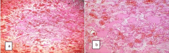
Hemangiomas are benign vascular neoplasms that have a characteristic clinical course marked by early proliferation and spontaneous involution. They occur in a number of organs including skin, lips, liver, colon, brain, etc.[1] [2] Primary thyroid hemangiomas are extremely uncommon with only a few cases have been reported. [3] We report a case of cavernous hemangioma presenting as solitary nodule thyroid. A 49-year-old male presented with one year history of slowly enlarging swelling on left side of the neck. There was no history of any associated pain, voice change or dyspnea. There was no history of any trauma or previous fine needle aspiration cytology (FNAC). There was no family history of thyroid disease. Local examination revealed an approximately 3x2.5 cm nontender swelling in front and left side of cricoid cartilage. The swelling was firm to hard with well defined margins and it moved with deglutition. Serum thyroid stimulating hormone was 3.8 mIU/L (normal range 0.4–4.0 mIU/L) and free T4 was 9 pmol/L (normal range 10.3–24.5 pmol/L). These hormonal levels were normal and no antithyroid antibodies were detected. Thyroid ultrasonography revealed a 2.5x2.8 cm isoechoic lesion in the left lobe of thyroid with a hypoechoic rim and a prominent vessel having arterial flow in its superior aspect. (Figure 1) Ultrasonography did not show any areas of calcification or cervical lymph nodes. Right lobe of thyroid was normal. The FNAC of the mass was attempted thrice but was inconclusive as only blood was aspirated. Patient underwent left hemithyroidectomy. Gross examination of the cut surface of the specimen revealed a well circumscribed nodular lesion measuring 2.5x2.8 cm. It appeared cystic and hemorrhagic in appearance Histological examination of the tumor confirmed a cavernous hemangioma. There were multiple, dilated, irregular anastomosing vascular channels of varying sizes. These vascular spaces were lined by bland endothelial cells. Amidst these were noted an occasional entrapped thyroid follicle. (Figure 2A-B) The patient was discharged from the hospital after five days with no signs of complication. In most cases, a cavernous hemangioma of the thyroid gland represents a secondary hemangioma occurring due to previous FNAC. These are formed due to vascular proliferation in organized hematoma following FNAC. [4] [5] Organization of the hematoma generally results in complete resolution, but it can give rise to vascular and fibroblastic proliferative changes that resemble a cavernous hemangioma. This is defined as secondary hemangioma. Primary hemangioma is a developmental anomaly resulting from inability of angioblastic mesenchyma to form canals. [6] Preoperative diagnosis of hemangioma of the thyroid is difficult since there are no pathognomic features on FNAC, ultrasonography or computed tomography scans. The presence of heterogenous signal intensity and serpentine pattern on magnetic resonance imaging (MRI) scan is considered highly suggestive of cavernous hemangioma. [7] In patients with a thyroid swelling who have a cold nodule on thyroid scan and only blood is aspirated on repeated FNACs, Technetium-99m erythrocyte blood pool imaging may be performed to diagnose hemangioma. Little or no increased activity is seen soon after injecting the label, and this appearance of poor perfusion and slow filling of the tumor is characteristic of cavernous hemangioma. Hemangioma should be considered in the diagnosis of any pulsatile mass involving the thyroid gland. Diagnosis before surgery is difficult and the definite diagnosis relies on histological findings of surgical specimen. |
|
|
|
|
|
|
|
References
|
|


|
[HTML Abstract]
[PDF Full Text]
|
|
Author Contributions:
Devika Gupta – Substantial contributions to conception and design, Acquisition of data, Analysis and interpretation of data, Drafting the article, Revising it critically for important intellectual content, Final approval of the version to be published Sunita Kakkar – Acquisition of data, Drafting the article, Revising it critically for important intellectual content, Final approval of the version to be published Pooja Gupta – Acquisition of data, Drafting the article, Revising it critically for important intellectual content, Final approval of the version to be published Vandana Rana – Acquisition of data, Drafting the article, Revising it critically for important intellectual content, Final approval of the version to be published |
|
Guarantor of submission
The corresponding author is the guarantor of submission. |
|
Source of support
None |
|
Conflict of interest
Authors declare no conflict of interest. |
|
Copyright
© Devika Gupta et al. 2013; This article is distributed the terms of Creative Commons Attribution License which permits unrestricted use, distribution and reproduction in any means provided the original authors and original publisher are properly credited. (Please see Copyright Policy for more information.) |
|
|