| |
|
|
|
Case Report
| ||||||
| Giant chylous cyst of liver: A case report | ||||||
| Mayank Baid1, Manoranjan Kar2, Sayak Roy1, Someshubhra Datta Roy1 | ||||||
|
1MBBS, MS, (PGT), Resident, Department of General Surgery, Medical College, Kolkata, India.
2MS, FAIS, FMAS, Professor, Department of General Surgery, Malda Medical College, West Bengal, India. | ||||||
| ||||||
|
[HTML Abstract]
[PDF Full Text]
[Print This Article]
[Similar article in Pumed] [Similar article in Google Scholar] 
|
| How to cite this article: |
| Baid M, Kar M, Roy S, Roy SD. Giant chylous cyst of liver: A case report. International Journal of Case Reports and Images 2013;4(8):444–447. |
|
Abstract
|
|
Introduction:
We present here a case of chylous cyst of liver with multiple cysts in mesentry, in a 14-year-old.
Case Report: A 14-year-old female presented in surgical outpatient unit with gradual swelling of abdomen for five months without any history of fever, jaundice, trauma or previous operation. Ultrasound and contrast enhanced computed tomography scan suggested a large multiseptate space occupying hypodense lesion. On laparotomy, a huge lobulated cyst containing clear fluid was found arising from left lobe of liver extending up to pelvis. Multiple small cysts were present in mesentery, mesoappendix and serosa of gallbladder. The giant cyst was excised completely with cystic left lobe of liver. Histopathology suggested chylous cyst of liver. There is no recurrence after two years of follow-up. Conclusion: We conclude that patient had chylous cysts at multiple places with largest from liver. | |
|
Keywords:
Chylous cyst, Cyst of liver, Multiple cysts
| |
|
Introduction
| ||||||
|
In 1842, von Rokitansky described a chylous mesenteric cyst. Since then, a few reports of chylous cysts are reported. We present here a case of chylous cyst of liver with multiple cysts in mesentry, in a 14-year-old female. As per our review, this may be a first reported giant chylous cyst of liver. | ||||||
|
Case Report
| ||||||
|
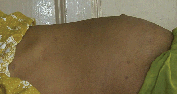
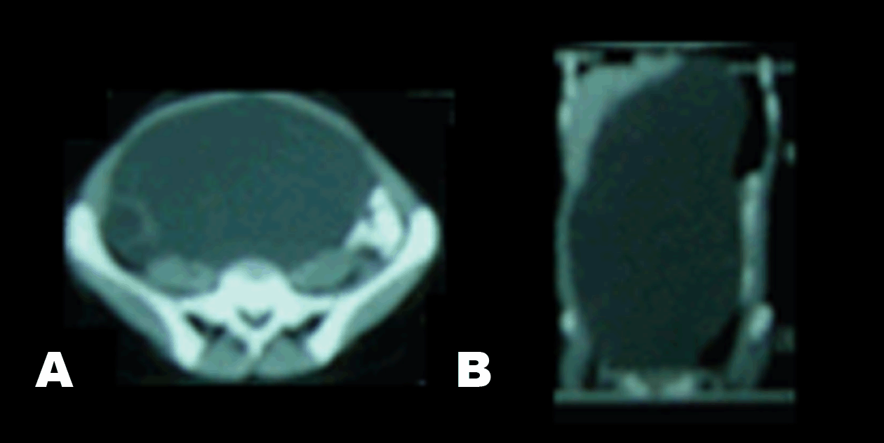
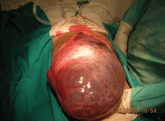
A 14-year-old female was presented at our surgical outpatient unit with complaint of gradual swelling of the upper abdomen and pain at the same site for five months. There was no history of fever, nausea, vomiting or jaundice. Bladder and bowel habits were normal. There was no history of loss of weight but slight decrease in appetite. There was no history of trauma to abdomen or any previous operative intervention in the abdomen. On examination patient had mild pallor, no jaundice, pulse 84 beats/minute, blood pressure 110/70 mmHg, respiratory rate 18/minute, no fever, and had no lymphadenopathy. There was a huge swelling in epigastrium extending to right hypochondrium laterally and extending beyond the umbilicus inferiorly. (Figure 1) On palpation swelling was cystic, non-tender, intra abdominal, margin was ill defined and local temperature was not raised. Liver and spleen were not separately palpable. There was no ascites. Air entry on both sides of chest, and spine were normal. Laboratory examination showed hemoglobin 10.4 g/dL, total count of white blood cell 6000/µL, Total bilirubin 1.1 mg/dL, SGPT 196 U/L, SGOT 173 U/L, and alkaline phosphate 386 U/L (Normal <98 U/L), serum amylase level 67 IU/dL. Ultrasonography and contrast enhanced computed tomography (CT) scan suggested a fairly large multiseptate space occupying lesion of size 20x12 cm in relation to left lobe of liver, abutting the liver and pancreas, with impression of resolving liver abscess or pancreatic pseudocyst. (Figure 2) During exploratory laparotomy, on opening the peritoneum a huge lobulated cyst with clear content was found arising from the left lobe of liver extending up to pelvis. (Figure 3) Multiple very small cystic lesions were also seen to be arising from mesentery of the intestine, (Figure 4) mesoappendix and serosa of the gallbladder. Right lobe of liver, kidney, spleen, intestinal wall and stomach appeared normal. After aspiration of content, the cyst completely collapsed and was excised in toto including the involved part of left lobe of liver. The nature of contained fluid was clear. Patient underwent cholecystectomy and appendicectomy. Cysts arising from mesentery were left intact. Postoperative period was uneventful. Histopathological report was cystic lesion lined by flattened endothelium, the cyst wall composed of fibrocollagenous tissue infiltrated with lymphocytes with impression of chylous cyst of liver, gallbladder wall infiltrated by chronic inflammatory cells and appendix showed lymphoid hyperplasia. After the operation the patient remained asymptomatic and there was no development of any detectable cyst on ultrasonography or recurrence in liver after two years on follow-up. | ||||||
| ||||||
| ||||||
| ||||||
| ||||||
|
Discussion
| ||||||
|
Classically, cystic disease of the liver is divided into nonparasitic and parasitic cyst. Nonparasitic cyst is more prevalent worldwide, although prevalence may vary significantly by geographic region. Generally, most hepatic cysts, regardless of type remain asymptomatic. Hepatic function is rarely affected by their presence [1]. Chylous cysts are rare pathological entities and relatively few cases have been reported in literature. These cysts are thought to represent benign proliferation of ectopic lymphatic that lack communication with the normal lymphatic system. [1] They are usually associated with mesentery of intestine. But have also been reported in colon, [2] retroperitoneum [3] and liver (in this case). Chylous cyst are often congenital but may be related to previous abdominal surgery, pelvic disease and trauma. [4] Mesenteric chylous cyst most commonly occur in second decade of life, but they have also been seen in first decade. Symptoms are usually poor and unspecific. Rarely, clinical presentation may be dramatic with acute abdominal pain and symptoms of intestinal obstruction [5] or simulating the rupture of aortic aneurysm. [6] The preoperative diagnosis requires all the common abdominal imaging techniques. Ultrasonography usually demonstrates, a cystic lesion, whose content may form a fluid-fluid level. A fat fluid interface on computed tomography is indicative of a chylous cyst. [7] But the correct diagnosis is usually, made in the operation theater or during histological examination. [4] Histologically, cysts are lined by a flattened endothelium with a fibrous wall; in which dilated lymphatics may be observed. Presence of cholesterol clefts further supports the pathological diagnosis of a chylous cyst. [8] Apart from being cause of an acute abdomen, these cysts may undergo rupture, hemorrhage or infection. [9] Malignant transformation is rare but not unknown. [10] Treatment of choice is complete surgical excision and is usually curative. This patient was asymptomatic apart from the swelling in the abdomen. On examination, she had an intra-abdominal, cystic, swelling in epigastrium. Preoperative diagnosis was inconclusive even after contrast enhanced computed tomography scan diagnosis was made during laparotomy which was confirmed by histopathological examination. Complete excision of the liver cyst was possible and the multiple cysts in the mesentry which were left intact. There is no evidence of development of symptomatic large cysts or recurrence in liver after two years on follow-up. | ||||||
|
Conclusion
| ||||||
|
We can conclude that patient had chylous cysts at multiple places with largest from liver. Most likely they are of separate origins but we cannot say conclusively. We have planned to follow-up this patient every six months with clinical examination and ultrasonography to look for development of any large symptomatic cyst. | ||||||
|
Acknowledgements
| ||||||
|
To patient for her acceptance and co-operation. | ||||||
|
References
| ||||||
| ||||||
|
[HTML Abstract]
[PDF Full Text]
|
|
Author Contributions
Mayank Baid – Substantial contributions to conception and design, Acquisition of data, Analysis and interpretation of data, Drafting the article, Revising it critically for important intellectual content, Final approval of the version to be published Manoranjan Kar – Substantial contributions to conception and design, Acquisition of data, Analysis and interpretation of data, Drafting the article, Revising it critically for important intellectual content, Final approval of the version to be published Sayak Roy – Substantial contributions to conception and design, Acquisition of data, Analysis and interpretation of data, Drafting the article, Revising it critically for important intellectual content, Final approval of the version to be published Someshubhra Datta Roy – Substantial contributions to conception and design, Acquisition of data, Analysis and interpretation of data, Drafting the article, Revising it critically for important intellectual content, Final approval of the version to be published |
|
Guarantor of submission
The corresponding author is the guarantor of submission. |
|
Source of support
None |
|
Conflict of interest
Authors declare no conflict of interest. |
|
Copyright
© Mayank Baid et al. 2013; This article is distributed the terms of Creative Commons Attribution License which permits unrestricted use, distribution and reproduction in any means provided the original authors and original publisher are properly credited. (Please see Copyright Policy for more information.) |
|
|