  |

|
 |
|
Clinical Image
| ||||||
| Pulmonary epithelioid hemangioendothelioma identified by computed tomography scan | ||||||
| Yoshiro Nakahara1, Tatsuru Okamura1, Kan Kato2, Tsunekazu Hishima3 | ||||||
|
1Department of Thoracic Oncology and Respiratory Medicine, Tokyo Metropolitan Cancer and Infectious Diseases Center Komagome Hospital, Tokyo, Japan.
2Department of Respiratory Medicine, Tokyo Kensei Hospital, Tokyo, Japan. 3Department of Pathology Tokyo Metropolitan Cancer and Infectious, Diseases Center, Komagome Hospital, Tokyo, Japan. | ||||||
| ||||||
|
[HTML Abstract]
[PDF Full Text]
[Print This Article]
[Similar article in Pumed] [Similar article in Google Scholar] 
|


| How to cite this article: |
| Nakahara Y, Okamura T, Kato K, Hishima T. Pulmonary epithelioid hemangioendothelioma identified by computed tomography. International Journal of Case Reports and Images 2013;4(7):396–398. |
|
Case Report
| ||||||
|
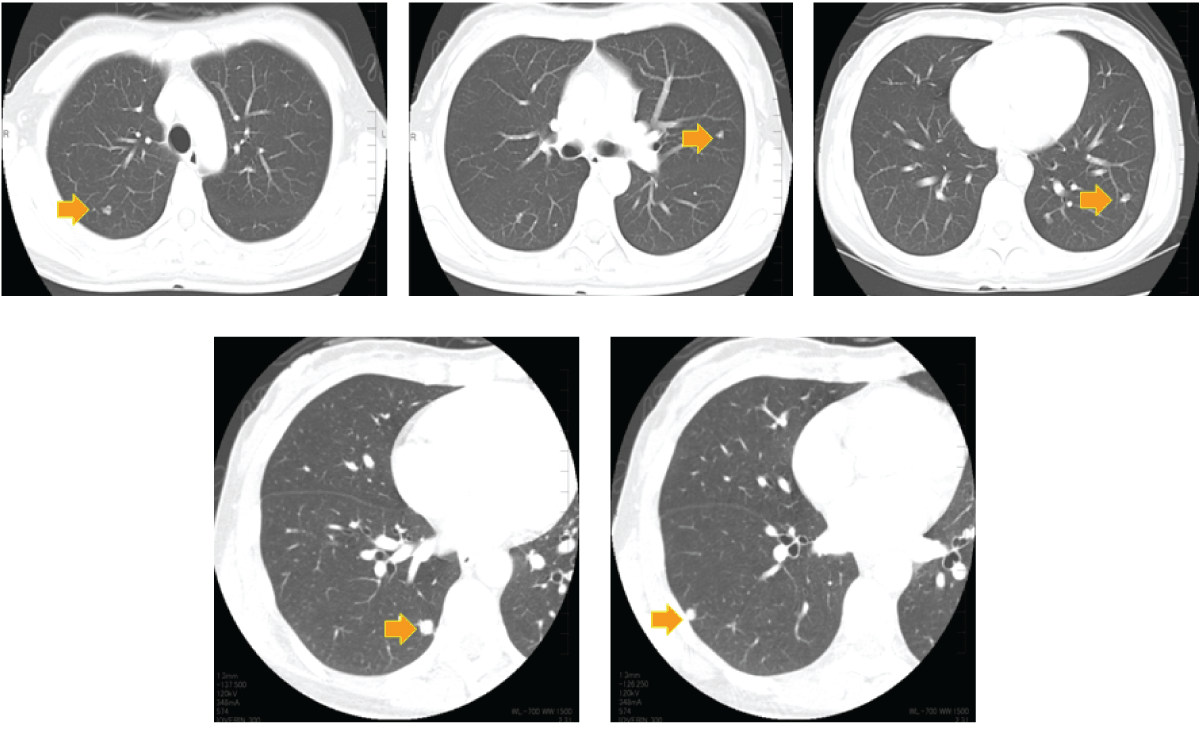
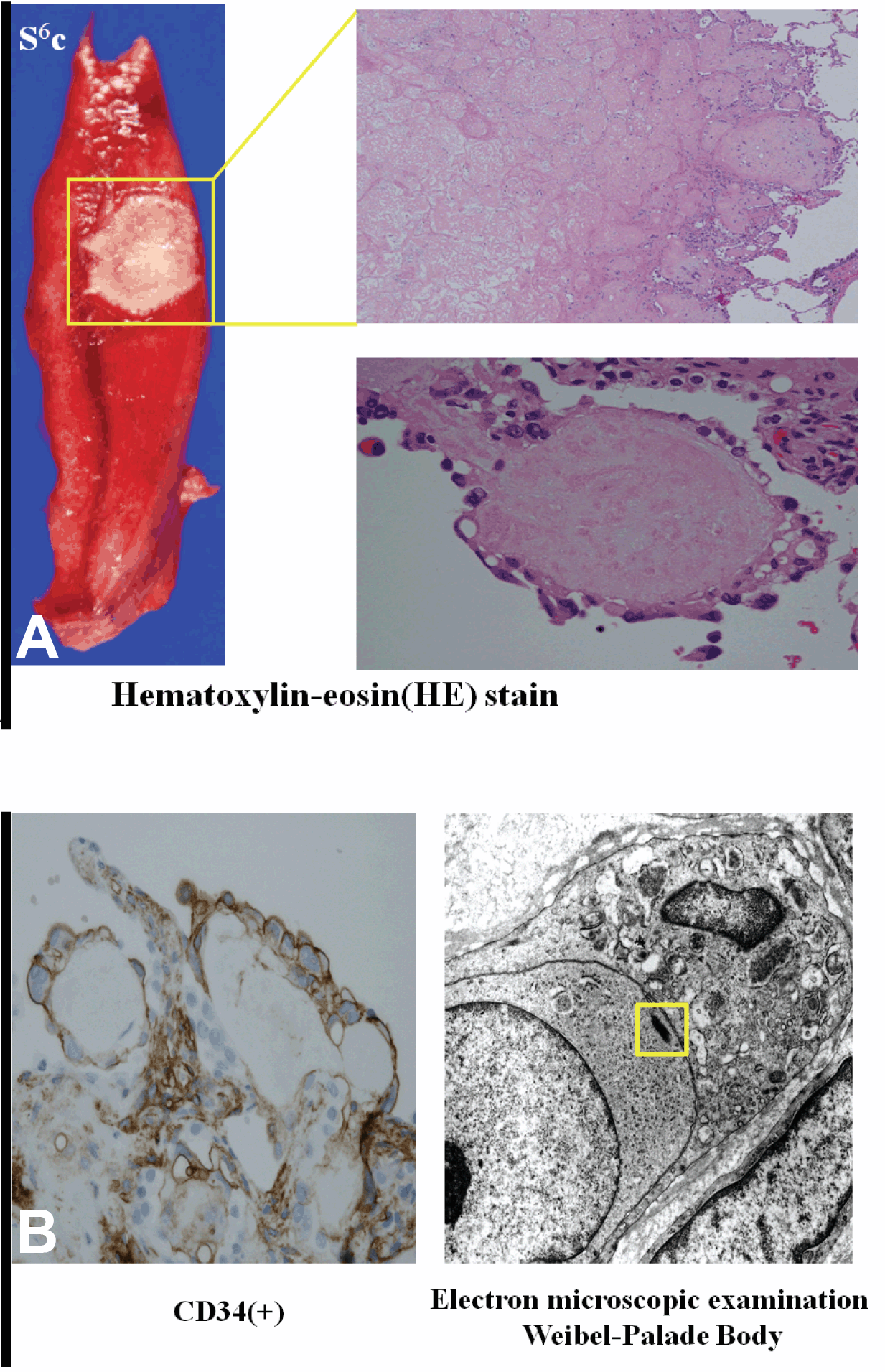
Computed tomography (CT) scan showed bilateral, nodular, lung shadows in an asymptomatic 38-year-old female. (Figure 1) Five nodules up to 10 mm each were identified in the right and left lungs. She was followed-up for four months, during which the size and density of these shadows remained unaltered. The patient requested a definitive diagnosis and underwent a video-assisted thoracoscopic lung biopsy of two lesions in the right lower lobe (S6c, S9a). A histopathological examination showed vacuolated tumor cells, immunohistochemical staining of which was positive for the endothelial marker CD34. Electron microscopy identified Weibel-Palade bodies. These findings supported a diagnosis of pulmonary epithelioid hemangioendothelioma (PEH). (Figure 2) All other organs were free of abnormalities and the patient was remained under observation for two years without treatment or disease progression. | ||||||
| ||||||
|
| ||||||
|
| ||||||
|
Discussion
| ||||||
|
The grade of malignancy of rare, vascular pulmonary epithelioid hemangioendothelioma (PEH) is low to intermediate. These tumors can simultaneously or sequentially arise from the lungs, liver, bone and soft tissue, which renders multicentric tumor growth difficult to distinguish from primary lesions with metastasis to other tissues. Most patients with pulmonary epithelioid hemangioendothelioma (PEH) are female (61–80%) and the median age is 36–50 years, which is relatively younger than those with lung cancer. No specific symptoms are associated with PEH, and 50–76% of patients are asymptomatic at the time the condition is detected by chest radiography. Our patient was also asymptomatic. Multiple pulmonary nodules, multiple pulmonary reticulonodular opacities, and diffuse infiltrative pleural thickening have been identified by computed tomography (CT) scan. [1] The most common feature of PEH on chest CT scan is the presence of multiple small discrete pulmonary nodules of up to 2 cm with well–defined margins in both lungs. However, most nodules are less than 1 cm in diameter. Dail et al. reviewed 20 patients and found that 20, 65% and 25% of them had < 10, 10–20 and > 20 nodules, respctively. [2] On the other hand, Kitaichi et al. reported the CT scan of 18 patients, among whom four and 14 had unilateral and bilateral opacities, respectively. Two of the four with unilateral opacities had a single nodular opacity and one patient had pleural effusion without nodular opacities. All 14 patients with bilateral opacities had plural nodules, and one patient had pleural effusion. None of them had lymphadenopathy. [3] Surgery is an option for unilateral nodules, but a single effective treatment has been suggested for multiple or bilateral involvement. [4] Although PEH usually grows very slowly, it can metastasize to other organs. The 5-year survival rate is around 60%. Respiratory failure (41.9%) and metastasis (38.7%) are common causes of death. [4] Hemorrhagic symptoms such as hemoptysis and pleural hemorrhagic effusion might indicate a poor prognosis. [4] | ||||||
|
Conclusion
| ||||||
|
Since multiple small lung nodules are likely be discovered more frequently with the increasing popularity of computed tomography (CT) screening, pulmonary epithelioid hemangioendothelioma (PEH) should be considered as a differential diagnosis. | ||||||
|
References
| ||||||
| ||||||

|
[HTML Abstract]
[PDF Full Text]
|
|
Author Contributions
Yoshiro Nakahara – Substantial contributions to conception and design, Acquisition of data, Analysis and interpretation of data, Drafting the article, Revising it critically for important intellectual content, Final approval of the version to be published Tatsuru Okamura – Substantial contributions to conception and design, Acquisition of data, Analysis and interpretation of data, Drafting the article, Revising it critically for important intellectual content, Final approval of the version to be published Kan Kato – Substantial contributions to conception and design, Acquisition of data, Analysis and interpretation of data, Drafting the article, Revising it critically for important intellectual content, Final approval of the version to be published Tsunekazu Hishima – Substantial contributions to conception and design, Acquisition of data, Analysis and interpretation of data, Drafting the article, Revising it critically for important intellectual content, Final approval of the version to be published |
|
Guarantor of submission
The corresponding author is the guarantor of submission. |
|
Source of support
None |
|
Conflict of interest
Authors declare no conflict of interest. |
|
Copyright
© Yoshiro Nakahara et al. 2013; This article is distributed the terms of Creative Commons Attribution License which permits unrestricted use, distribution and reproduction in any means provided the original authors and original publisher are properly credited. (Please see Copyright Policy for more information.) |
|
|