
  |

|
 |
|
Case Report
| ||||||
| Delayed splenic rupture presenting as atypical chest pain and right bundle branch block | ||||||
| Travelyan M Walker1, Omar K Danner2, Kenneth L Wilson3, L Ray Matthews4 | ||||||
|
1MD, General Surgery Resident, Department of Surgery, Morehouse School of Medicine, Atlanta, GA USA.
2MD, FACS, Assistant Professor, Department of Surgery, Morehouse School of Medicine, Atlanta, GA USA. 3MD, FACS, Assistant Professor and Director of Trauma, Department of Surgery, Morehouse School of Medicine, Atlanta, GA USA. 4MD, FACS, Assistant Professor and Director of Critical Care, Department of Surgery, Morehouse School of Medicine, Atlanta, GA USA. | ||||||
| ||||||
|
[HTML Abstract]
[PDF Full Text]
[Print This Article]
[Similar article in Pumed] [Similar article in Google Scholar] 
|


| How to cite this article: |
| Walker TM, Danner OK, Wilson KL, Matthews LR. Delayed splenic rupture presenting as atypical chest pain and right bundle branch block. International Journal of Case Reports and Images 2013;4(6):330–333. |
|
Abstract
|
|
Introduction:
Although splenic injury is the most frequent abdominal injury resulting from blunt trauma, delayed splenic rupture is a rare event. Traumatic rupture of the spleen has been well described in medical literature and is found in approximately 30% of patients undergoing surgery for blunt abdominal injury. Computed tomography negative delayed rupture of the spleen is a very rare clinical entity. This is in spite of computed tomography having a sensitivity and specificity for detection of splenic injuries as high as 96% and 100%, respectively. When a patient presents after blunt abdominal trauma, awareness of the problem of delayed splenic rupture, a high index of clinical suspicion and rapid diagnostic approach may help reducing the morbidity and mortality associated with unrecognized splenic injury and/or hemorrhage.
Case Report: We present a case report of a 61-year-old male restrained driver, involved in a motor vehicle crash, who presented with a normal spleen on initial computed tomography scan. Conclusion: Computed tomography - negative delayed rupture of the spleen is a very rare clinical entity. | |
|
Keywords:
Delayed splenic rupture, Splenic injury, Chest pain
| |
|
Introduction
| ||||||
|
Computed tomography (CT) negative delayed rupture of the spleen is a very rare clinical entity. [1] This is in spite of CT having a sensitivity and specificity for detection of splenic injuries as high as 96% and 100%, respectively. [2] [3] We present a case report of a 61-year-old male restrained driver, involved in a motor vehicle crash (MVC), who presented with a normal spleen on initial CT scan. Seven days later, he was rescanned due to a significant decrease in hemoglobin after having severe chest pain. The follow-up CT scan showed a large subcapsular hematoma. Although splenic injury is the most frequent abdominal injury resulting from blunt trauma, delayed splenic rupture is a rare event. Traumatic rupture of the spleen has been well described in medical literature and is found in approximately 30% of patients undergoing surgery for blunt abdominal injury. [4] Occult, spontaneous splenic rupture is rarely diagnosed, although the associated mortality/morbidity is high. From 1981 to 1990, 75 patients treated at St. Elizabeth Hospital Medical Center (Youngstown, OH) had blunt splenic injury. Splenic rupture was delayed in six (8%) of these patients. [1] Although there is no reliable symptom or sign during the latent period, abdominal pain occurs almost uniformly and Kehr's sign is quite common. [1] The astute clinician must have a high index of suspicion relying on clinical and roentgenographic study for rapid diagnosis and management. [5] Although, spontaneous splenic rupture is rare, it is vital that physicians consider this diagnosis when evaluating patients with abdominal pain and hypotension. Failure to consider splenic rupture could be catastrophic and early diagnosis is essential for a better outcome. The concept of delayed splenic rupture is fairly well established in literature. The term delayed splenic rupture is used to describe the situation in which early post-traumatic imaging of the spleen is normal and is followed by the diagnosis of splenic hemorrhage 48 or more hours after the initial insult. The entity of delayed splenic rupture represents an initially missed injury, a delayed presentation of the latter, or an actually delayed development of an initially latent, minor, splenic injury. Patients typically present after a mean lag time of five days after injury (range, 2–10 days). [6] Although post-traumatic splenic evaluation is indicated when there is unexplained hypovolemia, the mechanism of splenic injury is present, or the patient complains of left upper quadrant pain or tenderness, atypical chest pain or unstable angina is an unusual manifestation of occult splenic rupture. [7] | ||||||
|
Case Report
| ||||||
|
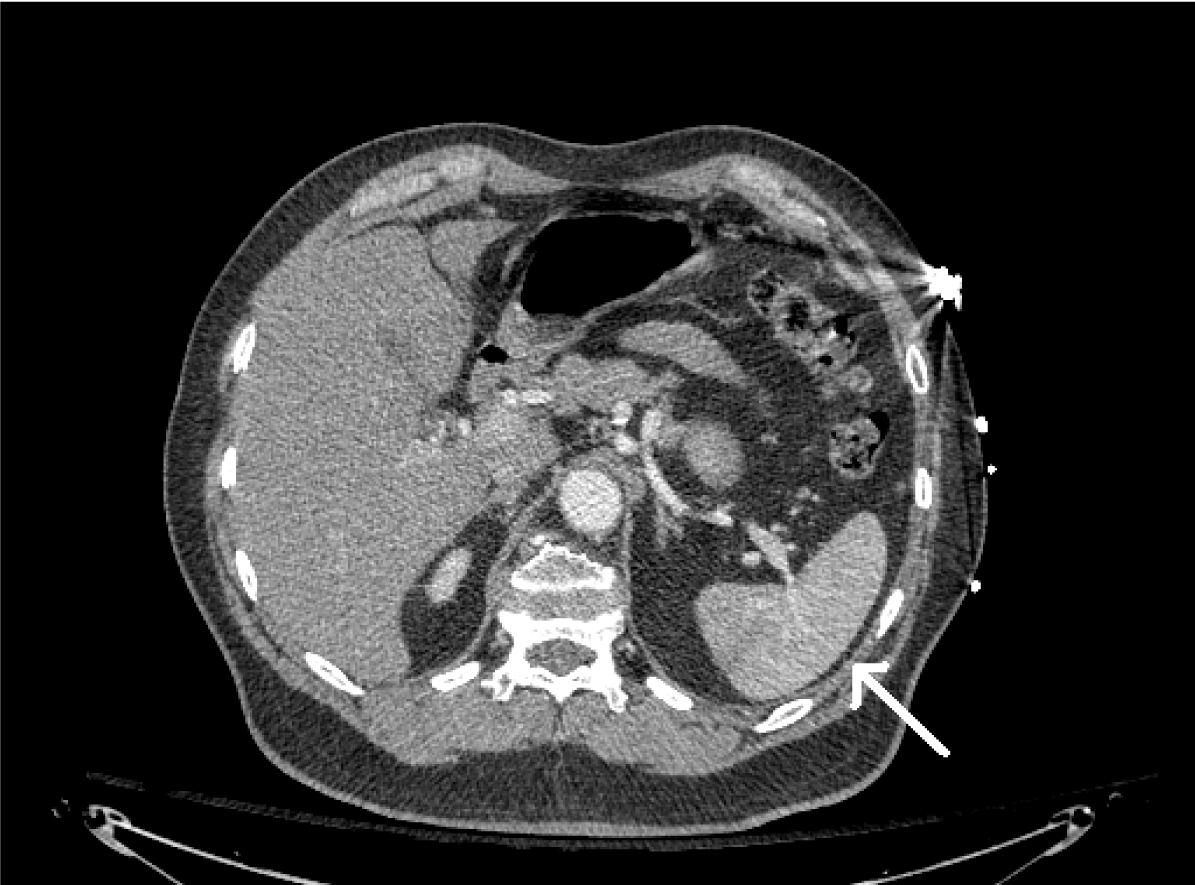
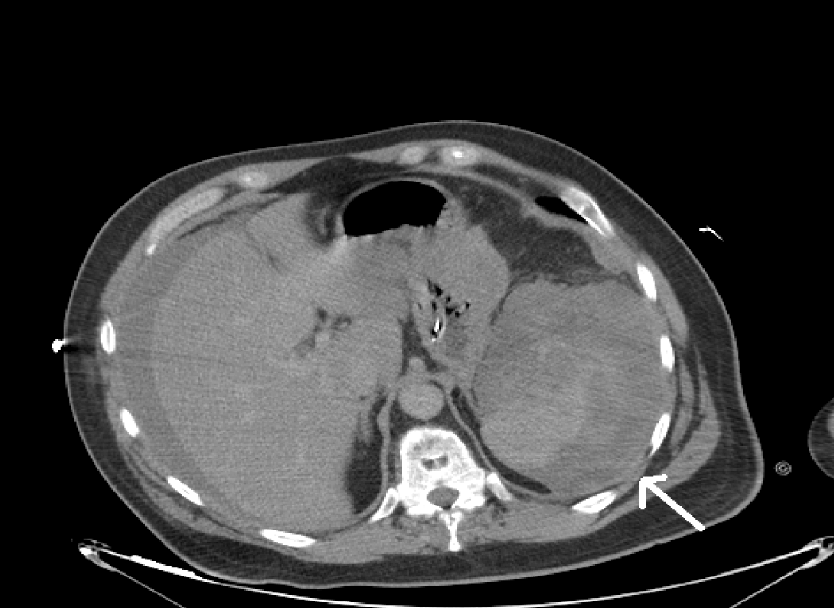
A 61-year-old male restrained driver was brought to the emergency room after being involved in a MVC. His Glasgow coma scale was 15. He had an 8-cm bleeding scalp laceration which was stapled in the trauma bay. The patient complained of right-sided chest wall pain. His abdominal exam was benign. His focused assessment with sonography for trauma (FAST) exam was negative. He was hemodynamically stable during his stay in the trauma bay. Patient's hemoglobin and hematocrit were 16.2 and 47.5, respectively. This coagulation parameters were normal. An electrocardiography revealed right bundle branch block for which the patient was being followed by a cardiologist. The CT scan of the abdomen revealed no free fluid, a spleen that was normal in size and enhances heterogeneously. (Figure 1) Computed tomography scan of the chest showed right 7th and 8th rib fractures and a right pneumothorax less than 10%. Patient was admitted to intensive care unit (ICU) for close monitoring for three days. A cardiac work-up was negative for any cardiac event. He was discharged to the surgical floor on hospital day-4. Patient was doing well up until hospital day-7 when he started to complain of severe sternal chest pain, shortness of breath, nausea and became diaphoretic and hemodynamically unstable with a systolic blood pressure in the 80s. His abdominal exam was nontender. Electrocardiography demonstrated right bundle branch block. Patient was resuscitated with intravenous fluids and was intubated for respiratory distress. Laboratory examination revealed hemoglobin 7.5g/dL. Cardiac enzymes were normal. Computed tomography scan of the abdomen revealed a large perisplenic hematoma measuring 12x14 cm and perisplenic fluid and hemorrhage. (Figure 2) Patient was then taken to interventional radiology (IR) where he underwent embolization of the splenic arterty. He was admitted to ICU for further resuscitation, treated for ventilator associated pneumonia and discharged home on hospital day-24 after completion of his antibiotic course for pneumonia. | ||||||
| ||||||
| ||||||
|
Discussion
| ||||||
|
Computed tomography scans are very sensitive in detecting splenic injuries after trauma. Delayed splenic rupture was defined as rupture of the spleen more than 48 hours after trauma. The first case of delayed splenic rupture documented by CT scan was reported in 1981 by Toombs. The time period is based on the "Latent Period of Baudet" of 48 hours which Baudet described as time from injury to rupture. [8] Prior to the use of CT scans the incidence of Delayed splenic rupture was estimated to range from 5–40% of all splenic injuries. At present with the use of thin slice CT scan the incidence is as low as 1–2%. The hallmark presentation of a patient with splenic rupture includes left upper quadrant pain, Kehr's sign (shoulder pain secondary to diaphragmatic irritation by the hemoperitoneum), [1] [2] as well as signs of generalized peritonitis such as guarding and rebound tenderness. [1] Our patient did not complain of abdominal pain during his hospital duration. He did report a history of cardiac disease and possibly a myocardial infarction. He was without palpitations, chest pain, dyspnea, orthopnea, or leg swelling prior to DSR. The patient’s chest pain was acute, nonspecific, and reported within minutes prior to decompensating secondary to his DSR. Advanced cardiac life support protocol was used to initially stabilize our patient. He was taken emergently to the interventional radiology suite where successful embolization of the splenic artery was performed. Although splenectomy remains the primary treatment for hemodynamically unstable patients with splenic injuries, nonsurgical management can safely be used in selected patients with DSR, especially for those with a good response to resuscitation. [9] When a patient presents after blunt abdominal trauma, awareness of the problem of delayed splenic rupture, a high index of clinical suspicion and rapid diagnostic approach may help reducing the morbidity and mortality associated with unrecognized splenic hemorrhage, either immediate or delayed. Emergency abdominal CT scan has become the 'gold standard' initial diagnostic modality. However, delayed splenic rupture can still develop in patients with low CT grades or scores. The choice between operative and nonoperative management of splenic trauma should be mainly based on clinical findings rather than CT scans. [10] Suitability of adult patients with blunt splenic injury for nonoperative management may be predicted at initial presentation, based on hemodynamic status and associated injuries. The quantity of hemoperitoneum and magnitude of splenic injury are predictive factors for failure of conservative treatment. Early definition of these factors may help identify those patients likely to be successfully treated without laparotomy. [10] Delayed splenic rupture is a rare manifestation of blunt splenic trauma, and splenectomy remains the primary treatment for patients with DSR. The success rate of nonsurgical therapy is 83%. Splenic artery embolization (SAE) can be used for splenic salvage with a success rate of 80%. The overall failure rate of DSR management is about 33%. Nonsurgical management can safely be used in selected patients with DSR, especially for those with a good response to resuscitation. SAE is as effective for DSR as it is for acute splenic injury. Physicians should consider SAE as an option for the treatment of DSR. [9] | ||||||
|
Conclusion
| ||||||
|
The accuracy of computed tomography for diagnosis of splenic injury is approximately 97% [3]. The most commonly injured organ in the setting of blunt trauma, splenic injuries must be excluded when evaluating patients who present when hemodynamic instability. Resuscitation, prompt diagnosis, and intervention remain vital when managing trauma patients. Although with an incidence of 1–2%, delayed splenic rupture (DSR) contributes to a significant mortality rate (5–15%) compared with that associated with acute splenic rupture (1%) [8]. Patients are presented most commonly with symptoms of abdominal pain, but may also be presented with chest pain and arrhythmias as demonstrated by our patient. Management of delayed splenic rupture is determined by the hemodynamic status of the patient and availability of interventional radiology services. | ||||||
|
References
| ||||||
| ||||||

|
[HTML Abstract]
[PDF Full Text]
|
|
Author Contributions
Travelyan M Walker – Analysis and interpretation of data, Drafting the article, Revising it critically for important intellectual content, Final approval of the version to be published Omar K Danner – Acquisition of data, Analysis and interpretation of data, Drafting the article, Revising it critically for important intellectual content, Final approval of the version to be published Kenneth L Wilson – Analysis and interpretation of data, Drafting the article, Revising it critically for important intellectual content, Final approval of the version to be published L Ray Matthews – Substantial contributions to conception and design, Acquisition of data, Analysis and interpretation of data, Drafting the article, Revising it critically for important intellectual content, Final approval of the version to be published |
|
Guarantor of submission
The corresponding author is the guarantor of submission. |
|
Source of support
None |
|
Conflict of interest
Authors declare no conflict of interest. |
|
Copyright
© Travelyan M Walker et al. 2013; This article is distributed the terms of Creative Commons Attribution License which permits unrestricted use, distribution and reproduction in any means provided the original authors and original publisher are properly credited. (Please see Copyright Policy for more information.) |
|
|