
  |

|
 |
|
Case Report
| ||||||
| Dengue hemorrhagic fever with multiple abscesses: An unusual presentation | ||||||
| Kunal Bhatia1, Manisha Thakur2, YC Porwal3, UC Kansra4, Arun Iyer5 | ||||||
|
1MBBS, PGY-II (MD Medicine), Department of Medicine, Vardhman Mahavir Medical College and Safdarjung Hosiptal, New Delhi, India.
2MD Medicine, Associate Professor, Department of Medicine, Vardhman Mahavir Medical College and Safdarjung Hospital, New Delhi, India. 3MD Medicine, Associate Professor, Department of Medicine, Vardhman Mahavir Medical College and Safdarjung Hospital, New Delhi, India. 4MD Medicine, FICP, FIACM, Professor, Department of Medicine, Vardhman Mahavir Medical College and Safdarjung Hospital, New Delhi, India. 5MBBS, PGY-III (MD Medicine), Department of Medicine,Vardhman Mahavir Medical College and Safdarjung Hospital, New Delhi, India. | ||||||
| ||||||
|
[HTML Abstract]
[PDF Full Text]
[Print This Article]
[Similar article in Pumed] [Similar article in Google Scholar] 
|


| How to cite this article: |
| Bhatia K, Thakur M, Porwal YC, Kansra UC, Iyer A. Dengue hemorrhagic fever with multiple abscesses: An unusual presentation. International Journal of Case Reports and Images 2013;4(6):326–329. |
|
Abstract
|
|
Introduction:
Dengue is a common arboviral illness endemic in India. Concurrent bacterial co-infection in dengue is rare and often underreported.
Case Report: We report a case of an 18-year-old male with dengue hemorrhagic fever presenting as multiple abscesses due to a concomitant bacterial co-infection. Surgical intervention was done and the pus aspirated isolated Escherichia coli, although blood cultures were negative. The patient responded favorably after starting an antimicrobial regimen based on pus culture sensitivity reports and was discharged thereafter. Conclusion: Clinicians should be aware of the possibility of a concurrent bacterial co-infection in patients with dengue virus infection/dengue hemorrhagic fever so as to initiate a timely additional antimicrobial treatment. | |
|
Keywords:
Dengue fever, Dengue hemorrhagic Fever, Unusual presentation, Abscesses
| |
|
Introduction
| ||||||
|
Dengue is the most rapidly spreading mosquitoborne viral disease in the world with an estimated 50 million dengue infections occurring annually and approximately 2.5 billion people live in dengue endemic countries. [1] The clinical spectrum of dengue is highly variable, ranging from a mild flu-like syndrome to severe disease, with shock and hemorrhage. However, in the recent years various authors around the globe have reported atypical manifestations associated with dengue fever. Some unusual manifestations of dengue fever that have been previously described include fulminating liver failure, rhabdomyolysis, acute renal failure, myocarditis, neurological disturbances of encephalopathy, Gullian-Barré syndrome and transverse myelitis. [2] [3] [4] [5] [6] Here we are reporting a case of dengue hemorrhagic fever associated with multiple abscesses. | ||||||
|
Case Report
| ||||||
|
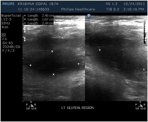
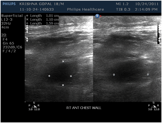
A 18-year-old male, mason by profession, presented with high grade fever with chills, myalgia, joint pains and prostration for two days followed by pain over the right shoulder, right anterior chest wall and the left gluteal region. On the 3rd day of fever, patient started complaining of painful swellings over the above mentioned areas along with black tarry loose stools (2–3 episodes/day) with abdominal distension which was gradual in onset and progressive. The patient got admitted to a private hospital in Agra where he was found to be dengue NS1Ag positive. He also underwent needle aspiration of the swelling present over the right shoulder and was given broad spectrum antibiotics (intravenous amoxicillin-clavulanic acid, azithromycin and teicoplanin) and supportive care for three days. However, the patient’s general condition did not improve and he presented to us in the emergency room with a diagnosis of dengue hemorrhagic fever complicated by multiple abscesses. Further history taking revealed that the patient continued to have fever for the past seven days along with passage of black tarry loose stools (2–3 episodes/day), abdominal distension, painful swellings involving the right shoulder, right anterior chest wall and left gluteal region for the last five days. No history of petechial rashes or bleeding from any other site could be elicited. History of trauma, intramuscular injections, intravenous drug abuse, and diabetes mellitus were negative. On physical examination, patient was pale, febrile with a temperature of 102°F, pulse rate of 98/min, blood pressure 124/60 mmHg and Hess test/tourniquet test was negative. On local examination, a swelling 1.5×1 cm over the right shoulder, a 4×3 cm swelling over the left gluteal region and another swelling of 3×3 cm over the right anterior chest wall were seen, which were hot, tender, firm with diffuse margins, showing fluctuation and surrounding induration; suggestive of abscesses. Respiratory, cardiovascular and neurological examination were within normal limits. Per abdomen examination revealed distended abdomen with presence of free fluid and no organomegaly. Laboratory investigations (on the day of admission) revealed hemoglobin of 7.2 g/dL with peripheral smear showing normocytic normochromic anemia and no malarial parasite, total leucocyte count of 7400/mm3 (neutrophils 65%, lymphocytes 31%, monocytes 2%, eosinophils 2%), platelet count 1.9x10⁴/mm3, PCV 31.6%, serum bilirubin 1.1 mg/dL, aspartate transaminase 153 IU/L, alanine transaminase 65 IU/L, alkaline phosphatase 267 IU/L. Renal functions were within normal limits. Chest roentgenography showed minimal bilateral pleural effusion. Ultrasonography of abdomen revealed partially distended gallbladder with edematous and thickened wall, mild hepatosplenomegaly and moderate ascites suggestive of polyserositis. Samples for blood culture and urine culture were withdrawn and patient was started on broad spectrum intravenous antibiotics (ceftazidime + vancomycin). Ultrasonography of left gluteal region showed a lobulated complex cystic lesion with internal echoes with approximate size of 3.9×1.9×3.3 cm (13.5 cm3) located in the intramuscular plain with adjacent soft tissue edema. (Figure 1) Another abscess with approximate size of 3.54×1.52×2.1 cm was located on the right anterior chest wall with similar features. (Figure 2) On the 9th day of the illness, incision and drainage of the abscesses was done after transfusing platelet rich plasma; and the pus aspirated was sent for microbiological examination, culture and sensitivity. No organisms were identified in the blood and urine cultures. Dengue serology was positive for IgM antibodies (MAC-ELISA) and HIV-ELISA was nonreactive. Microbiological investigations showed gram negative bacilli with a background of inflammatory cells and necrotic debris. Culture and sensitivity of the aspirate from all the three sites showed a growth of Escherichia coli which was sensitive to ertapenem and meropenem. The patient continued to have fever, malena and generalized weakness with hemoglobin dropping to 6.5 g/dL with a PCV of 24.7% and platelet count decreased to 10,000/mm3 on day 10. Therefore, in accordance with the 2009 WHO guidelines for management of dengue hemorrhagic fever, patient was transfused with 2 units of fresh whole blood and 4 units of platelet rich plasma. Following blood transfusion the patients’ hemoglobin increased to 7.9 g/dL with platelet count of 1.9x10⁴/mm3 and a PCV of 29.1%; but fever persisted. Subsequently, the patient was shifted onto intravenous meropenem based on pus culture sensitivity report. Antimicrobial therapy with antiseptic dressing of the drainage sites was continued for the next few days along with platelet count and hematocrit monitoring daily. On the 14th day of the illness, patient started showing signs of improvement with platelet count increasing to 3.9x10⁴/mm3 and a PCV of 37.5%. Patient became afebrile with no signs of bleeding from any site and had persistently increasing platelet counts thereafter. Hence, the patient was discharged after completing 7 days of antimicrobial therapy. | ||||||
| ||||||
| ||||||
|
Discussion
| ||||||
|
Dengue infection is an arboviral disease endemic in many parts of the world including India. Dengue infections are usually asymptomatic, but can present with classic dengue fever, dengue hemorrhagic fever or dengue shock syndrome. In the last few years there has been an increased spread of the dengue fever disease worldwide which has allowed the observation of a large number of patients with atypical clinical presentations. [2] [3] [4] [5] [6] The occurrence of bacterial co-infection in patients with dengue has been noted by some authors, but the available information comes from anecdotic reports. The microorganisms that have been identified to occur simultaneously with dengue virus infection include Gram-negative Enterobacteriaceae species, Enterococcus faecalis, Mycobacterium tuberculosis, Klebsiella pneumonia, Klebisella ozaenae, Moraxella lacunata and Staphylococcus aureus. [7] [8] Our patient had dengue hemorrhagic fever complicated by formation of multiple abscesses involving different regions of the body. The isolation of Escherichia coli in the pus aspirates can be explained by the disintegration of intestinal mucosal barrier, possibly through endothelial damage or intestinal hemorrhage, resulting in creation of a portal of entry for pathogens that normally inhabit the intestinal tract to enter the circulation. [9] However, isolation of E. coli in the pus with blood and urine cultures being negative can be explained by the use of broad-spectrum antibiotics empirically before taking blood and urine samples for culture. The occurrence of bacterial co-infection in our patient can also be explained with the state of immunosuppression caused by the virus, as acute dengue infection modulates the immune system by causing impaired T cell proliferation and cell mediated responses leading to immunosuppression and increased risk of acquiring other complicating infections.[10] The possibility of co-infection was considered in our patient, as the fever persisted beyond the anticipated period characteristic of dengue fever along with the presence of multiple soft tissue abscesses. Hence, it is important for clinicians to recognize dual infection in patients presenting with dengue and non-resolving fever as concurrent bacteremia can be easily overlooked especially in dengue endemic areas. | ||||||
|
Conclusion
| ||||||
|
Dengue fever with a concurrent bacterial infection is rare and often underreported. As the spread of dengue virus increases, clinicians need to be vigilant about the unusual manifestations of the disease and the importance of recognizing them early. The possibility of a concomitant bacterial infection in a patient with dengue virus infection should be taken into consideration when the fever lasts for more than 5–7 days so as to initiate timely appropriate antimicrobial therapy. | ||||||
|
References
| ||||||
| ||||||

|
[HTML Abstract]
[PDF Full Text]
|
|
Author Contributions
Kunal Bhatia – Conception and design, Acquisition of data, Analysis and interpretation of data, Drafting the article, Critical revision of the article, Final approval of the version to be published Manisha Thakur – Conception and design, Acquisition of data, Analysis and interpretation of data, Drafting the article, Critical revision of the article, Final approval of the version to be published YC Porwal – Conception and design, Acquisition of data, Analysis and interpretation of data, Drafting the article, Critical revision of the article, Final approval of the version to be published UC Kansra – Conception and design, Acquisition of data, Analysis and interpretation of data, Drafting the article, Critical revision of the article, Final approval of the version to be published Arun Iyer – Conception and design, Acquisition of data, Analysis and interpretation of data, Drafting the article, Critical revision of the article, Final approval of the version to be published |
|
Guarantor of submission
The corresponding author is the guarantor of submission. |
|
Source of support
None |
|
Conflict of interest
Authors declare no conflict of interest. |
|
Copyright
© Kunal Bhatia et al. 2013; This article is distributed the terms of Creative Commons Attribution License which permits unrestricted use, distribution and reproduction in any means provided the original authors and original publisher are properly credited. (Please see Copyright Policy for more information.) |
|
|