| |
|
|
|
Case Report
| ||||||
| Postoperative Stevens-Johnson syndrome secondary to piperacillin-tazobactam: A case report | ||||||
| Mihir M Shah1, Kelsey Larson1, Neilendu Kundu1, R Matthew Walsh2 | ||||||
|
1MD, Resident, General Surgery, Cleveland Clinic Foundation, Cleveland, Ohio, USA.
2MD, Faculty, General Surgery, Cleveland Clinic Foundation, Cleveland, Ohio, USA. | ||||||
| ||||||
|
[HTML Abstract]
[PDF Full Text]
[Print This Article]
[Similar article in Pumed] [Similar article in Google Scholar] 
|
| How to cite this article: |
| Shah MM, Larson K, Kundu N, Walsh RM. Postoperative Stevens–Johnson syndrome secondary to piperacillin–tazobactam: A case report. International Journal of Case Reports and Images 2013;4(6):316–320. |
|
Abstract
|
|
Introduction:
Toxic epidermal necrolysis (TEN) and Stevens–Johnson syndrome (SJS) are severe, potentially fatal cutaneous reactions to medications affecting only 1–2 per million individuals each year. They present as tenderness, erythema and blistering of both the skin and mucus membranes. We describe a case of Stevens–Johnson syndrome linked to piperacillin–tazobactam treatment, which is a previously unreported cause and a rare occurrence in postoperative patients.
Case Report: A 37-year-old female with chronic pancreatitis post-total pancreatectomy with auto-islet transplant was re-admitted on postoperative day 12 with abdominal pain and leukocytosis. Her workup revealed a small intra-abdominal fluid collection suspicious for an abscess and she was empirically started on piperacillin–tazobactam. She developed hives on her extremities with associated itching after four days of treatment. Piperacillin–tazobactam was discontinued, diphenhydramine was administered for symptomatic relief, and she was discharged on an oral regimen of ciprofloxacin and metronidazole. She returned the following day with a rash extending onto her chest, abdomen, feet, face, lips and mouth, with newly appeared bullae covering approximately 35–50% of body surface. A skin biopsy was consistent with SJS, with piperacillin–tazobactam identified as the likely causative agent. Conclusion: The most common medications linked to TEN/SJS are sulfonamide antibiotics (especially sulfamethoxazole/trimethoprim), cephalosporins, quinolones, allopurinol, carbamazepine, phenytoin, phenobarbital, and NSAIDs (oxicam-type). While medications the most common cause, mycoplasma pneumoniae and herpes simplex virus (HSV) have also been linked to SJS. The mortality rate of biopsy-confirmed SJS ranges from 1–5%, whereas the mortality of TEN ranges from 25–35%. Though many patients experience pruritis and/or rashes as side-effects to medications given post-operatively, it is important to keep in mind more serious reactions such as TEN and SJS as these conditions as they have a substantial associated morbidity and even mortality. | |
|
Keywords:
Stevens-Johnson syndrome, Toxic epidermal necrolysis, Piperacillin–tazobactam
| |
|
Introduction
| ||||||
|
Stevens-Johnson syndrome (SJS) and toxic epidermal necrolysis (TEN) are rare, but severe, potentially fatal exfoliating dermatitis conditions affecting only 1–2 per million individuals annually. [1] They typically present as tenderness, erythema, and blistering of both skin and mucus membranes. [1] We report a case of SJS linked to piperacillin-tazobactam treatment, which is a previously unreported cause and a rare occurrence in postoperative patients. | ||||||
|
Case Report
| ||||||
|
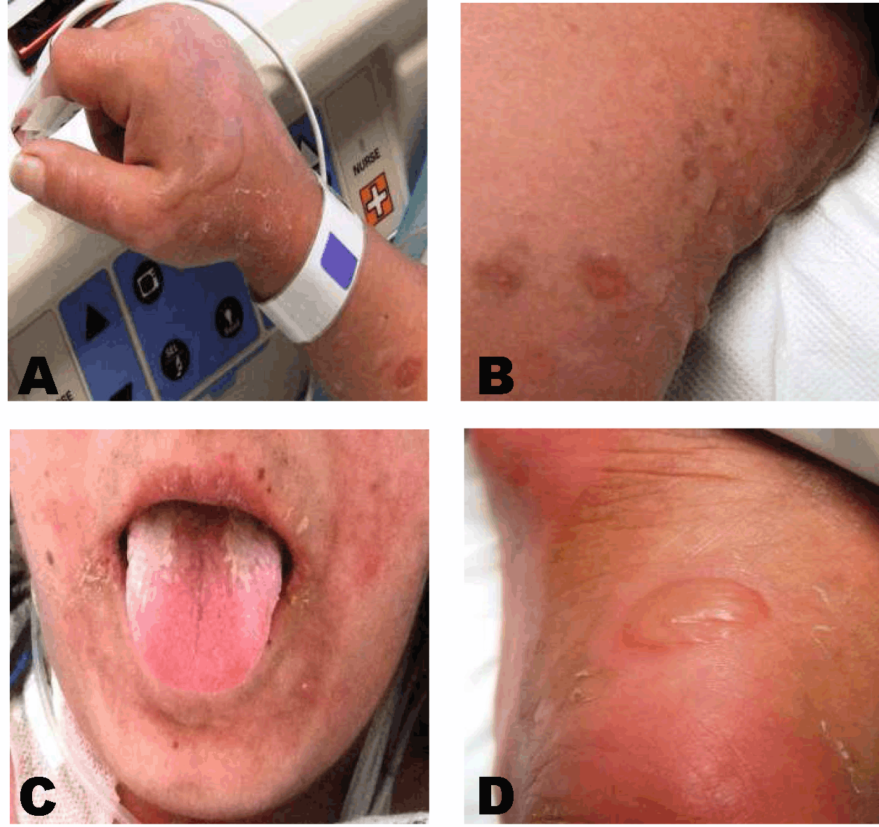
A 37-year-old female with a past medical history significant for Turner's syndrome, type-II diabetes mellitus and chronic pancreatitis, had undergone pancreatic duct drainage and partial pancreatic head resection (Frey procedure) 18 months prior to this admission. She had persistent pancreatic pain requiring narcotics and repeated hospitalizations. She presented to the clinic with chronic, epigastric abdominal pain. A discussion was held and she elected to proceed with total pancreatectomy with auto-islet transplant, which was subsequently performed at our institution. Her postoperative course was unremarkable and she was discharged on postoperative day (POD) 11 in satisfactory condition. The patient returned the day after discharge with worsening abdominal pain, mild distention, and worsened leukocytosis. A computed tomography (CT) scan was performed demonstrating a 4x2 cm fluid collection in the mesentery. She was empirically started on piperacillin-tazobactam 3.37 mg IV Q6 hrs after obtaining samples for blood and urine cultures. Her blood and urine cultures were negative, she remained afebrile, and her white blood cell count continued to decrease. Prior to discharge four days later, she developed hives and itching on her extremities. Piperacillin-tazobactam was stopped and she was administered diphenhydramine for relief of symptoms. Her symptoms were controlled and she was discharged home in stable condition on an oral regimen of ciprofloxacin 250 mg BID and metronidazole 500 mg daily. On the evening of discharge (POD 16), the patient noted spreading of her rash onto her chest, abdomen, feet, mouth and lips with the appearance of bullae. She developed fever, chills, rigors as well as dysuria and oropharyngeal burning. She returned to the emergency department the next morning. Upon evaluation, she was noted to have 35–50% of her body surface area covered with a rash and bullous lesions. (Figure 1A–D) Initial concern was for TEN versus SJS. Dermatology and ophthalmology were promptly consulted, and a skin biopsy was obtained. IgA levels and blood cultures were negative. Her skin lesions were treated with Vaseline gauze (Covidien Mansfield, MA) and Kerlix (Covidien Mansfield, MA), and she was started on solumedrol 60 mg IV BID. Skin biopsy sections demonstrated interface dermatitis characterized by detached epidermis with numerous dyskeratotic keratinocytes and scant perivascular infiltrate within the dermis, consistent with SJS. (Figure 2A–C) Piperacillin-tazobactam was suspected as the likely causative agent after a thorough review by all teams involved. She did not have any ophthalmologic issues. After stabilization and initial treatment, she was transferred to the regional burn center for continuation of management. She was initially managed with local skin care and IV antibiotics with significant improvement. She was subsequently lost to follow-up after discharge from the burn center. Unfortunately she is now listed as deceased in electronic medical record system, with unknown date and cause of death. | ||||||
|
| ||||||
|
| ||||||
|
Discussion
| ||||||
|
Epidemiology: TEN and SJS occur in approximately 1–2 per million individuals each year, with relatively similar rates reported in multiple countries around the world. [1] [2] Small differences in the rates of SJS and TEN can likely be attributed to genetics, medication availability, prescribing practices and other concurrent conditions, including cancers and infection. One notable population with a significantly increased risk for SJS and TEN includes those infected with HIV, as the annual incidence is 1000 times higher than the general population (1/1000). [1] [3] [4] Clinical Evaluation: Initial presentation of the disease typically begins several days prior to cutaneous manifestations. Symptoms include fevers, keratitis sicca, odynophagia and malaise making it difficult to differentiate from routine drug reactions. [1] [2] [5] When skin lesions first appear, they often begin on the trunk and face in addition to the palms and soles and appear as dusky red, flat, poorly delineated areas. They then continue to spread outward. [1] [5] Evidence of involvement of the buccal, genital, and/or ocular mucosa should raise suspicion of SJS or TEN and occurs in >90% of patients. [1] Ocular involvement early is common, and can include conjunctivitis, edema, erythema, crusting, discharge erosions and corneal ulcerations. [1] [6] [7] Epidermal detachment usually occurs as a slightly later secondary manifestation. The patent can be tested by applying lateral mechanical pressure and observing for detachment (Nikolsky’s sign) or visualizing bullae extending to surrounding skin when vertical pressure is applied (Asbor–Hansen sign). While not pathognomonic for SJS or TEN, these exam findings are helpful for ascertaining the danger of the reactions. In addition to epidermal involvement, the respiratory, gastrointestinal (including the liver), and urinary systems can be involved. [8] [9] [10] The TEN survivors have a 50% risk of long-term sequelae including changes in pigmentation of the skin and ocular complications ranging from simple (keratitis sicca, 46%) to severe (visual loss 5%, corneal ulceration 2%). [1] [11] Etiology: The pathophysiology of SJS and TEN remains unclear. Current theories support immune-mediated phenomena, resulting in keratinocyte apoptosis followed by necrosis. The generalized mechanism for this reaction is MHC-1 drug presentation leading to clonal expansion of cytotic T-cells, found in high numbers in the blisters of SJS/TEN patients. [1] However, additional mediators including FasL and granulysin (cytotoxic molecule), are likely to play primary roles in the actual generalized apoptosis of the keratinocytes. [1] [5] The Fas-FasL pathway has been linked to apoptosis in SJS/TEN via triggered intracellular DNA degradation [5]. The use of IVIG as treatment for SJS/TEN is based on this link between the disease and FasL. [1] [5] The exact pathway by which a medication leads to secretion or activation of Fas-FasL and/or ganulysin and thus keratinocyte apoptosis remains to be explained. Genetics: The link between SJS and genetic HLA class was first reported in the Han Chinese population with a strong association between HLA-B*1502 and carbamazepine. [1] [12] This association was confirmed in other Asian populations. However, this was not valid in European populations, leading to the conclusion that genetic markers are not specific in predicting SJS/TEN susceptibility in the general population. [1] Drugs: Numerous medications have been identified as causative agents for SJS and TEN. The most common medications linked to SJS/TEN over short-term use include trimethoprim-sulfamethoxazole and other sulfonamides, aminopenillins, cephalosporins, quinolones, and chlormezaone. Some medications are linked to SJS/TEN following long-term use, such as carbamazepine, phenytoin, phenobarbital, valproic acid, NSAIDs (oxicam-type), nevirapine, lamotrigine, sertraline, and allopurinol. With these medications, SJS/TEN usually occurs within two months after beginning treatment. [1] [3] Disease: While medications, the primary cause of SJS and TEN, mycoplasma pneumoniae and HSV have also been linked to development of these diseases without medication exposure. [1] HIV patients are at significantly higher risk for development of SJS/TEN compared to the general population. Diagnosis: The diagnosis of SJS versus TEN relies on clinical vigilance and pathologic confirmation. SJS is defined by mainly isolated lesions with <10% body surface area skin detachment, while TEN is a more severe and wide-spread form with widely confluent lesions and skin detachment covering >30% of the body surface area. [1] Skin biopsy is the only way to confirm definitive diagnosis and rule-out other autoimmune blistering diseases, fixed drug reactions, and staphylococcal scaled skin syndrome. Histopathologic analysis reveals necrosis of the epidermis involving all layers, with evidence of keratinocyte apoptosis. Direct immunofluorescent staining is also carried out to rule-out autoimmune causes. [1] Given the severe nature of SJS/TEN and its initial presentation, a rapid diagnostic test to predict SJS versus benign drug reaction is being developed. [13] Patients who are diagnosed with SJS have increased systemic levels of FasL as well as increased granulysin levels. While the FasL serum levels are too low to measure, the granulysin serum levels can be measured in rapid fashion to yield positive versus negative results in 15 minutes, similar to a rapid strep test. This method had sensitivity of 80% and specificity of 95.8% for SJS/TEN versus benign drug reaction. While not widely available for clinical use, further development of fast, efficient, early diagnostic tools is important for improved patient outcomes in the future. Management: Initial management when a diagnosis of SJS/TEN is suspected begins with immediate cessation of all new medications as well as urgent consultation to dermatology and ophthalmology. Universally accepted treatment includes monitoring volume status, evidence of sepsis, and conservative wound treatment without debridement. [1] The role of IVIG in treatment in addition to corticosteroids is generally accepted, although controversy remains regarding the exact details of the treatment. [14] Prognosis: The SCORTEN scoring system was developed as a validated means for predicting disease severity and mortality associated with SJS/TEN. (Table 1) (Table 2) [1] This score is useful for determination of appropriate treatment (all scores >3 should be treated in ICU and transferred to a regional burn center as soon as possible) and patient/family counseling. The leading cause of death in most patients is sepsis leading to acute renal failure. [1] The patient in this case report had a SCORTEN score of 4 (tachycardia + >10% body area involvement + glucose elevation + bicarbonate >20), corresponding to a 58.3% mortality. | ||||||
| ||||||
| ||||||
|
| ||||||
|
Conclusion
| ||||||
|
While many patients experience pruritis and/or rashes as side-effects to medications given post-operatively, it is important to keep in mind more serious reactions such as toxic epidermal necrolysis and Stevens–Johnson syndrome as these conditions can be rare causes of post-operative morbidity and mortality. If there is any concern for possible toxic epidermal necrolysis or Stevens–Johnson syndrome, prompt consultations to dermatology and ophthalmology should be obtained, possible causative agents should be immediately discontinued, and early transfer to a burn center should be initiated following confirmed diagnosis. | ||||||
|
References
| ||||||
| ||||||
|
[HTML Abstract]
[PDF Full Text]
|
|
Author Contributions
Mihir Shah – Conception and design, Acquisition of data, Analysis and interpretation of data, Drafting the article, Critical revision of the article, Final approval of the version to be published Kelsey Larson – Conception and design, Acquisition of data, Analysis and interpretation of data, Drafting the article, Critical revision of the article, Final approval of the version to be published Neilendu Kundu – Acquisition of data, Analysis and interpretation of data, Drafting the article, Critical revision of the article, Final approval of the version to be published R Matthew Walsh – Conception and design, Acquisition of data, Analysis and interpretation of data, Drafting the article, Critical revision of the article, Final approval of the version to be published |
|
Guarantor of submission
The corresponding author is the guarantor of submission. |
|
Source of support
None |
|
Conflict of interest
Authors declare no conflict of interest. |
|
Copyright
© Mihir Shah et al. 2013; This article is distributed the terms of Creative Commons attribution 3.0 License which permits unrestricted use, distribution and reproduction in any means provided the original authors and original publisher are properly credited. (Please see Copyright Policy for more information.) |
|
|