
  |

|
 |
|
Case Report
| ||||||
| Simultaneous Dieulafoy's lesion and marginal ulcer in gastric bypass patient | ||||||
| Vinay S Gundlapalli1, Arunkumar Baskara1, Susan Suozzo2, Brandon Oconnell3 | ||||||
|
1MD MRCS, Mercy Catholic Medical Center, Philadelphia.
2MA, Kansas City University of Medicine and Biosciences. 3FACS, Mercy Catholic Medical Center, Philadelphia. | ||||||
| ||||||
|
[HTML Abstract]
[PDF Full Text]
[Print This Article]
[Similar article in Pumed] [Similar article in Google Scholar] 
|


| How to cite this article: |
| Gundlapalli VS, Baskara A, Suozzo S, Oconnell B. Simultaneous Dieulafoy’s lesion and marginal ulcer in gastric bypass patient. International Journal of Case Reports and Images 2013;4(6):299–302. |
|
Abstract
|
|
Introduction:
Dieulafoy's lesion is one of the rare causes for gastrointestinal hemorrhage. Marginal ulcer is an anastamotic ulcer in the gastric pouch of a gastric bypass patient is a common cause gastrointestinal hemorrhage.
Case Report: We report a case of 27-year-old female who had undergone laparoscopic gastric bypass surgery who presented with gastrointestinal hemorrhage. Upper gastrointestinal endoscopy demonstrated a Dieulafoy’s lesion and a marginal ulcer in the gastric pouch. The Dieulafoy’s lesion was successfully managed with epinephrine injection and clip application. The marginal ulcer was not the bleeding source. Conclusion: To our knowledge this is the first reported case of simultaneous Dieulafoy lesion and marginal ulcer in a gastric pouch of a gastric bypass patient. | |
|
Keywords:
Dieulafoy's lesion, Marginal ulcer, Gastric bypass, Gastrointestinal hemorrhage
| |
|
Introduction
| ||||||
|
Dieulafoy's lesion is a rare cause of gastrointestinal hemorrhage and may be difficult to recognize. It consists of an arteriole that protrudes through a solitary, tiny mucosal defect, usually within 6 cm of the gastroesophageal junction on the lesser curve of the stomach. Marginal ulcer is a common reason for gastrointestinal hemorrhage in gastric bypass patients. We report an interesting case of gastrointestinal hemorrhage secondary to a Dieulafoy’s lesion in the gastric pouch of a Roux-en-Y gastric bypass at the same time this patient also had a marginal ulcer in the gastric pouch. | ||||||
|
Case Report
| ||||||
|
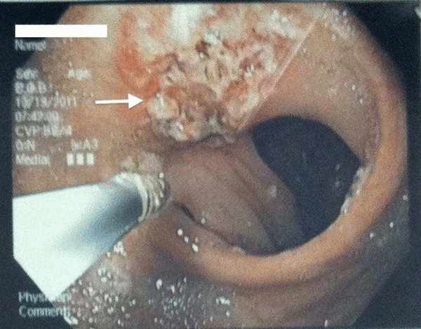
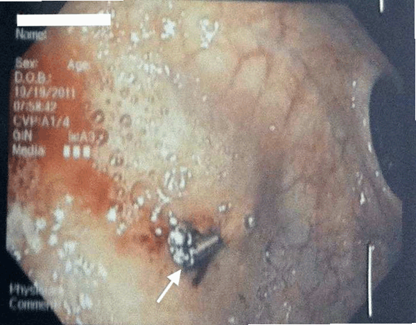
A 27-year-old African-American female with a history of morbid obesity, she underwent laparoscopic gastric Roux-en-Y bypass surgery one year ago, presented to the emergency room with a two-day history of melena associated with abdominal pain, nausea, and hematemesis. She did not have history of smoking or alcohol abuse. Her only medication was acetaminophen/diphenhydramine (Excedrin) for migraine headaches. Her hemoglobin (Hb) on admission was 7.7 mg/dL. She was symptomatic with tachycardia and weakness. She received a unit of packed red cells. Her hemoglobin increased to 8.4 mg/dL after transfusion. The next day she had further episodes of hematemesis and melena with a hemoglobin drop to 7.0 mg/dL. She had an upper gastrointestinal endoscopy and antral biopsy prior to the bypass procedure, which showed erosive esophagitis. During the current admission, she underwent an upper gastrointestinal endoscopy, which showed a marginal ulcer (Figure 1) of about 1.5 cm in diameter on the jejunal side of gastrojejunal anastomosis and a bleeding Dieulafoy’s lesion of about 3 mm (Figure 2) in the gastric pouch. The bleeding Dielufoy’s lesion was controlled with epinephrine injection, electrocautry and endoclip application. A total of 18 mL of 1:10,000 epinephrine solution was injected around the lesion and then the lesion was cauterized with bipolar electrocoagulation (BICAP) electrocautery at 17 watts setting. (Figure 3) There was still some mild ooze, so two endoclips were applied onto this area with excellent hemostasis. Her hemoglobin was stable at 9 mg/dL after the procedure and was started on diet which she tolerated. She was discharged home with proton pump inhibitors. Her follow-up visit in the clinic after couple weeks was unremarkable with no abdominal pain, hematemesis or melena with a stable hemoglobin of 9.4 mg/dL. Patient had uneventful hospital course after the procedure. | ||||||
| ||||||
| ||||||
| ||||||
|
Discussion
| ||||||
|
Dieulafoy's lesion was first described by Galland in 1884, and he described three cases of these lesions in 1898. [1] Dieulafoy called the lesion "exulceratio simplex" because of the small size and the large, histologically normal underlying artery. In approximately 4–9% of massive upper gastrointestinal hemorrhage, no demonstrable cause can be found. [2] [3] [4] [5] Dieulafoy’s lesion is thought to be the cause of 1–2% of such cases. A Dieulafoy’s lesion is a difficult lesion to recognize, especially when bleeding is inactive. Therapeutic endoscopy is the initial treatment of choice. The first endoscopic intervention is successful in 85% of patients, while 10% of patients need a repeat endoscopy, and 5% of patients may require surgical intervention. [6] [7] Marginal ulcer is an anastmotic ulcer seen at the gastrojejunostomy site of the gastric bypass. This is one of the frequently occurring complications after gastric bypass surgery. The incidence of marginal ulcer has been quoted between 2–20 % in literature. The etiology is not very well understood. The presence of certain risk factors such as smoking, NSAID, steroids, increased acid and H. pylori infection have been suggested to increase the incidence. Patients may present with epigastric pain, nausea, vomiting or upper gastrointestinal bleeding. The treatment of marginal ulcer is usually conservative with mucosal protection, proton pump inhibitors and H. pylori eradication. Endoscopic evaluation is diagnostic as well as therapeutic. Surgical intervention is required for failure of conservative management and perforation. This case demonstrates the diagnosis and successful endoscopic management of these rare lesions. Although Dieulafoy’s lesions are rare, they are an important and potentially life-threatening cause of gastrointestinal hemorrhage. There has been no documented association between Roux–en-Y gastric bypass and Dieulafoy’s lesions so far but if there is any, will be a question that remains to be answered. | ||||||
|
Conclusion
| ||||||
|
To our knowledge this is the first time the combination of Dieulafoy’s lesion and marginal ulcer has ever been reported in a gastric bypass patient. Careful evaluation of a gastric pouch is very important during an endoscopy for upper gastrointestinal bleeding as small Dieulafoy’s lesions can be very easily missed. | ||||||
|
Acknowledgements
| ||||||
|
We wish to express our sincere thanks to Dr Steven Litchenstein, Head, Department of Gastroenterology, Mercy Catholic Medical Center, Philadelphia. | ||||||
|
References
| ||||||
| ||||||


|
[HTML Abstract]
[PDF Full Text]
|
|
Author Contributions
Vinay S Gundlapalli – Substantial contributions to conception and design, Acquisition of data, Analysis and interpretation of data, Drafting the article, Revising it critically for important intellectual content, Final approval of the version to be published Arun Baskara– Acquisition of data, Analysis and interpretation of data, Drafting the article, Revising it critically for important intellectual content, Final approval of the version to be published Susan Souzzo– Substantial Acquisition of data, Analysis and interpretation of data, Drafting the article, Revising it critically for important intellectual content, Final approval of the version to be published Brandon Oconnell– Acquisition of data, Analysis and interpretation of data, Drafting the article, Revising it critically for important intellectual content, Final approval of the version to be published |
|
Guarantor of submission
The corresponding author is the guarantor of submission. |
|
Source of support
None |
|
Conflict of interest
Authors declare no conflict of interest. |
|
Copyright
© Vinay S Gundlapalli et al. 2013; This article is distributed the terms of Creative Commons Attribution License which permits unrestricted use, distribution and reproduction in any means provided the original authors and original publisher are properly credited. (Please see Copyright Policy for more information.) |
|
|