
  |

|
 |
|
Case Report
| ||||||
| Feeding dystonia: A classical presentation of neuroacanthocytosis | ||||||
| Suman Kushwaha1, Akhila Panda2, Vachan Mehta2, Seema Malik2, Ishita Pant3 | ||||||
|
1Associate Professor Neurology, DM Neurology, Department of Neurology, Institute of Human Behavior and Allied Sciences, Delhi, India.
2Senior Resident Neurology, Institute of Human Behaviour and Allied Sciences, Delhi, India. 3Assistant Professor, Pathology, Institute of Human Behavior and Allied Sciences, Delhi, India . | ||||||
| ||||||
|
[HTML Abstract]
[PDF Full Text]
[Print This Article]
[Similar article in Pumed] [Similar article in Google Scholar] 
|


| How to cite this article: |
| Kushwaha S, Panda A, Mehta V, Malik S, Pant I. Feeding dystonia: A classical presentation of neuroacanthocytosis. International Journal of Case Reports and Images 2013;4(1):37–40. |
|
Abstract
|
|
Introduction:
Neuroacanthocytosis (NA) is a heterogeneous neurodegenerative genetic disorder caused by disease specific genetic mutation. Being an extremely rare disorder, only a few thousand cases have been reported till date. This clinical entity was described by Citchley et al. and was initially named Levine-Citchley syndrome. It is characterized by movement disorder due to degeneration of the basal ganglia along with cognitive and behavior changes. The classical clinical presentation includes the troublesome abnormal involuntary movements in form of chorea, dystonia and dyskinesia. Self mutilation of the lips and tongue is characteristic of choreoacanthocytosis. The NA syndromes have been broadly divided into two subtypes, (i) core NA syndromes, and (ii) conditions with alterations in the lipoprotein metabolism. The genes of the different NA syndromes have been identified but the mechanism of these genetic mutations is not known. The management of NA is primarily symptomatic and rehabilitative.
Case Report: We are describing a case of neuroacanthocytosis with typical phenotype of choreoacanthocytosis. The phenomenon of feeding dystonia is classically being discussed in this report. Neuroimaging demonstrated the atrophy of the caudate nucleus resembling Huntington's chorea. Acanthocytic red blood cells were seen in peripheral smear in our patients. Conclusion: The recognition of neuroacanthocytosis is improved due to better characterization of the clinical symptoms and investigations of this heterogeneous entity. The presented case describes the typical clinical characteristics and investigations supporting the diagnosis of this under diagnosed clinical syndrome. | |
|
Keywords:
Feeding dystonia, Chorea, Acanthocytes, Neuroacanthocytosis
| |
|
Introduction
| ||||||
|
Neuroacanthocytosis (NA) is an uncommon, heterogeneous, genetic neurodegenerative disorder. This clinical entity was described by Citchley et al. and was initially named Levine-Citchley syndrome. The clinical manifestations include variety of movement disorders and cognitive and behavioral changes closely resembling Huntington's disease. The orofaciolingual dyskinesia is prominent and characteristic of neuroacanthocytosis. There is presence of thorny deformation in the lipid membrane of the circulating red blood cells called acanthocytosis. These acanthocytes are distinct and unique for this syndrome. The recognition of neuroacanthocytosis is improved due to better characterization of the clinical symptoms and investigations of this heterogeneous entity. The presented case describes the typical clinical characteristics and investigations supporting the diagnosis of this under diagnosed clinical syndrome. | ||||||
|
Case Report
| ||||||
|
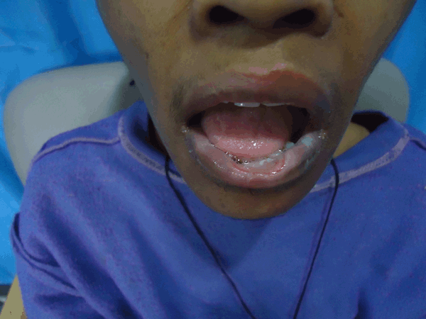
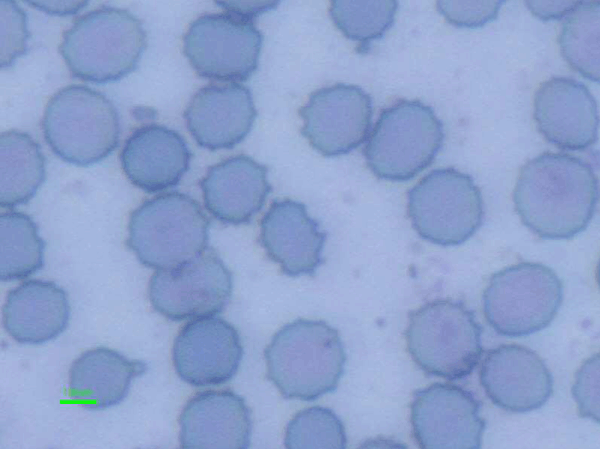
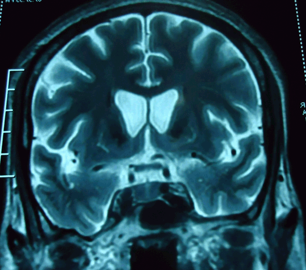
A 30-year-old female presented with three years history of progressive difficulty in eating. She attributed it to abnormal involuntary movement of tongue and sustained spasmodic movement of the neck. She used different maneuvers during eating like pushing the bolus with the fingers inside the mouth and drinking water and food in supine position. She sometimes developed choking sensation and cough while eating food. Occasionally, she had nasal regurgitation while drinking water. She had abnormal, repetitive, non-patterned, involuntary, arrhythmic, non-purposeful movements of the whole body including head and neck. These movements disappeared in sleep and were not suppressed with voluntary action. Since last nine months she had developed abnormal behavior in the form of excessive and non-purposeful talking, repeating the sentences and motor acts. The caregivers reported the habit of self-mutilation. She repeatedly chewed her lips which lead to severe injury of lower lip. There was no history of visual problems, limb weakness, sensory, cerebellar or autonomic disturbance. She had significant weight loss. Her personal history revealed poor maintenance of hygiene and history of secondary amenorrhea. Family history and past medical history were non-contributory. General physical examination showed poor self care and cachexia. The vital parameters were normal. A significant lip biting mark was present in inner aspect of lower lip (Figure 1) suggestive of self-mutilation and repeated friction of the lower lip due to orofacial dyskinesia. Other systemic examination was normal. On neurological examination, she was conscious and oriented. Marked behavior changes in form of repetition of words and perseveration of motor acts were present. Neuropsychological assessment shows high frontality on frontal lobe assessment by frontal assessment battery (FAB) battery. Memory functions were normal. She had features of obsessive compulsive behavior and anxiety. Cranial nerve examination was normal. The limbs were hypotonic with power of 5/5 on Medical Research Council (MRC) scale in all the limbs. Generalized areflexia with flexor plantar response was present. All the primitive reflexes were absent. Sensory, cerebellar and autonomic system examination revealed no abnormality. Extra pyramidal system examination showed combination of abnormal involuntary movements. Chorea was prominent involving orolingual, neck, trunk and limbs. Tongue had classical jack in box phenomenon. In addition to chorea, head and neck shows intermittent dystonic movements. Due to combination of these abnormal movements of tongue and neck the patient had classical feeding difficulties described as feeding dystonia. She walked with bizarre gait due to abnormal movements of the trunk. The routine investigations, complete blood count, kidney and liver function tests, serum electrolytes, lipid profile, copper studies, HIV ELISA, blood VDRL were within normal limits. Creatinine phosphokinase was raised to 614 U/L. Peripheral blood acanthocytes were visible on screening of three consecutive samples. (Figure 2) Nerve conduction studies showed sensorimotor axonal neuropathy. Neuroimaging of brain using 3 Tesla MRI showed diffuse cerebral atrophy with significant caudate atrophy. (Figure 3) | ||||||
| ||||||
| ||||||
| ||||||
|
Discussion
| ||||||
|
Neuroacanthocytosis is a rare movement disorder syndrome. The prevalence is estimated to be less than 1–5 million. Neuroacanthocytosis has varied clinical presentations ranging from involuntary hyperkinetic movements, neuromuscular involvement to cognitive and behavioral changes. Onset of symptoms is usually in adulthood. The core NA syndrome includes autosomal recessive choreoacanthocytosis and X-linked Mcleod syndrome. The other subgroup includes neuroacanthocytosis with lipoprotein disorders e.g. abetalipoproteinemia (Bassen, Kornzweig disease). This symptom variation in presentation is responsible for the under diagnosis of this clinical entity. Chorea acanthocytosis is caused by mutation of 73 exon gene on chromosome 9, VPS13A which codes for Chorein. [1] Our patient had characteristic phenotype of neuroacanthocytosis. She had presented in adulthood with typical orofacial and lingual dyskinesia troubling her in eating. Due to abnormal posturing of the neck and repeated tongue protrusion she had characteristic feeding dystonia which has been described in neuroacanthocytosis. [2] Weight loss due to poor nutritional status is a major concern in these patients. Lingual and orofacial dyskinesia causes irritation of lower lip in combination with lip biting resulting in severe lower lip injury. The patient had bizarre movements involving the whole body in form of violent trunk spasms and head thrusting which were evident on walking. This typical gait is described as "rubber man" gait. [3] As the disease progresses the hyperkinetic movement disorder evolves into the hypokinetic or bradykinetic state. These patients have major disability due to combination of movement disorders like dystonia and chorea. Neuropsychiatric symptoms are prominent in neuroacanthocytosis and may appear before the movement disorder. eizure were absent in our patient while they are usually seen in approximately 40% patients. [4] The patients have elevated creatinine kinase levels and axonal neuropathy. The neuromuscular involvement in choreoacanthocytosis includes myopathy and axonal sensory-motor neuropathy. [5] McLeod syndrome should be considered as a differential diagnosis of choreoacanthocytosis as neuromuscular involvement is more common in this subtype of neuroacanthocytosis. [6] Hepatosplenomegaly and cardiac involvement is seen in McLeod syndrome. Panthothenate kinase-associated neurodegeneration (PKAN) an autosomal recessive disorder is another differential diagnosis. It is a rapidly progressive childhood disorder. Dystonia along with cognitive and behavior changes are the prominent clinical manifestation. Huntington's disease is the closest differential diagnosis as the phenotype includes chorea, behavior and cognitive deficits. The pattern of caudate atrophy on neuroimaging is similar in both these disorders. The genetic testing for Huntington's disease is diagnostic. The classical phenotype of uncommon movement disorder, neuroacanthocytosis presents with combination of abnormal movements and feeding dystonia along with self mutilatilating behavior and cognitive deficits in our patient. The clinical diagnosis of sporadic variant of choreoacanthocytosis is supported by classical neuroimaging and presence of acanthocytosis in the peripheral smear. Due to lack of facility for analysis of VPS13a or Chorein gene the confirmation of diagnosis could not be reached in our case. The management remains mainly symptomatic. The incapacitating movements can be reduced with typical antipsychotics and dopamine depletors like tetrabenezine. Choreoacanthocytosis progresses over 15–30 years. Long term outcome of patient is poor. Neurorehabilitation and behavior therapy plays important role in functional improvement as well as maintainance of better quality of life. Our patient is maintaining well on treatment and regularly followed up in outpatient department. | ||||||
|
Conclusion
| ||||||
|
Our case demonstrates the classical phenotype of neuroacanthocytosis. The variation in symptomatology causing the different phenotypes with significant overlap makes the diagnosis difficult. The important clinical differences exist among the phenotypes and should be emphasized while making the clinical diagnosis. The better recognition and understanding of the phenomenology in neuroacanthocytosis will facilitate us in diagnosis of this entity. | ||||||
|
References
| ||||||
| ||||||

|
[HTML Abstract]
[PDF Full Text]
|
|
Author Contributions
Suman Kushwaha – Conception and design, Analysis and interpretation of data, Critical revision of the article, Final approval of the version to be published Akhila Panda – Acquisition of data, Drafting the article, Final approval of the version to be published Vachan Mehta – Acquisition of data, Drafting the article, Final approval of the version to be published Seema Malik – Acquisition of data, Drafting the article, Final approval of the version to be published Ishita Pant – Analysis and interpretation of data, Drafting the article, Final approval of the version to be published |
|
Guarantor of submission
The corresponding author is the guarantor of submission. |
|
Source of support
None |
|
Conflict of interest
Authors declare no conflict of interest. |
|
Copyright
© Suman Kushwaha et al. 2013; This article is distributed the terms of Creative Commons Attribution License which permits unrestricted use, distribution and reproduction in any means provided the original authors and original publisher are properly credited. (Please see Copyright Policy for more information.) |
|
|