| |
|
|
|
Case Report
| ||||||
| Unusual case of ascites | ||||||
| Guido Poggi1, Benedetta Montagna1, Pamela Di Cesare1, Erica Quaquarini1, Emma Pozzi1, Carlo Aprile2 | ||||||
|
1Department of Oncology II, IRCCS Maugeri Foundation, Pavia, Italy.
2Consultant, Department of Nuclear Medicine, IRCCS S. Matteo Hospital, Pavia, Italy. | ||||||
| ||||||
|
[HTML Abstract]
[PDF Full Text]
[Print This Article]
[Similar article in Pumed] [Similar article in Google Scholar] 
|
| How to cite this article: |
| Poggi G, Montagna B, Di Cesare P, Quaquarini E, Pozzi E, Aprile C. Unusual case of ascites. International Journal of Case Reports and Images 2013;4(1):32–36 |
|
Abstract
|
|
Introduction:
Clostridium difficile infection is very commonly related to antibiotic therapy. The spectrum of clinical manifestation of C. difficile infection may include, in an increasing order of severity, absence of symptoms, colitis without formation of pseudomembranes and pseudomembranous colitis (PMC). PMC is a severe but rare complication of the infection. It is related to the bacterial production of enterotoxin A and B. Its clinical features include diarrhea, abdominal tenderness and fever. In the worst case, it may progress to toxic megacolon and colonic perforation. Ascites is an infrequent direct complication of most severe cases of PMC.
Case Report: We report a case of ascites arising two weeks after the resolution of C. difficile infection, in which hypoalbuminemia, caused by protein losing enteropathy, was the most likely pathogenetic mechanism. The patient recovered completely after human albumin intravenous support and diuretics. Conclusion: Protein losing syndrome represents a group of disorders in which hypoproteinemia and edema occur in the absence of either proteinuria or defects in protein synthesis. It is a consequence of the intestinal epithelial cell damage and increased mucosal permeability induced by C. difficile enterotoxin. The clinical manifestations are ascites and low serum albumin levels. The treatment requires human albumin intravenous integrations and diuretics. Ascites could be a direct complication of antibiotic-associated pseudomembranous colitis, but it could also be the manifestation of a protein-losing enteropathy, caused by inflammatory damage of gastrointestinal mucosa. | |
|
Keywords:
Clostridium difficile, Infection, Complications, Protein losing enteropathy, Ascites, MSSA infection
| |
|
Introduction
| ||||||
|
Clostridium difficile is a Spore forming, gram positive bacillus. It is a well-recognized cause of antibiotic associated diarrhea. The spectrum of clinical manifestation of C. difficile infection may include, in an increasing order of severity, absence of symptoms, antibiotic-associated colitis without the formation of pseudomembranes and pseudomembranous colitis (PMC). PMC is a severe but rare complication of the infection, related to the bacteria production of enterotoxin A and B. Its clinical features include diarrhea, abdominal tenderness, fever, dehydration and leukocytosis. In the worst case, it may progress to toxic megacolon and colonic perforation resulting from full-thickness colonic wall necrosis. The diagnosis of PMC depends on the demonstration of C. difficile toxins in the stool or on endoscopic evidence of adherent yellow plaques along colonic wall. Computed tomography (CT) scan is also useful for PMC diagnosis, showing colonic wall thickening, low-attenuation mural thickening corresponding to mucosal and sub-mucosal edema and pericolonic stranding. [1] [2] [3] Early treatment with oral metronidazole or oral vancomycin usually leads to a clinical improvement within 48–72 hours. Surgical intervention is mandatory in perforation and may be required in severe cases where medical treatment is not sufficient. Ascites is an infrequent direct complication of most severe cases of PMC. [4] We describe a case of ascites arising two weeks after the resolution of C. difficile infection, in which hypoalbuminemia, secondary to protein losing enteropathy was the most likely pathogenic mechanism. | ||||||
|
Case Report
| ||||||
|
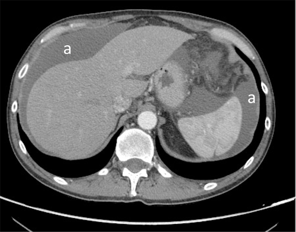
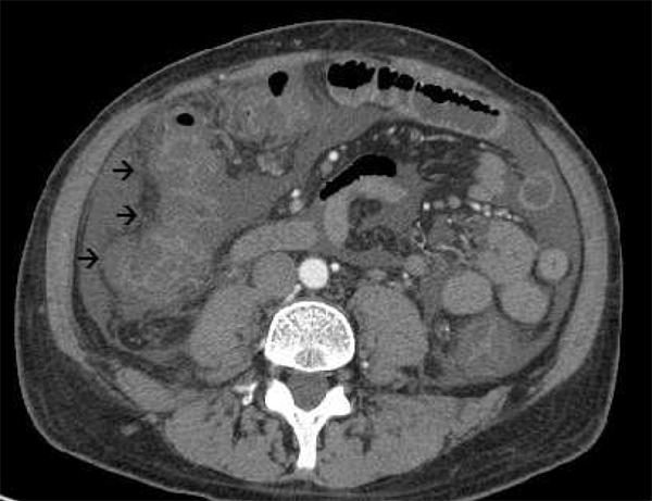
A 54-year-old man was admitted to our hospital with complaints of abdominal distension and bilateral peripheral edema. He had no previous history of such illness. Ten weeks before his admission, he developed a soft tissue infection at the back of his left hand which rapidly evolving to a necrotising fasciitis of the left forearm and required surgical decompression followed by necrotic tissue debridements. Microbiological culture of the tissue samples taken from the wound identified a growth of a methicillin sensitive staphylococcus aureus (MSSA) producing panton-valentine leukocidin toxin. Blood cultures were negative. The patient was treated with intravenous ciprofloxacin (200 mg/bid) and clindamycin (500 mg/bid) for ten days. After the treatment, he was discharged home and continued therapy with oral ciprofloxacin (500 mg/bid) and oral rifampicin (600 mg/day). After one month of treatment, he developed non-bloody, watery diarrhea consisting of 8-12 stools daily, fever and abdominal pain. The patient was re-hospitalized. Stool cultures were negative for shigella, salmonella, campylobacter and yersinia bacteria while they were positive for C. difficile toxins. The EIA-test for C. difficile was also negative. The patient was treated with oral vancomycin (500 mg every 6 hours for 5 days), with progressive clinical benefit and resolution of diarrhea, and he was discharged home. Due to the subsequent appearance of abdominal swelling and peripheral edema, occurring two weeks after the resolution of abdominal symptoms, the patient was re-admitted to our hospital. On physical examination, the patient was alert and had little discomfort. Vital signs were normal and the patient had no fever. Abdominal examination showed diffuse abdominal distension with active bowel sounds, a dull percussion note and mild, diffuse tenderness without jaundice. The patient had pitting edema of lower extremity. Lymphadenopathy was not present. Neurological and cardiac examinations were normal. Laboratory tests revealed normal hemoglobin values, normal renal function tests, no electrolytes abnormalities, normal hepatic and coagulation function. Erythrocyte sedimentation rate and C-reactive protein were negative. Total serum protein was 5.0 g/dL with marked hypoalbuminemia (2.5 g/dL). Mild lymphopenia and hypogammaglobulinemia were also detected. Hepatitis B surface antigen and hepatitis C antibody were negative. Computed tomography scan showed thickened colonic wall and massive ascites without evidence of hepatosplenomegaly, portal or hepatic vein thrombosis or peritoneal thickening (Figures 1, 2). Abdominal paracentesis was performed. Asatic fluid examination showed WBC count of 400 cells/mm3 with 65% polymorphonuclear leukocytes, total protein 3.0 g/dL, albumin 1.4 g/dL. Serum-ascites albumin gradient (SAAG) was 1.1 and ascitic fluid/serum LDH ratio was 0.78. Cultures of ascitic fluid for bacteria and mycobacteria were negative and cytological examination did not find malignant cells. The C. difficile toxin was not detected in ascitic fluid. Urine collection over 24 hours failed to show proteinuria. A stool collection for fecal fat was normal. Serologic testing for celiac disease was negative. Fecal a-1 antitrypsin (A1AT) clearance was found to be elevated (288 mg per 100 mL; normal value <30 mg/dL) suggesting that the severe hypoalbuminemia might be caused by protein losing enteropathy. Tc-99m labeled human serum albumin scintigraphy demonstrated an abnormal excretion of Tc-99m albumin into the large bowel confirming the diagnosis of protein loosing enteropathy (Figure 3). During the hospitalization, patient was treated with salt restriction, human albumin intravenous infusion and diuretics. The patient gradually recovered and he was discharged. Three weeks later, ascites had completely disappeared as shown by ultrasound examination. At a follow-up visit one month later, he reported having no diarrhea and serum albumin levels were normal. After three months, an abdominal CT scan showed complete resolution of increased thickness of the colonic wall and there was no evidence of ascites. | ||||||
| ||||||
| ||||||
| ||||||
|
Discussion
| ||||||
|
Ascites is a clinical condition that may represent the initial manifestation of a systemic disease or of otherwise unsuspected abdominal disease. In most cases, ascites appears as part of a well-recognized illness, for example cirrhosis, congestive heart failure, nephrosis or disseminated carcinomatosis. On occasion, ascites may develop as an isolated finding in the absence of a clinically evident disease. In such cases, a careful analysis of ascitic fluid may suggest the etiology: the serum ascites-albumin gradient (SAAG) > or < 1 g/dL can differentiate between a transudate or an exudate fluid. Serum ascite-albumin gradient >1 is usually related to uncomplicated chirrosis, alcoholic hepatitis, congestive heart failure and Budd-Chiari syndrome; while SAAG <1 is related to other causes, such as peritoneal carcinomatosis, tuberculous peritonitis, pancreatitis, serositis, pyogenic peritonitis, and nephritic syndrome. [5] In our case, ascites was the late complication of a C. difficile associated diarrhea following a prolonged antibiotic therapy for a soft tissue infection. The patient had no other systemic disease that could have justified the occurrence of ascites, such as hepatic or cardiac failure. SAAG was 1.1 g/dL with mild leukocytosis in the fluid and proteinuria was absent. A literature review revealed that ascites occurs more frequently as an indirect complication of severe cases of PMC. Systemic capillary leak is considered the most likely mechanism of ascites, occurring in the worst cases of PMC. [6] [7] [8] However, our patient did not developed a severe form of colitis and the onset of ascites occurred after a few days from the complete resolution of gastrointestinal symptoms. On investigating we found that, in this case, the prevalent mechanism of formation of ascites was a protein-losing enteropathy. Protein-losing enteropathy is not a specific disease but rather a group of gastrointestinal and non-gastrointestinal disorders occurring with hypoproteinemia and edema in the absence of either proteinuria or defects in protein synthesis. Diseases characterized by excess protein loss into the gastrointestinal tract are caused by mucosal ulceration (e.g., ulcerative colitis, gastrointestinal carcinomas, and peptic ulcer), by damage to mucosa without ulceration (e.g., celiac sprue and Ménétrier's disease) or by lymphatic dysfunction; either primary lymphatic disease or secondary to partial lymphatic obstruction (e.g. intestinal lymphangiectasia, mesenteric nodes or lymphoma, cardiac disease). [9] C. difficile enterotoxins A and B induce intestinal epithelial cell damage, increase mucosal permeability, stimulate interleukin (IL)-8 synthesis, and cause an acute inflammatory response characterized by neutrophil recruitment and tissue damage. [10] This condition can lead to a protein losing syndrome with severe hypoalbuminemia that can result in peripheral edema and ascites, as we described in this case report. The management of ascites involves, apart from the specific antibiotic therapy for C. difficile infection, human albumin intravenous integrations and diuretics. | ||||||
|
Conclusion
| ||||||
|
In summary, ascites could be a direct complication of antibiotic-associated pseudomembranous colitis but it could also be the manifestation of a protein losing enteropathy, caused by the inflammatory damage of the gastrointestinal mucosa. | ||||||
|
References
| ||||||
| ||||||
|
[HTML Abstract]
[PDF Full Text]
|
|
Author Contributions
Guido Poggi – Substantial contributions to conception and design, Acquisition of data, Analysis and interpretation of data, Drafting the article, Final approval of the version to be published Benedetta Montagna – Substantial contributions to conception and design, Acquisition of data, Analysis and interpretation of data, Drafting the article, Final approval of the version to be published Pamela Di Cesare – Substantial contributions to conception and design, Acquisition of data, Analysis and interpretation of data, Drafting the article, Final approval of the version to be published Erica Quaquarini – Substantial contributions to conception and design, Acquisition of data, Analysis and interpretation of data, Drafting the article, Final approval of the version to be published Emma Pozzi – Substantial contributions to conception and design, Acquisition of data, Analysis and interpretation of data, Drafting the article, Final approval of the version to be published Carlo Aprile – Substantial contributions to conception and design, Acquisition of data, Analysis and interpretation of data, Drafting the article, Final approval of the version to be published |
|
Guarantor of submission
The corresponding author is the guarantor of submission. |
|
Source of support
None |
|
Conflict of interest
Authors declare no conflict of interest. |
|
Copyright
© Guido Poggi et al. 2013; This article is distributed the terms of Creative Commons Attribution License which permits unrestricted use, distribution and reproduction in any means provided the original authors and original publisher are properly credited. (Please see Copyright Policy for more information.) |
|
|