| |
|
|
|
Case Report
| ||||||
| Spinal tuberculomas mimicking spinal dural arteriovenous fistula: A case report | ||||||
| Jyoti Sureka1, Varsha Mary Khalkho2, Binita Riya Chacko3 | ||||||
|
1Associate Professor, Department of Radiology, Christian Medical College and Hospital, Vellore, Tamil Nadu, India.
2Tutor, Department of Radiology, Christian Medical College and Hospital, Vellore, Tamil Nadu, India. 3Assistant Professor, Department of Radiology, Christian Medical College and Hospital, Vellore, Tamil Nadu, India. | ||||||
| ||||||
|
[HTML Abstract]
[PDF Full Text]
[Print This Article]
[Similar article in Pumed] [Similar article in Google Scholar] 
|
| How to cite this article: |
| Sureka J, Khalkho VM, Chacko BR. Spinal tuberculomas mimicking spinal dural arteriovenous fistula: A case report. International Journal of Case Reports and Images 2013;4(1):28–31. |
|
Abstract
|
|
Introduction:
Tuberculosis is a very common disease in developing countries and has been found to affect almost all the parts of the body. We report the case of a patient who had spinal cord tuberculomas without evidence of symptoms of systemic tuberculosis. The lesions were located at the surface of lower thoracic cord and mimicked a spinal-dural arteriovenous fistula (SDAVF) on magnetic resonance imaging (MRI).
Case Report: The patient was 45-year-old man who presented with a history of progressive paraparesis with a clinical suspicion of intramedullary tumor. First diagnosis was made as SDAVF on MRI. Then he underwent diagnostic and therapeutic digital substraction angiogram which was negative for the same. Again MRI was reviewed by a senior radiologist and a final diagnosis of spinal cord pial surface tuberculomas was made, confirmed by cerebrospinal fluid analysis and treated by appropriate anti-tubercular therapy. Conclusion: Tuberculosis can mimic a number of disease entitiles. It is important to be familiar with various a typical radiological presentations of tuberculosis. | |
|
Keywords:
Tuberculoma, Spinal dural, Arteriovenous malformation
| |
|
Introduction
|
|
Tuberculosis (TB) is a very common disease in developing countries and has been found to affect almost all the parts of the body. It can affect any organ or organ system of the body. Tuberculosis primarily affects the chest and can involve multiple extrapulmonary sites like heart, bones, joints, gastrointestinal system, genitourinary system, central nervous system, eyes, etc. Spinal TB accounts for more than half of the musculoskeletal TB. The extradural form of spinal TB is most common. [1] Uncommonly, it can present as arachnoidal, intradural extramedullary and intramedullary form. Spinal intradural-intramedullary tuberculoma is extremely rare entity can give rise to a variety of clinical and radiologic features which can mimic a number of other spinal cord lesions particularly intramedullary tumors. [2] [3] We report the case of a patient who had spinal cord tuberculomas without evidence of symptoms of systemic tuberculosis. The lesions were located at the surface of lower thoracic cord and mimicked a spinal dural arteriovenous fistula (SDAVF) on magnetic resonance imaging (MRI). |
|
Case Report
|
|
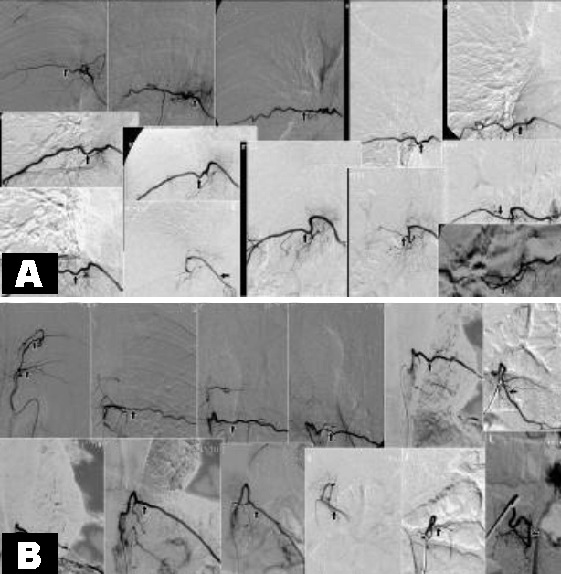
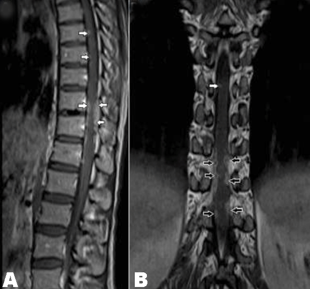
A 45-year-old male presented to the neurology out patient department (OPD) with complaints of back pain and progressive lower limb weakness since six months. There were no bowel or bladder difficulties. On physical examination, there was spine tenderness over the mid and lower thoracic spine, muscle weakness and abnormal reflexes in lower limbs. Laboratory investigations revealed positive results for human immunodeficiency virus (HIV) and hepatitis B virus (HBV). However, complete blood picture, ESR, C-reactive protein and other routine blood examinations were within normal limits. Chest X-ray was also normal. Based on clinical findings possibility of intramedullary tumor was considered. Patient underwent gadolinium-enhanced MRI of spine. Sagittal and coronal T2-weighted MRI of thoracolumbar spine showed multiple tortuous flow voids along the surface of mid and lower thoracic spinal cord. The lower thoracic cord was also slightly enlarged and showed increased intramedullary signal intensity (Figure 1). Diagnosis of SDAVF with edema or ischemia of cord secondary to venous hypertension was made. For further management, patient underwent diagnostic and therapeutic selective spinal DSA. The angiogram did not reveal any feeding vessels or nidus to suggest arteriovenous fistula (AVF) (Figure 2A–B). MRI images were further reviewed by a senior radiologist with a referring doctor. The coronal gadolinium-enhanced T1-weighted images showed multiple, small, almost similar sized ring and nodular enhancing lesions along the pial surface of the cord with central T2 hypointensity which appeared as a flow void on T2-weighted sagittal and coronal images (Figure 3). There was no focal enhancing lesion or abnormal enhancement in the region of high signal intensity of the lower thoracic spinal cord. Considering the immunocompromised status, a final diagnosis of spinal cord pial surface tuberculomas with associated cord edema or myelitis was made. MRI of the brain did not reveal any lesion. Patient underwent CSF analysis which grew acid-fast bacilli and isolated species was Mycobacterium tuberculosis and hence the diagnosis was confirmed and first line anti-tubercular therapy (ATT) was started. The patient responded well to ATT. |
|
|
|
|
|
|
|
Discussion
|
|
Central nervous system TB is commonly seen in tropical countries. [4] In immunocompromised individuals, the presentation of tubercular lesions may be atypical and can result in delayed diagnosis. [5] [6] Imaging plays an important role in recognition of these atypical cases that mimic other neurologic conditions thus helps in early diagnosis and treatment, which otherwise may result in irreversible neurological sequel. [4] [5] [6] A few case reports talk about the isolated meningeal or spinal tuberculoma mimicking spinal tumor. However, spinal TB mimicking a SDAVF not yet reported in literature. Extradural form is the most common while arachnoiditis, intradural and intramedullary tuberculomas are uncommon presentation of spinal TB. [4] [7] This atypical form of TB can occur as a direct extension from the vertebrae, as a downward extension of intracranial tubercular meningitis, and less commonly as tuberculous lesions primarily developing in spinal meninges. [5] [8] MRI is the imaging modality of choice for these lesions. Spinal cord TB, generally, present as intramedullary tuberculomas with or without myelitis and syrinx. Clinically as well as radiologically, intramedullary tuberculomas may be difficult to differentiate from space-occupying lesions such as primary and metastatic intramedullary spinal tumors and other chronic granulomatous diseases (sarcoidosis, histiocytosis and brucellosis). [9] Commonly the intramedullary tuberculomas have specific MR features and can be diagnosed on imaging. It has a typical "target sign" on T2-weighted imaging, demonstrating low signal center (caseous material) surrounded by high signal rim (peripheral infective granulation tissue), which helps to differentiate tuberculoma from other intramedullary lesions. [10] [11] On intravenous contrast study, these lesions show rounded nodular and ring like peripheral enhancement with non-enhancing center of the lesion. If the lesion has a typical appearance on MRI, and if the patient has systemic tuberculosis, diagnosis of tuberculoma can be made easily. [9] [10] [11] If the patient does not have systemic tuberculosis and has immunocompromised status, MRI features can be atypical as seen in our case and diagnosis can be difficult. In our case, all the lesions were of almost similar size and appeared as hypointense dots on the surface of cord on T2-weighted images and thus mimicked SDAVF. Further cord swelling, hyperintensity and involvement of lower thoracic cord again supported the diagnosis of SDAVF. During the initial MRI evaluation, gadolinium enhanced imaging was overlooked and thus the lesion was mistakenly diagnosed as SDAVF and patient had to undergo DSA. Gadolinium enhanced images clearly demonstrated ring and nodular enhancement of all the T2 hypointense lesions thus helped in making the final diagnosis of tuberculomas. |
|
Conclusion
|
|
Tuberculosis has a variety of clinical and radiologic features and can mimic a number of other disease entities. Therefore it is important to be familiar with the various atypical radiological presentations of TB particularly in immunocompromised patients to ensure early, accurate diagnosis and treatment and to avoid unnecessary invasive and costly investigation like DSA. Spinal cord tuberculomas can mimic SDAVF if the lesions are multiple, very small and similar in size and are located at the pial surface of the cord. The associated cord swelling and hyperintensity can simulate venous hypertension associated with SDAVF. Gadolinium enhanced imaging is extremely important. The coronal T1-weighted post contrast imaging can show enhancing periphery and non-enhancing center of the lesion which is not seen in hypertrophied vessels of SDAVF. |
|
References
|
|
|
[HTML Abstract]
[PDF Full Text]
|
|
Author Contributions
Jyoti Sureka – Substantial contributions to conception and design, Acquisition of data, Analysis and interpretation of data, Drafting the article, Revising it critically for important intellectual content, Final approval of the version to be published Varsha Mary Khalkho – Analysis and interpretation of data, Revising it critically for important intellectual content, Final approval of the version to be published Binita Riya Chacko – Analysis and interpretation of data, Revising it critically for important intellectual content, Final approval of the version to be published |
|
Guarantor of submission
The corresponding author is the guarantor of submission. |
|
Source of support
None |
|
Conflict of interest
Authors declare no conflict of interest. |
|
Copyright
© Jyoti Sureka et al. 2013; This article is distributed the terms of Creative Commons Attribution License which permits unrestricted use, distribution and reproduction in any means provided the original authors and original publisher are properly credited. (Please see Copyright Policy for more information.) |
|
|