|
|
|
|
Letter to Editors
| ||||||
| Collision tumors of ovary: A rare phenomenon | ||||||
| Shaista Choudhary1, Shankar Adisesha2, | ||||||
|
1MD, Pathology, Associate Professor, Pathology, B R Ambedkar Medical College, Bangalore, Karnataka, India.
2MD, Pathology, Head of Department, Pathology, Annapurna Medical College, Salem, Tamilnadu, India. | ||||||
| ||||||
|
[HTML Abstract]
[PDF Full Text]
[Print This Article]
[Similar article in Pumed] [Similar article in Google Scholar] 
|
| How to cite this article: |
| Choudhary S, Adisesha S. Collision tumors of ovary: A rare phenomenon. International Journal of Case Reports and Images 2012;3(10):68–70. |
|
Letter to Editor's
| ||||||
|
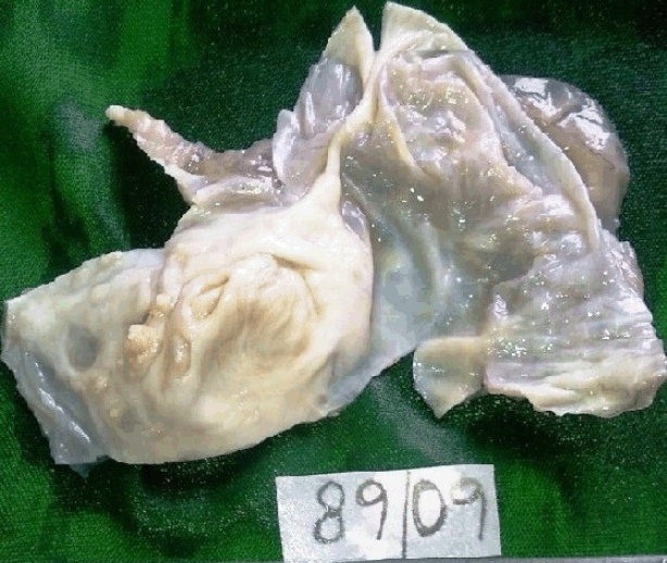
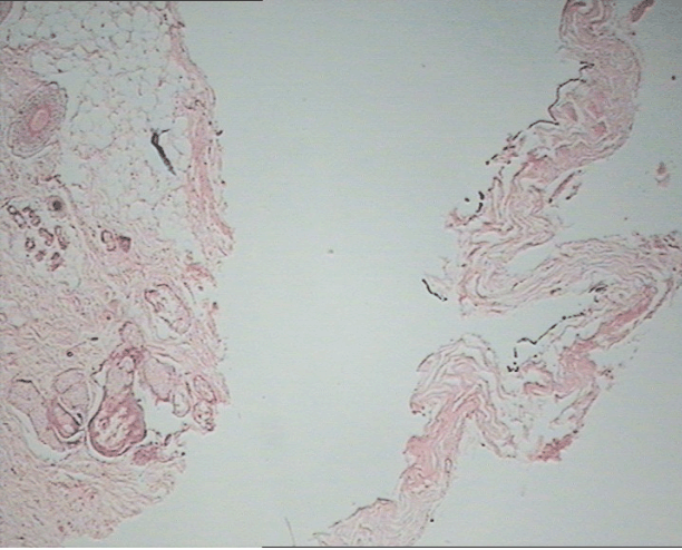
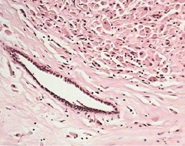
Collision tumors represent a coexistence of two adjacent but histologically distinct tumors without admixture in the same tissue or organ. Though such tumors have been reported often in various organs, their occurrence in ovary is rare. We report here a rare case of collision tumor of ovary comprising serous cystadenoma and mature cystic teratoma. A 55-year-old woman was presented with palpable mass in the lower abdomen associated with pelvic pain and dysfunctional uterine bleeding. Patient underwent right salphingo oophorectomy following ultrasound diagnosis of ovarian tumor. Grossly the tumor measured 6 cm in diameter. Cut surface revealed a trilocular cyst with two locules containing clear fluid. Inner wall of one of the locules exhibited few tiny polypoidal excrescences (Figure 1). The other locule had a gelatinous appearance. Microscopy revealed a combination of morphologic features. Sections from two locules showed the cyst wall lined by unilayer of low cuboidal epithelium which at places exhibited presence of cilia. A few papillary processes lined by ciliated/cuboidal epithelium were also noted. Sections from locule with gelatinous appearance revealed cyst wall lined by well differentiated stratified squamous epithelium (Figure 2), beneath which was seen adipose tissue, sebaceous glands, sweat glands, hair follicles, ganglion cells (Figure 3) and a focus of colloid filled area. Based on the microscopy, a diagnosis of collision tumor of ovary was made. We present this case because collision tumors in ovary are a rare entity and combination of serous cystadenoma with teratoma is rarer. [1] Though collision tumors have been reported earlier, combined serous cystadenoma with mature cystic teratoma is rarely reported. With the available literature, there is just one similar case reported in a non child bearing young woman. [2] There are instances of collision tumors consisting of teratoma with serous cystadenocarcinoma, mucinous cystadenocarcinoma and/or granulosa cell tumor. [3] In a study conducted at Seoul national University college of medicine, the authors reviewed seven pathologically proven cases of collision tumors of ovary associated with teratoma. Ovarian teratomas were co-existent with mucinous cystadenoma (4 cases), borderline mucinous tumor (1 case), mucinous cystadenocarcinoma (1 case) and dysgerminoma (1 case). [4] There is a single case report of collision tumor composed of a colonic adenocarcinoma arising in a sigmoid diverticulosis coexisting with recurrent ovarian granulosa cell tumor. [5] Though mature cystic teratoma of ovary contains derivatives of all three embryonic germ cell layers, it rarely presents with ovarian epithelial or sex cord stromal tumors. Rare cases of ovarian cystic teratoma in association with surface epithelial tumors have been reported in literature and occurrence of serous cystadenomas with mature cystic teratoma is even rarer. [3]Collision tumors have been described in various organs including oesophagus, stomach, liver, bone, kidney, brain and lung. Such tumors involving ovary are rare. In conclusion, we would like to emphasize upon the fact that multiloculated cysts have to be extensively examined, so as not to miss any component which might have a bearing on prognosis of the patient. Such cases need to be documented for academic as well as prognostic purpose. | ||||||
| ||||||
|
| ||||||
| ||||||
|
| ||||||
| ||||||
|
References
| ||||||
| ||||||
|
[HTML Abstract]
[PDF Full Text]
|
|
Author Contributions:
Shaista Choudhary – Conception and design, acquisition of data, Drafting the article, Final approval of the version to be published Shankar Adisesha – Analysis and interpretation of data, Critical revision of the article, Final approval of the version to be published |
|
Guarantor of submission:
The corresponding author is the guarantor of submission. |
|
Source of support:
None |
|
Conflict of interest:
Authors declare no conflict of interest. |
|
Copyright:
© Shaista Choudhary et al. 2012; This article is distributed the terms of Creative Commons Attribution License which permits unrestricted use, distribution and reproduction in any means provided the original authors and original publisher are properly credited. (Please see Copyright Policy for more information.) |
|
|