|
|
|
|
Case Report
| ||||||
| Gallstone ileus | ||||||
| Sherif Monib1, Ahmed Farghaly1, Andrew Ritchie1, Mustafa Halawa1 | ||||||
|
1Department of General Surgery, West Hertfordshire Hospitals NHS Trust, Hertfordshire, UK.
| ||||||
| ||||||
|
[HTML Abstract]
[PDF Full Text]
[Print This Article]
[Similar article in Pumed] [Similar article in Google Scholar] 
|
| How to cite this article: |
| Monib S, Farghaly A, Ritchie A, Halawa M. Gallstone ileus. International Journal of Case Reports and Images 2012;3(10):54–57. |
|
Abstract
|
|
Introduction:
Gallstone ileus is an uncommon complication of cholelithiasis but an established cause of mechanical bowel obstruction in the elderly. Perforation of the small intestine proximal to the obstructing gallstone is rare, and only a handful of cases have been reported.
Case Report: We report the case of a 74-year-old man who presented with a clinical picture of small bowel obstruction secondary to a gall stone impacted in the jejunum. Conclusion: The diagnosis of gallstone ileus should be always kept in mind when dealing with any elderly patient presenting with abdominal pain, distension and a previous history of gall bladder stones. | |
|
Key Words:
Gallstone, Ileus, Bowel obstruction
| |
|
Introduction
| ||||||
|
Gallstone ileus is an uncommon condition that may result when a gallbladder stone enters into the intestinal tract. The pathogenesis in the majority of cases is a cholecystoduodenal fistula, but rarely, cholecystogastric and cholecystocolonic fistulas can form and produce a gallstone ileus. Gallstone ileus is a rare cause of intestinal obstruction, accounting for only 1–4% of all cases. [1] Whilst only 0.3–0.5% of patients with cholelithiasis will go on to develop gallstone ileus, the mortality ranges from 12–18%. [2] [3] Mortality increases in those with multiple co-morbidities and the elderly | ||||||
|
Case Report
| ||||||
|
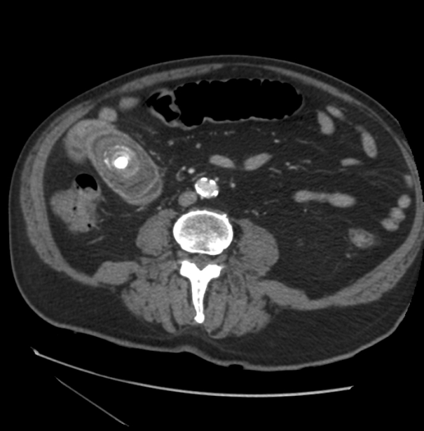
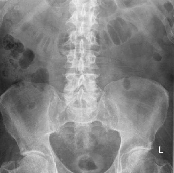
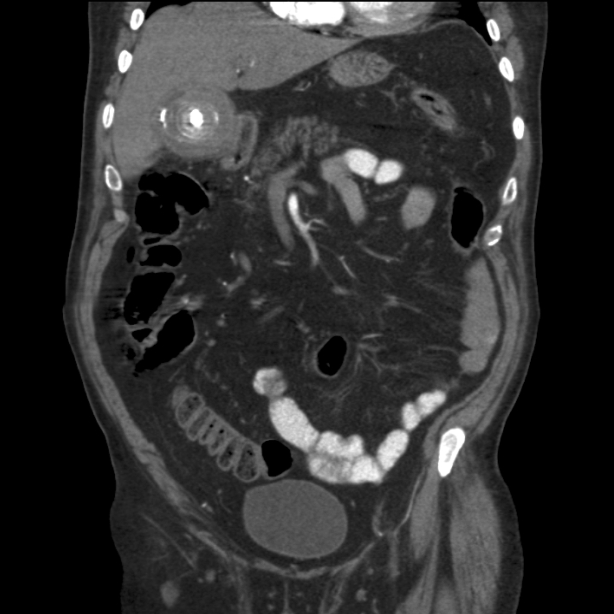
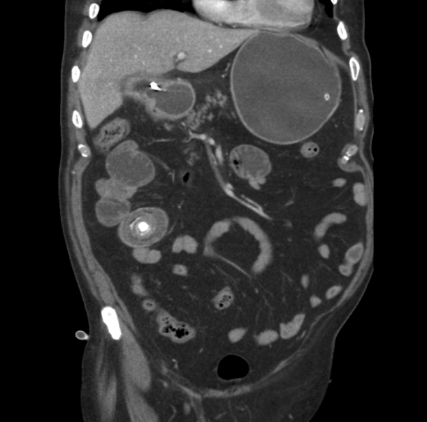
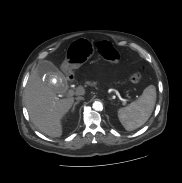
This is a case of a 74-year-old male seen in the Accident and Emergency department with a two day history of vomiting, dehydration, abdominal distension and pain. Following an early assessment by the on call medical team, a surgical opinion was sought as his clinical picture was consistent with an intestinal obstruction. His medical history included type II diabetes mellitus but there was no history of previous abdominal surgery. Two months prior to admission, he attended the same accident and emergency department with abdominal pain. On that occasion he underwent a computed tomography (CT) scan and was diagnosed with acute calcular cholecystitis (Figures 1 and 2). On this occasion, he was treated conservatively with plans to carry out an elective laparoscopic cholecystectomy three months later. On physical examination, the patient appeared generally well. He was apyrexial and his observations were all stable. On abdominal examination he had marked abdominal distension (mainly central) with tenderness in the right upper quadrant and epigastric region. There was no rebound tenderness and Murphy's sign was negative. Digital rectal examination revealed an empty rectum. Initial laboratory investigations revealed a mild hyponatraemia and hyperglycaemia. Liver function tests were not deranged and amylase was normal: Sodium 128 mmol/L and Potassium 3.8 mmol/L, Hb 16.2g/dL, white blood cells (WBCs) 7.0x10⁹ /L, Platelets 141x109/L, PT 10.4 s, APTT 23.9 s, Amylase 30 U/L, T. Bil 20 mg/dL, ALT 30 U/L, ALP 94 U/L, Total Calcium 2.27 mmol/L, Alb 42 g/L, random blood sugar 15.2 mmol/L. Plain abdominal radiograph of the patient demonstrated distended loops of small bowel (Figure 3). Abdominopelvic CT scan showed that a very large gallstone had migrated and impacted in the jejunum with proximally dilated and distally collapsed jejunal loops (Figures 4 and 5). Based on the history, physical examination and CT scan findings, our nominal diagnosis was of gallstone ileus. After fluid resuscitation the patient underwent an exploratory laparotomy, approximately six hours after confirming mechanical small bowel obstruction. At laparotomy, there was a transition zone at the point of impaction, however, the bowel segment appeared healthy with no perforation. The gallbladder was adherent to the duodenum and no attempt was made to separate the two. A longitudinal enterotomy followed by successful extraction of the stone and primary transverse closure of the enterotomy was carried out without complications. Postoperatively, the patient was transferred to a general surgical ward where he recovered well. On the second postoperative day, the patient was able to tolerate fluids and a light diet. He mobilized well and was discharged home the following day. After one month, the patient was seen in the out patient department. He had continued to recover well and an elective cholecystectomy was arranged for three months later. | ||||||
| ||||||
| ||||||
| ||||||
| ||||||
| ||||||
|
Discussion
| ||||||
|
Gallstone ileus is an uncommon cause of small bowel obstruction that occurs almost exclusively in the elderly. [2] [3] [4] [5] [6] In the population over 65-years-old, gallstone ileus is thought to account for up to 25% of cases of mechanical small-bowel obstruction. In this subsection of the general public it carries a mortality of 12-50%. [4] As with the pattern of incidence in cholelithiasis, gallstone ileus occurs three to five times more frequently in women than in men. [2] The gallstone enters the intestinal tract through a fistula formed between the gallbladder and the duodenum, stomach or colon. The terminal ileum is the most frequent site of obstruction. [1] However, as with our patient, there are other obstruction points, including jejunum (30%), colon (2.5%) and duodenum which is eponymously known as Bouveret syndrome. [2] Gallstone ileus remains a diagnostic challenge despite advances in imaging techniques, and pre-operative diagnosis is often delayed. Plain abdominal radiographs may reveal signs of small bowel obstruction and concomitant aerobilia to suggest the diagnosis. [7] [8] Whilst the classic radiological triad, or Rigler triad, of pneumobilia, small bowel obstruction and ectopic gallstone is specific for the disease, it is unfortunately only present in 9–14% of cases. [7] Therefore, the current definitive investigation of choice is CT scan. The principal goal in management of gallstone ileus is a quick effective relief of mechanical bowel obstruction. When appropriate, it is possible to avoid surgery, for example, if the stone is in within reach of an endoscope, either in the proximal small bowel or in the colon, there is the option to treat by lithotripsy and removal of the fragments. [2] Spontaneous passage of gallstones large enough to cause impaction has been reported, but most patients require surgical intervention and the prominent risk of perforation underlines why a conservative approach is rarely adopted. Extracorporeal shockwave lithotripsy can be successful, but this method is limited by the presence of bowel gas. The majority of patients require surgery either for bowel obstruction or perforation. Surgical options include enterotomy and removal of the stones (enterolithotomy) with subsequent elective cholecystectomy or enterolithotomy plus cholecystectomy and repair of the fistula in the same sitting. [3] Whilst the most appropriate surgical intervention remains unclear, authors mostly favor enterolithotomy to relieve the ileus, followed by subsequent elective Cholecystectomy. Reports suggest this method of management causes fewer complication and there is a 50% chance of spontaneous fistula closure. [7] In addition, enterolithotomy alone minimises operating time in the emergency setting and is technically less demanding. [9] This is especially advantageous in the high risk surgical candidates. For the frail patient, enterolithotomy alone is often adequate treatment and subsequent Cholecystectomy is not mandatory. [10] Zuegal et al. (1997) propose that the one stage procedure significantly decreases morbidity and mortality because removing the gallbladder and biliary-enteric fistula prevents future recurrence of gallstone ileus and recurrent biliary symptoms. [11] It also avoids the need for a second operation. In cases of perforation secondary to pressure necrosis of impaction or a perforated jejunal diverticulum, due to increased intra-luminal pressure proximal of the obstructing gallstone, the only way of management is segmental bowel resection and primary anastomosis. [12] | ||||||
|
Conclusion
| ||||||
|
Although it is a rare cause of bowel obstruction, gallstone ileus should be kept in mind when dealing with a case of small bowel obstruction, especially in elderly patient as early surgical intervention is the mainstay of treatment. | ||||||
|
References
| ||||||
| ||||||
|
[HTML Abstract]
[PDF Full Text]
|
|
Author Contributions:
Sherif Monib – Substantial contributions to conception and design, Acquisition of data, Analysis and interpretation of data, Drafting the article, Revising it critically for important intellectual content, Final approval of the version to be published Ahmed Farghaly – Substantial contributions to conception and design, Acquisition of data, Analysis and interpretation of data, Drafting the article, Revising it critically for important intellectual content, Final approval of the version to be published Andrew Ritchie – Substantial contributions to conception and design, Acquisition of data, Analysis and interpretation of data, Drafting the article, Revising it critically for important intellectual content, Final approval of the version to be published Mustafa Halawa – Substantial contributions to conception and design, Acquisition of data, Analysis and interpretation of data, Drafting the article, Revising it critically for important intellectual content, Final approval of the version to be published |
|
Guarantor of submission:
The corresponding author is the guarantor of submission. |
|
Source of support:
None |
|
Conflict of interest:
Authors declare no conflict of interest. |
|
Copyright:
© Sherif Monib et al. 2012; This article is distributed the terms of Creative Commons Attribution License which permits unrestricted use, distribution and reproduction in any means provided the original authors and original publisher are properly credited. (Please see Copyright Policy for more information.) |
|
|