|
|
|
|
Case Report
| ||||||
| Traumatic brachiocephalic artery pseudoaneurysm following penetrating injury on the contralateral side | ||||||
| Victor Kong1, Damon Jeetoo2, Grant Laing3, Damian Clarke4 | ||||||
|
1Surgical Registrar, Edendale Hospital - Private Bag X 509, Plessislaer, 3216, Pietermaritzburg, South Africa.
2Radiology Registrar, Edendale Hospital - Private Bag X 509, Plessislaer, 3216, Pietermaritzburg, South Africa. 3Consultant Surgeon, Edendale Hospital - Private Bag X 509, Plessislaer, 3216, Pietermaritzburg, South Africa. 4Senior Consultant Surgeon, Edendale Hospital - Private Bag X 509, Plessislaer, 3216, Pietermaritzburg, South Africa. | ||||||
| ||||||
|
[HTML Abstract]
[PDF Full Text]
[Print This Article]
[Similar article in Pumed] [Similar article in Google Scholar] 
|
| How to cite this article: |
| Kong V, Jeetoo D, Laing G, Clarke D. Traumatic brachiocephalic artery pseudoaneurysm following penetrating injury on the contralateral side. International Journal of Case Reports and Images 2012;3(10):21–23. |
|
Abstract
|
|
Introduction:
Trauma to the brachiocephalic artery is uncommon, but is associated with significant morbidity and mortality. The development of pseudoaneurysm following both penetrating and blunt trauma have been well documented, mostly in the setting of blunt trauma.
Case Report: A highly unusual case of a patient who developed a pseudoaneurysm of the brachiocephalic artery on the side contralateral to the side of the original penetrating injury has been reported. Conclusion: Pseudoaneurysm of the brachiocephalic artery following penetrating neck injury in the contralateral side is exceeding rare. It may remain symptomatic and present long after the initial injury. Clinician must have a high index of suspicion for such potentially serious injury. | |
|
Key Words:
Penetrating, Trauma, Brachiocephalic, Pseudoaneurysm
| |
|
Introduction
| ||||||
|
We present the case of a 21-year-old male with a penetrating neck injury who subsequently developed a large pseudoaneurysm of the brachiocephalic artery on the contralateral side of the original injury. | ||||||
|
Case Report
| ||||||
|
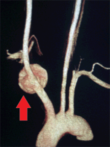
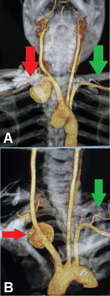
A 21-year-old male was referred to the trauma unit with a single penetrating wound caused by a screwdriver, and located in the lateral, infraclavicular region on the left side. He had no other significant medical history. His blood pressure was normal and equal in both arms. No pulsatile mass was noted. Three weeks before, he initially assessed at another hospital, and a confirmed hemothorax had been treated with an intercostal chest drain. He was subsequently discharged; however, he presented again with an infected wound in the drain-insertion site, with pus discharge. He was hemodynamically stable, with tachycardia (heart rate 90/min), low-grade pyrexia (temperature, 37.6°C), leucocytosis (white cell count, 16x103) and a chest radiograph showing a marked opacity at the left base. An empyema thoracis secondary to a retained hemothorax was suspected and treatment with intravenous antibiotics was initiated. A contrast CT scan of the thorax obtained on the following day and revealed a consolidation in the left lower lobe of the lung, with adjacent air collection. A large round mass was also noted on the right side of the neck, adjacent to the brachiocephalic trunk suspicious of a pseudoaneurysm. Subsequent CT Angiography (64-slice MDCT) of the neck vessels confirmed a large pseudoaneurysm (38.5 mm sagittal, 31.6 mm transverse and 33.2 mm longitudinal), located on the right brachiocephalic trunk, just prior to the bifurcation into the right subclavian and common carotid arteries (Figures 1 and 2). A decision was made to manage the empyema non-operatively. Once it has subsided, he underwent an open repair of the pseudoaneurysm and made an uneventful recovery. | ||||||
| ||||||
| ||||||
|
Discussion
| ||||||
|
Trauma to the brachiocephalic artery is uncommon. The exact incidence of this injury is unknown, as most cases with significant injury tend to exsanguinate prior to reaching the hospital. [1] Isolated pseudoaneurysm of the brachiocephalic artery as a result of penetrating and blunt trauma is a well-documented complication, [2] but still relatively rare. Many cases reported in the literature were related to blunt trauma, although penetrating trauma is relatively more common. [3] A wide range of presentations is seen, but many can remain asymptomatic and present many years after the initial injury. [4] Mortality in treated patients can be as high as 38%, and the further increases if mutiple vessels are involved. [5] Once the diagnosis is confirmed, patients must undergo definitive management. Traditionally, open surgical repair via median sternotomy and occasionally with cardiopulmonary bypass, [6] has been practiced. Recent advances in endovascular techniques have enabled treatment of selected cases with excellent results. [7] The anatomical location of the pseudoaneurysm was highly unusual (as an incidental finding on thorax CT), because it was located on the contralateral side of the original injury. The patient had no other previous injuries and was asymptomatic after the injury. To our knowledge, this is the first case in literature where presentation of the pseudoaneurysm was present on the side contralateral to the injury. The only other case with a similar site of injury was reported by Erkut et al. [8] where a pseudoaneurysm of the right common carotid artery was observed. The trajectory of penetration from the left infraclavicular area to the brachiocephalic artery on the right with no other associated injury can be difficult to appreciate. Anatomically, the most likely explanation was that the penetrating object followed an anterior course in the upper mediastinum. The common carotid artery and left subclavian artery tend to lie slightly posterior in the sagittal plane to the brachiocephalic trunk. Thus it possible for it to miss the left side vessels and injury the brachiocephalic artery. The alternative was the presence of the pseudoaneurysm prior to injury, but in view of the clear history, seems unlikely. | ||||||
|
Conclusion
| ||||||
|
Although pseudoaneurysm of brachiocephalic artery resulting from penetrating trauma is a rare presentation, it remains an important entity because of its implication in subsequent management. Clinicians must remain vigilant of such injury, particularly with delayed presentations. Thorough clinical assessment is mandatory, with judicious use of imaging studies to identify such an injury that has the potential of causing serious adverse outcomes. | ||||||
|
References
| ||||||
| ||||||
|
[HTML Abstract]
[PDF Full Text]
|
|
Author Contributions:
Victor Kong – Substantial contributions to conception and design, Acquisition of data, Analysis and interpretation of data, Drafting the article, Revising it critically for important intellectual content, Final approval of the version to be published Damon Jeetoo – Substantial contributions to conception and design, Acquisition of data, Analysis and interpretation of data, Drafting the article, Revising it critically for important intellectual content, Final approval of the version to be published Grant Laing – Substantial contributions to conception and design, Acquisition of data, Analysis and interpretation of data, Drafting the article, Revising it critically for important intellectual content, Final approval of the version to be published Damian Clarke – Substantial contributions to conception and design, Acquisition of data, Analysis and interpretation of data, Drafting the article, Revising it critically for important intellectual content, Final approval of the version to be published |
|
Guarantor of submission:
The corresponding author is the guarantor of submission. |
|
Source of support:
None |
|
Conflict of interest:
Authors declare no conflict of interest. |
|
Copyright:
© Victor Kong et al. 2012 This article is distributed the terms of Creative Commons Attribution License which permits unrestricted use, distribution and reproduction in any means provided the original authors and original publisher are properly credited. (Please see Copyright Policy for more information.) |
|
|