|
|
|
|
Case Report
| ||||||
| Primary serous carcinoma of peritoneum: A case report | ||||||
| Shelly Sehgal1, Reena Agarwal1, Prashant Goyal2, Sompal Singh1 Vinita Kumar3 Ruchika Gupta4 | ||||||
|
1MD, Department of Pathology, Swami Dayanand Hospital, Shahdara, New Delhi, India.
2DCP, Department of Pathology, Swami Dayanand Hospital, Shahdara, New Delhi, India. 3MD, Department of Gynecologic Oncology, Delhi State Cancer Institute, Dilshad Garden, New Delhi, India. 4MD, Department of Pathology, Chacha Nehru Bal Chiktsalaya, New Delhi, India. | ||||||
| ||||||
|
[HTML Abstract]
[PDF Full Text]
[Print This Article]
[Similar article in Pumed] [Similar article in Google Scholar] 
|
| How to cite this article: |
| Sehgal S, Agarwal R, Goyal P, Singh S, Kumar V, Gupta R. Primary serous carcinoma of peritoneum: A case report. International Journal of Case Reports and Images 2012;3(10):16–20. |
|
Abstract
|
|
Introduction:
Primary serous papillary carcinoma of the peritoneum (PSPCP) is a rare malignant epithelial tumor that is histologically indistinguishable from papillary serous carcinoma of the ovary (PSCO). It is defined as primary tumor of peritoneum that diffusely involves the peritoneal surface but spares or only superficially invades the ovaries. Better recognition of this entity in recent years has contributed to an increasing diagnostic frequency.
Case Report: A case of 50-year-old female who was presented with abdominal distension and pain is reported. Ascitic fluid cytology showed malignant cells favoring papillary serous adenocarcinoma. Preoperative serum CA-125 was markedly elevated. CECT scan showed omental thickening with normal uterus and ovaries. Exploratory laparotomy revealed massive ascites with extensive peritoneal deposits and normal sized ovaries. Histopathology confirmed diagnosis of PSPCP with surface involvement of both the ovaries. Conclusion: PSPCP is a rare neoplasm, histologically indistinguishable from PSCO. We presented this case report to emphasise that peritoneum, can also be a primary site of malignancy and that it presents and is managed just like primary ovarian cancer. Pre-op diagnosis of this entity is difficult. Histopathology is mandatory to confirm the diagnosis. | |
|
Key Words:
Papillary, Serous, Peritoneum, Ovary, CA-125
| |
|
Introduction
| ||||||
|
Primary serous papillary carcinoma of the peritoneum (PSPCP) is a rare malignant epithelial tumor that is histologically indistinguishable from papillary serous carcinoma of the ovary (PSCO). It is considered to originate from embryonic nests of mullerian cells in the peritoneum. This case report describes the clinical, radiological, pathological findings of the entity and to emphasize that peritoneum can also be primary site of malignancy and should be considered in the differential diagnosis of peritoneal carcinomatosis. | ||||||
|
Case Report
| ||||||
|
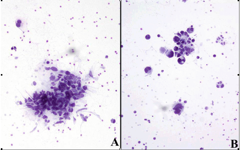
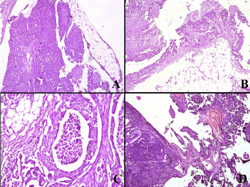
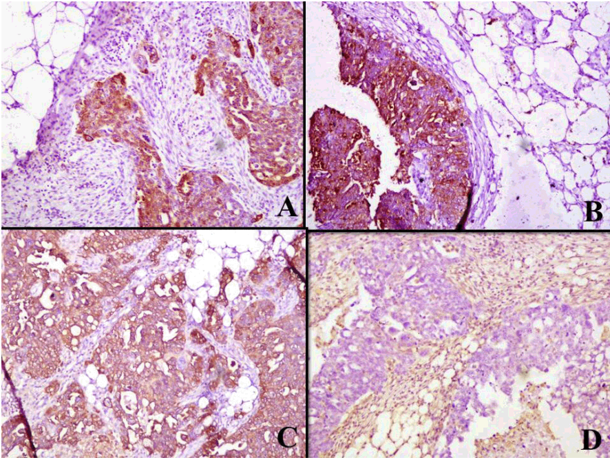
Case History: A 50-year-old postmenopausal female who was presented with abdominal distension and pain. On examination, abdomen was distended tensely with ascites, but there were no lymph node enlargement, or that of liver and spleen. On pelvic examination, cervix was normal with no mass palpable in fornices but some nodularity and thickening was palpable in pouch of douglas. Preoperative serum CA-125 was elevated to 1850 U/mL (reference range 0–30 U/mL), while CEA had normal value of 0.479 ng/mL. Ascitic fluid cytology was reported as having malignant cells favoring papillary serous adenocarcinoma. Upper and lower gastrointestinal endoscopy were normal. CECT scan showed omental thickening with normal uterus and ovaries (Figure 1). Preoperative diagnosis of PSPCP was made on basis of cytologic and radiologic findings and no evidence of primary disease elsewhere. Exploratory laparotomy revealed massive 3–4 L of ascitic fluid. Extensive peritoneal deposits were seen all over the abdominal and pelvic peritoneum, including large confluent deposits on under surface of diaphragm. Uterus and both ovaries were normal in size. Omentum showed small deposits though there was no caking. Tumor nodules were seen in pelvic peritoneum, pouch of douglas, and uterovesical peritoneum. She underwent panhysterectomy along with infragastric omentectomy and removal of other deposits. Residual disease was multiple small deposits all over parietal peritoneum, and over undersurface of diaphragm. Post- operative CA-125 was 127 U/mL. Histopathology revealed PSPCP with surface involvement of both the ovaries. Final diagnosis was stage 3C primary serous papillary adenocarcinoma of peritoneum. Post- operatively, she was placed on three weekly regime of paclitaxel, carboplatin and ifosfamide with mesna as part of adjuvant treatment. She has received three cycles and her latest CA-125, six months postoperative was 23 U/mL. Patient was doing well at the time of last follow up. Pathological findings: Ascitic fluid cytology showed moderately cellular smears showing predominantly singly dispersed and few 3-dimensional clusters (Figure 2A) of tumor cells. These cells had peripherally placed nuclei, at places showing glandular arrangement (Figure 2B). Tumor cells showed marked pleomorphism with high N/C ratio. The cells had irregular nuclear margin, coarse chromatin, prominent nucleoli, moderate cytoplasm, often showing cytoplasmic vacuolation and cytoplasmic protrusions. Many bizarre and multinucleated tumor giant cells were seen. Few cells with atypical mitosis were also observed. The findings were consistent with diagnosis of malignant cytology favoring serous adenocarcinoma. Subsequently, panhysterectomy specimen along with omentum was received. Grossly, specimen of uterus with cervix along with bilateral fallopian tubes was unremarkable and both the ovaries were normal in size. Omentum was received as fibrofatty tissue measuring 45x30x5 cm and serial sectioning showed many tiny nodules. Extensive sampling was done from these areas and showed an invasive tumor arranged in solid and complex glandular structures which were partly papillary (Figure 3A). Intervening areas showed extensive desmoplastic response along with proliferation of tumor cells over the omentum surface (Figure 3B). Focal areas of comedo necrosis were also identified (Figure 3C). The papillary structures were lined by several layers of cells with nuclear crowding and high N/C ratio. Cytologically, nuclei were of high grade with vesicular chromatin and prominent nucleoli with frequent mitosis (6-8/10 hpf). The surface of both the ovaries showed tumor deposits involving only ovarian surface epithelium (Figure 3D). The hysterectomy specimen along with both the fallopian tubes did not show any significant pathology. A panel of immunostains was applied comprising CA-125, CK-7, EMA, Vimentin, S-100. Tumor cells showed positivity for CA-125, CK-7 and EMA and rest of the markers were negative (Figure 4A-D). Final diagnosis of PSPCP was made. | ||||||
| ||||||
| ||||||
|
| ||||||
|
| ||||||
|
Discussion
| ||||||
|
PSPCP is a rare malignant epithelial tumor that is histologically indistinguishable from PSCO. It is defined as primary tumor of peritoneum that diffusely involves the peritoneal surface but spares or only superficially invades the ovaries. [1] This entity was first described by Swerdlow [2] in his case report in 1959 as mesothelioma of pelvic peritoneum. Since then several studies [3] [4] [5] have established PSPCP as a separate entity and has been reported under different names. The true incidence of PSPCP remains unknown although an estimated relative frequency to ovarian cancer is 1:10. [3] Better recognition of this entity in recent years has contributed to an increasing diagnostic frequency approaching 18% of laparotomies performed for ovarian carcinoma. [3] We presented this case report to emphasise that peritoneum, can also be a primary site of malignancy and that it presents and is managed just like primary ovarian cancer. Preoperative diagnosis of this entity is difficult but normal sized ovaries radiologically and positive serous cytology in ascitic fluid can be helpful to recognize this lesion. However, histopathology is still mandatory to confirm the diagnosis. Since, Lauchlan [6] first included the female peritoneum in the definition of secondary mullerian system in 1972; PSPCP is better understood as neoplasm that arises from mesothelial cells under mullerian influence. [1] This theory, therefore, explains why PSPCP behaves like PSCO in many ways with similar clinical, radiological and immunohistochemical findings with similar sensitivity to platinum based chemotherapy. PSPCP is mostly reported in elderly females. Like its ovarian counterpart the tumor often presents with abdominal distention and pain. [5] CT and MRI scans suggest omental caking, peritoneal nodules or enhancement with ascitis without ovarian enlargement in most of the patients. [1] CA-125 is markedly elevated like in ovarian malignancy and can be used for monitoring the efficacy of therapy and for early detection of recurrence. [1] [3] The diagnostic inclusion criteria of PSPCP were defined by gynecologic oncology group [7] in 1993 to differentiate it from PSCO as (1) both ovaries must be either physiologically normal in size or enlarged by a benign process, (2) involvement at extra-ovarian sites must be greater than the involvement on the surface of either ovary, (3) the ovarian component must be non-existent or confined to surface epithelium or less than 5x5 mm within the stroma, and (4) the histological and cytological characteristics of the tumor must be predominantly of serous type that is similar or identical to any grade of ovarian serous papillary adenocarcinoma. Cytologically, closest differential of papillary serous cytology in ascitic fluid is malignant mesothelioma (MM) (Table 1) and poorly differentiated squamous cell carcinoma. Squamous cell carcinoma is relatively rare in effusions and shows dispersed cells with well demarcated cytoplasm as compared to adenocarcinoma. Histologically, PSPCP must be differentiated from MM, benign papillary mesothelioma, metastatic peritoneal carcinomatosis, borderline primary peritoneal serous tumor, endosalpingiosis, psammocarcinoma of peritoneum and pseudomyxoma peritonei. [5] [8] MM is closely related to long term asbestos exposure, has a male predominance, frequent spindle cell component, cytoplasmic eosinophilia and sometimes extensive cell vacuolization. Benign papillary mesothelioma has well formed papillae, mostly lined by one layer of single cell type showing little or no anaplasia or mitosis and absence of invasion into the peritoneum. Morphologically, PSPCP cannot be differentiated from metastatic peritoneal carcinomatosis; diagnosis of later rests on recognizing a primary tumor usually in ovary, fallopian tube or endometrium. Serous psammocarcinoma of peritoneum is a less virulent variation of PSPCP that has been described. It has proportionately larger number of psammoma bodies and less aggressive cytologic appearance. The treatment of PSPCP consists of debulking surgery including hysterectomy, salpingo-oopherectomy, omentectomy followed by platinum based chemotherapy. [9] Surgery remains critically important for both diagnosis and treatment of PSPCP. Once diagnosis is established and extend of disease documented, maximum cytoreduction becomes primary goal of management. Prognosis of PSPCP is poor. Medial survival time varies between 7 and 28 months while 5 year survival rate ranges from 0 to 26.5%. [1] In few of the case matched control studies, prognosis of PSPCP was similar to that of stage III or IV serous ovarian papillary carcinoma when the same treatment was performed. [10] | ||||||
| ||||||
|
| ||||||
|
Conclusion
| ||||||
|
PSPCP is a rare neoplasm, histologically indistinguishable from papillary serous carcinoma of the ovary, which diffusely involves the peritoneum but spares or minimally invades the ovaries. The clinical presentation and behavior is also similar to its ovarian counterpart. Ascitic fluid cytology combined with radiological and clinical data allows a presumptive preoperative diagnosis of PSPCP. However, one needs to histologically study the ovaries before rendering a final diagnosis. We suggest that PSPCP should be included in differential diagnosis when ascites, omental caking, and peritoneal nodules or enhacement are observed in a postmenopausal women with or without ovarian enlargement. | ||||||
|
References
| ||||||
| ||||||
|
[HTML Abstract]
[PDF Full Text]
|
|
Author Contributions:
Shelly Sehgal – Conception and design, Acquisition of data, Analysis and interpretation of data, Drafting the article, Critical revision of the article, Final approval of the version to be published Reena Agarwal – Conception and design, Acquisition of data, Analysis and interpretation of data, Drafting the article, Critical revision of the article, Final approval of the version to be published Prashant Goyal – Conception and design, Acquisition of data, Analysis and interpretation of data, Drafting the article, Critical revision of the article, Final approval of the version to be published Sompal Singh – Drafting the article, Critical revision of the article, Final approval of the version to be published Vinita Kumar – Conception and design, Acquisition of data, Analysis and interpretation of data, Drafting the article, Critical revision of the article, Final approval of the version to be published Ruchika Gupta – Drafting the article, Critical revision of the article, Final approval of the version to be published |
|
Guarantor of submission:
The corresponding author is the guarantor of submission. |
|
Source of support:
None |
|
Conflict of interest:
Authors declare no conflict of interest. |
|
Copyright:
© Shelly Sehgal et al. 2012; This article is distributed the terms of Creative Commons Attribution License which permits unrestricted use, distribution and reproduction in any means provided the original authors and original publisher are properly credited. (Please see Copyright Policy for more information.) |
|
|