|
|
|
|
Case Report
| ||||||
| Isolated intracranial relapse of hodgkin lymphoma in a child | ||||||
| Harriet Holme1, Niharendu Ghara2, Thomas S Jacques3, Paul Humphries4, Stephen Daw1, Ananth Shankar1 | ||||||
|
1Department of Paediatric and Adolescent Oncology, University College, London NHS Foundation Trust Hospitals, 6th Floor central, 250 Euston Road, London, NW1 2PG.
2Department of Paediatric and Adolescent Oncology, University College London NHS Foundation Trust Hospitals, 6th Floor central, 250 Euston Road, London, NW1 2PG. 3Neural Development Unit, UCL Institute of Child Health and Department of Histopathology Great Ormond Street NHS Foundation Trust, London. 3Department of Radiology, University College London NHS Foundation Trust Hospitals, 3rd Floor west 250 Euston Road, London, NW1 2PG. | ||||||
| ||||||
|
[HTML Abstract]
[PDF Full Text]
[Print This Article]
[Similar article in Pumed] [Similar article in Google Scholar] 
|
| How to cite this article: |
| Holme H, Ghara N, Jacques TS, Humphries P, Daw S, Shankar A. Isolated intracranial relapse of hodgkin lymphoma in a child. International Journal of Case Reports and Images 2012;3(10):12–15. |
|
Abstract
|
|
Introduction:
Although involvement of the central nervous system (CNS) is well described in non-Hodgkin lymphoma (NHL) at diagnosis and/or at relapse, there are few reports in the pediatric literature of primary or relapsed CNS Hodgkin lymphoma (HL).
Case Report: We report dural disease as the only site of relapse of HL in an immunocompetent seven-year-old child, presenting with diplopia. Imaging demonstrated a well-circumscribed dural-based mass at the base of the skull involving the clivus and enveloping the pituitary. Restaging did not show disease elsewhere, with normal bone marrow trephine biopsies and cytological examination of the cerebrospinal fluid. Biopsy of the dural-based tumor confirmed classical HL. Eleven months before, he was diagnosed with stage IIIA HL nodular sclerosis subtype and underwent four courses of combination chemotherapy with a complete metabolic response on early response assessment. Forty-eight months after completing treatment for relapsed HL, he remains well in complete remission, although future relapse of HL cannot be excluded. Conclusion: There were no specific risk factors predictive of relapse in this child. He had responded well to standard chemotherapy as early response assessment after two courses showed complete metabolic response. Treatment of this child at relapse was challenging, since standard salvage chemotherapy regimens comprise drugs not well known to traverse the blood brain barrier. This case highlights the rarity of intracranial HL in children and difficulty with regards to the diagnosis, evidence based treatment and overall prognosis. We suggest that irrespective of any imaging abnormality, histological confirmation should be sought before commencement of definitive treatment. | |
|
Key Words:
Lymphoma, CNS relapse, Pediatric hematology/oncology, Hodgkin lymphoma
| |
|
Introduction
| ||||||
|
Hodgkin lymphoma (HL) involving the central nervous system (CNS) is a rare. Involvement of the CNS is well described in non-Hodgkin lymphoma (NHL) at both diagnosis and/or at relapse. However, there are few reports in the pediatric and adult literature of primary or relapsed CNS HL. [1] [2] [3] [4] [5] The management of this child including a review of literature is discussed. | ||||||
|
Case Report
| ||||||
|
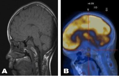
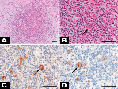
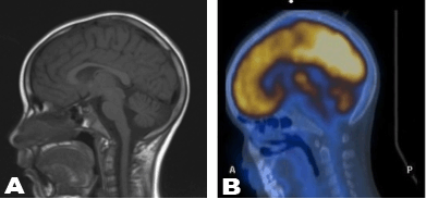
A seven-year-old boy was presented with diplopia. Clinical examination was normal except for a left convergent squint. Magnetic resonance imaging (MRI) examination of the brain was normal. Eleven months before, he was diagnosed with stage IIIA HL nodular sclerosis subtype and underwent four courses of combination chemotherapy [Vincristine/Oncovin, Etoposide, Prednisolone, and Doxorubicin/Adriamycin (OEPA) x 2 and Cyclophosphamide, Vincristine/Oncovin, Prednisolone and Procarbazine (COPP) x 2]. Since early response assessment (ERA) with 18 fluoro-2-deoxy-D-glucose (FDG) positron emission tomography (PET) after two courses of OEPA showed a complete metabolic response, he did not receive radiotherapy. However, his diplopia progressed over two months, and a generalized seizure prompted another MRI brain scan; this showed a well-circumscribed dural-based mass at the base of the skull involving the clivus and enveloping the pituitary (Figure 1A). This lesion was also FDG-avid (Figure 1B). Restaging with whole body MRI and FDG PET did not show disease elsewhere. Bone marrow trephine biopsies and cytological examination of the cerebrospinal fluid were normal. Serologic testing for human immunodeficiency virus was negative. He had no family history of malignancy or immunodeficiency. Full evaluation of the integrity of the hypothalamic pituitary axis prior to the commencement of relapse treatment was normal. Biopsy of the dural-based tumor confirmed classical HL. Figure 2 shows nodules containing Hodgkin cells and occasional Reed-Sternberg (RS) cells set against mixed inflammatory cell infiltrate. The Hodgkin cells and RS cells were positive for cluster of differentiation (CD) 30, CD15 and Epstein-Barr virus-encoded ribonucleic acid (EBER) but negative for CD3 and CD20. Histological examination showed clear evidence of residual epithelium, consistent with anterior pituitary gland, and glial tissue both infiltrated by HL as shown by cytokeratin immunohistochemistry (using MNF116), and Glial fibrillary acidic protein (GFAP) immunohistochemistry, respectively. Clinical examination prior to the commencement of relapse chemotherapy showed left 6th and 7th cranial nerve palsies. He had no palpable lymphadenopathy or hepatosplenomegaly. He received two cycles of salvage chemotherapy comprising high dose cytarabine/Ara C, dexamethasone and cisplatin/planitol (DHAP). An ERA was performed 14 days after the 2nd DHAP course and included whole body MRI and PET scans; MRI scan showed almost complete resolution of the dural lesion (Figure 3A) while the PET scan showed complete metabolic response (Figure 3B). He had 2 further courses of DHAP followed by 30 Gy radiotherapy to the clivus and pituitary region. Forty-eight months after completing the treatment for relapsed HL, he remains well in complete remission, although future relapse of HL cannot be excluded. | ||||||
|
| ||||||
|
| ||||||
| ||||||
|
Discussion
| ||||||
|
Hodgkin lymphoma unlike NHL rarely involves the CNS and occurrence of intracranial disease is usually with disseminated relapse. [2] [6] [7] We report dural disease as the only site of relapse of HL in an immunocompetent child. There were no specific risk factors predictive of relapse in this case report; the child had responded well to OEPA chemotherapy as the ERA by FDG PET after two courses showed a complete metabolic response. [8] It is arguable that without radiotherapy, his initial treatment was sub-optimal but this approach was according to the European pediatric Hodgkin treatment strategy and there are no suggestions that non-irradiated patients with intermediate risk HL and good early response have a worse outcome. [9] [10] Although the striking feature of the MRI scan at relapse was the dural-based lesion involving the pituitary, there was also abnormal signal within the clivus. The mechanism of leptomeningeal involvement is unknown, but thought to originate from systemic hematogenous spread. [11] [12] [13] However, in our patient the dural lesion was the only site of relapse. It is difficult to know whether the clivus was the site of the actual relapse with dural involvement as an extension from the clivus or vice versa (Figure 1). Review of whole body MRI and FDG PET scans at first presentation did not show any evidence of bone involvement. Since standard salvage chemotherapy regimens comprise drugs not well known to traverse the blood brain barrier (BBB), treatment of this child at relapse was challenging. We chose chemotherapy that included high dose cytarabine and cisplatin, both reported to penetrate the CNS. [14] [15] Success of the relapse treatment strategy is reflected in the ERA MRI and FDG PET scans (Figure 3A–B). It is possible initial biopsy was beneficial, with surgery disrupting the BBB helping with CNS penetration of chemotherapy. [16] | ||||||
|
Conclusion
| ||||||
|
This case highlights the rarity of intracranial HL in children and difficulty with regards to the diagnosis, evidence based treatment or overall prognosis. Considering this case report, we suggested that irrespective of any imaging abnormality, histological confirmation should be sought before commencement of definitive treatment. | ||||||
|
References
| ||||||
| ||||||
|
[HTML Abstract]
[PDF Full Text]
|
|
Author Contributions:
Harriet Holme – Substantial contributions to conception and design, Acquisition of data, Analysis and interpretation of data, Drafting the article, Revising it critically for important intellectual content, Final approval of the version to be published Niharendu Ghara – Acquisition of data, Analysis and interpretation of data, Final approval of the version to be published Thomas S Jacques – Acquisition of data, Analysis and interpretation of data, Final approval of the version to be published Paul Humphries – Acquisition of data, Analysis and interpretation of data, Final approval of the version to be published Stephen Daw – Acquisition of data, Analysis and interpretation of data, Final approval of the version to be published Ananth Shankar – Acquisition of data, Analysis and interpretation of data, Final approval of the version to be published |
|
Guarantor of submission:
The corresponding author is the guarantor of submission. |
|
Source of support:
None |
|
Conflict of interest:
Authors declare no conflict of interest. |
|
Copyright:
© Harriet Holme et al. 2012; This article is distributed the terms of Creative Commons Attribution License which permits unrestricted use, distribution and reproduction in any means provided the original authors and original publisher are properly credited. (Please see Copyright Policy for more information.) |
|
|