|
|
|
|
Case Report
| ||||||
| Cystadenoma of the appendix presenting with an intestinal obstruction of the ileum: A case report | ||||||
| Tadahiro Nozoe1, Katsuo Sueishi2, Mayuko Kohno1, Tomohiro Iguchi1, Takashi Maeda1, Takahiro Ezaki1 | ||||||
|
1Department of Surgery, Fukuoka Higashi Medical Center, 1-1-1, Chidori, Koga, 811-3195, Japan.
2Department of Pathology, Fukuoka Higashi Medical Center, 1-1-1, Chidori, Koga, 811-3195, Japan. | ||||||
| ||||||
|
[HTML Abstract]
[PDF Full Text]
[Print This Article]
[Similar article in Pumed] [Similar article in Google Scholar] 
|
| How to cite this article: |
| Nozoe T, Sueishi K, Kohno M, Iguchi T, Maeda T, Ezaki T. Cystadenoma of the appendix presenting with an intestinal obstruction of the ileum: A case report. International Journal of Case Reports and Images 2012;3(9):13–16. |
|
Abstract
|
|
Introduction:
Small bowel obstruction due to an appendiceal mucocele is comparatively rare.
Case Report: A rare case of a small bowel obstruction due to a mucocele of the appendix, which had been histopathologically diagnosed as a mucinous cystadenoma. Partial resection of the ileum and appendectomy through traditional laparotomy relieved the patient from abdominal pain and congestion from the intestinal obstruction. Conclusion: Small bowel obstruction due to an appendiceal mucocele should be considered among the differential diagnosis of intestinal obstruction for patients who have no previous history of laparotomy. | |
|
Key Words:
Appendix, Mucocele, Cystadenoma, Small bowel obstruction
| |
|
Introduction | ||||||
|
Intestinal tumors, [1] mucosal edema of the intestine due to enteric anisakiasis, [2] and malrotation of the bowel [3] are often the causal lesions in patients with small bowel obstruction and no previous history of laparotomy or intestinal tumors. Appendiceal mucocele is a comparatively a rare disease and intestinal obstruction other than intussusception of the bowel caused by appendiceal mucocele is quite atypical. A case of small bowel obstruction caused by compression by a mucinous cystadenoma of the appendix has been given here. | ||||||
|
Case Report
| ||||||
|
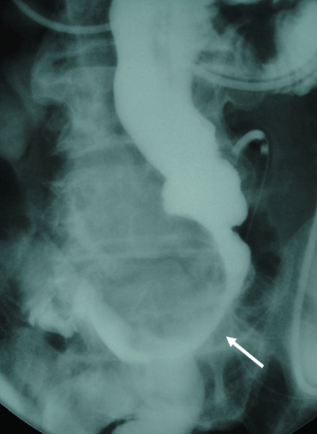
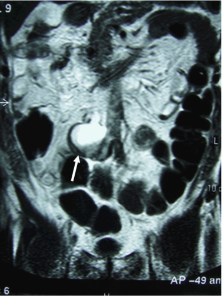
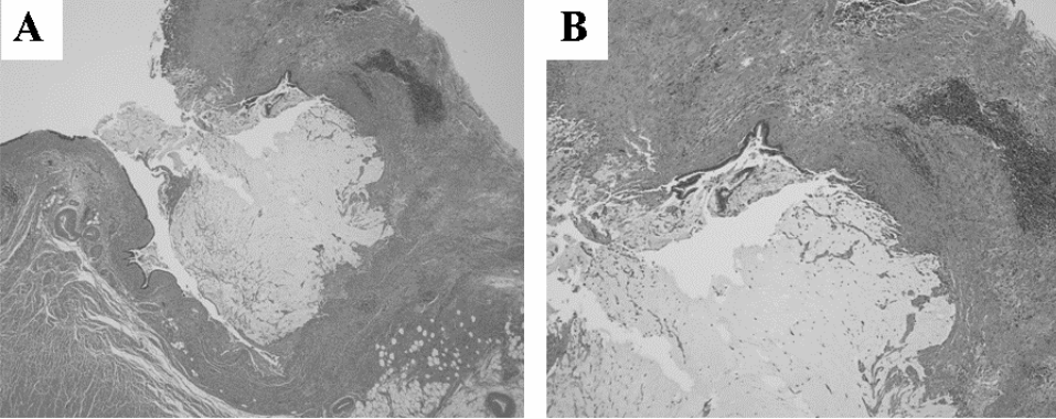
A 74-year-old male came to our institute with complains of abdominal pain and vomiting. He had no previous history of laparotomy. A long tube was inserted to reduce the symptoms of abdominal fullness and a small bowel series demonstrated a complete obstruction of the ileum (Figure 1). Magnetic resonance imaging (MRI) suggested a cystic lesion that might be correlated with the small bowel obstruction (Figure 2). Laparotomy revealed a cystic lesion located in the ileum, 80 cm from the terminal ileum causing a complete compression of the small bowel. Moreover, the entire cystic lesion was continuous with the appendix, a partial resection of small bowel and appendectomy was performed. The cystic lesion was found on the edge of the appendix (Figure 3A). Although the lumen of the ileum was completely occluded by the cystic lesion, the mucosa of the ileum was not involved with the tumor (Figure 3B). The cystic lesion contained the transparent mucin (Figure 3C). The wall of the cystic lesion was lined with intestinal epithelium without atypia associated with chronic inflammation of the appendiceal mucosa and there was mucin secreting epithelium lining the appendix (Figure 4). The lesion demonstrated the pathological features of mucinous cystoadenoma of the appendix. | ||||||
| ||||||
| ||||||
|
| ||||||
|
| ||||||
|
Discussion
| ||||||
|
Appendiceal mucoceles are defined as a group of lesions in which the appendiceal lumen becomes distended with mucus. The lesions are histopathologically divided into three subtypes: mucosal hyperplasia, mucinous cystoadenoma, and mucinous cystadenocarcinoma. [4] Appendiceal mucocele is a comparatively rare disease and it often remains asymptomatic during the clinical course, and the disease itself has been accepted to be a neoplasm with low grade malignant potential, other than cystadenocarcinoma associated with pseudomyxoma peritonei. Tumorous lesions including GIST, carcinoid tumor, lymphoma, primary cancer, and metastasized carcinoma usually metastasize from extra-intestinal melanoma; lung cancer and breast cancer have been reported as causal lesions for obstruction of the small intestine. [5] [6] Although there are a few reports of intussusception caused by appendiceal mucocele, [7] [8] small bowel obstruction caused by a simple compression of appendiceal mucocele has been rarely presented. [9] Magnetic resonance imaging demonstrated a cystic lesion obstructing the ileum in the current patient, and a GI series showed the intestinal obstruction was far from the terminal ileum. These findings suggested that a cystic tumor in the small intestine or an appendiceal mucocele. While no definitive diagnosis could be made preoperatively and the latent possibility of cystadenocarcinoma could be also considered as a differential diagnosis. An absolutely perfect dissection of the cystic lesion without any injury is required to avoid peritoneal dissemination, [10] especially in case of appendiceal cystadenocarcinoma in association with an aggressive pseudomyxoma peritonei. [11] A conventional laparotomy was performed in the current case, and the lesion was eventually diagnosed as cystadenoma of the appendix. The patient recovered with no adverse events. | ||||||
|
Conclusion
| ||||||
|
Appendiceal mucocele should be considered one of the causal lesions for small bowel obstruction in patient who does not have any history of laparotomy. | ||||||
|
References
| ||||||
| ||||||
|
[HTML Abstract]
[PDF Full Text]
|
|
Author Contributions:
Tadahiro Nozoe – Substantial contributions to conception and design, Acquisition of data, Analysis and interpretation of data, Drafting the article, Revising it critically for important intellectual content, Final approval of the version to be published Katsuo Sueishi – Substantial contributions to conception and design, Acquisition of data, Drafting the article, Final approval of the version to be published Mayuko Kohno – Substantial contributions to conception and design, Drafting the article, Final approval of the version to be published Tomohiro Iguchi – Substantial contributions to conception and design, Drafting the article, Final approval of the version to be published Takashi Maeda – Substantial contributions to conception and design, Drafting the article, Final approval of the version to be published Takahiro Ezaki – Substantial contributions to conception and design, Drafting the article, Final approval of the version to be published |
|
Guarantor of submission:
The corresponding author is the guarantor of submission. |
|
Source of support:
None |
|
Conflict of interest:
Authors declare no conflict of interest. |
|
Copyright:
© Tadahiro Nozoe et al. 2012; This article is distributed the terms of Creative Commons Attribution License which permits unrestricted use, distribution and reproduction in any means provided the original authors and original publisher are properly credited. (Please see Copyright Policy for more information.) |
|
|