| Table of Contents |  |
|
Case Report
|
| A rare case of heteropagus twin managed in his preadolescence at a rural tertiary level medical institute |
| Shailendra Pal Singh1, Subhash Singh Rajput2 |
|
1MS, Associate Professor, Department of Surgery, UP Rural Institute of Medical Sciences & Research, Saifai, Etawah, Uttar Pradesh, India.
2Mch, Professor and Head, Department of CVTS, UP Rural Institute of Medical Sciences & Research, Saifai, Etawah, Uttar Pradesh, India. |
|
doi:10.5348/ijcri-2012-08-162-CR-9
|
|
Address correspondence to: Dr. Shailendra Pal Singh B203 TYPE 4 Doctor's Residence UP Rural Institute of Medical Sciences & Research Saifai, Etawah, Uttar Pradesh, Pin code: 206301 India Phone: +919457879726 Fax: +91 5688 276501 Email: drspsinghsaifai@gmail.com |
|
[HTML Abstract]
[PDF Full Text]
|
| How to cite this article: |
| Singh SP, Rajput SS. A rare case of heteropagus twin managed in his preadolescence at a rural tertiary level medical institute. International Journal of Case Reports and Images 2012;3(8):31–33. |
|
Abstract
|
|
Introduction:
Parasitic or heteropagus twins are asymmetric conjoined twins in whom the severely defective twin (parasite) is depended on intact twin (autosite) for its vascular supply and growth. It is an extremely rare condition with estimated incidence of less than one per million live births.
Case Report: A case of this rare entity which presented to us late in his preadolescence. The parasite had partially developed pelvis, lower limbs, nipples, hairs, nails and loops of the intestine. The case was successfully managed at a rural tertiary level institute. Conclusion: Parasitic or heteropagus twin is a extremely rare condition which require detailed evaluation to diagnose the degree of vascular, organ and soft tissue attachments before successful surgical excision. | |
|
Key Words:
Parasitic twin, Heteropagus twin, Malformation, Exoparasitic twin
| |
|
Introduction
| ||||||
|
Parasitic or heteropagus twinning is an extremely rare condition, with an estimated incidence of less than 0.1 in 100,000 live births. [1] The earliest credible described case of parasitic twin is perhaps way back in 16th century by a French surgeon Ambroise Pare. [2] [3] As defined by Spencer Parasitic twins are conjoined, in which one of the pair is severely defective (Parasite) which is depended on its growth and survival on the other twin (autosite). [4] Most of the reported cases are from developing countries and were managed in their infancy or early childhood. [5] Here we are reporting a case of this rare entity which presented to us in his preadolescence and was successfully managed at a rural tertiary level medical institute. | ||||||
|
Case Report
| ||||||
|
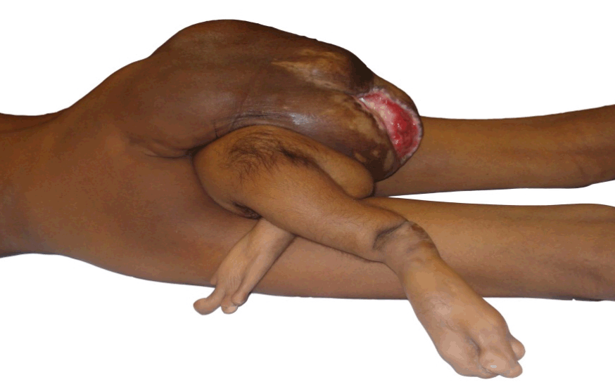
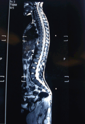
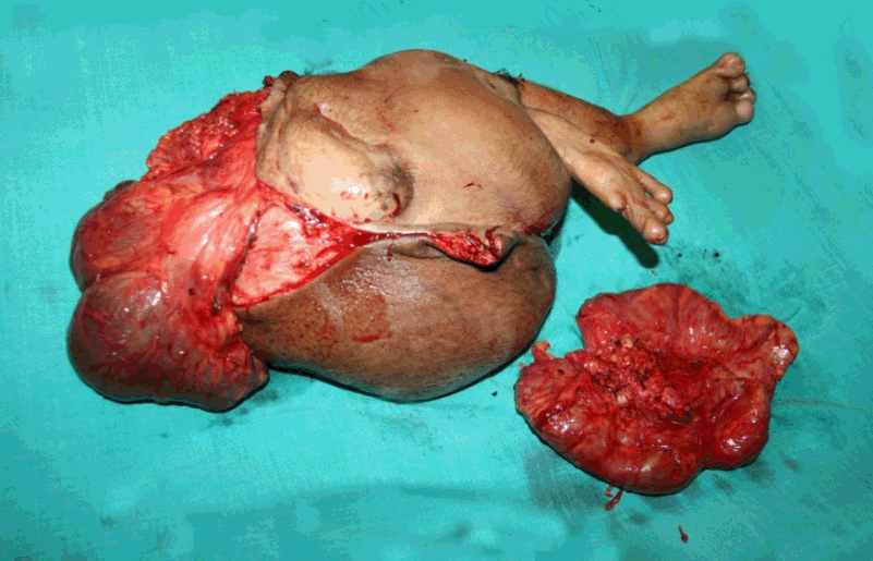
A 16-year-old boy presented in the outdoor department of our institute with history of malformation attached to his lower back since birth. The boy was born 16 years back, with history of vaginal breech delivery at home with prolonged labor. At birth the infant had irregular mass in his lower back covered by a membrane which ruptured in few weeks to reveal a malformation initially interpreted as supernumerary limbs which has grown in size with the growth of child . The boy had difficulty in performing daily routine activities including unable to dress properly and lie in supine. The patient was of average built and nutrition; vitals were normal and had mild pallor. The sacral portion of the spine was deformed protruding backward at an angle of about 45 degrees, underneath to which was attached a malformation. This malformed structure had well appreciable lower limbs with toes and nails although no movements were recorded in these limbs either spontaneous or upon stimulation. There were nipples, hairs, and pubic hair area and skin ulceration of size 6x7 cm noticed over malformation (Figure 1). The patient was undertaken for detailed radiological examination including ultrasound abdomen, Contrast enhanced computed tomography of abdomen and MRI spine. These investigations did not reveal any gross communication of malformation with the abdominal cavity or spine of the patient (Figure 2). The surgical excision of the malformation was performed without any significant complication. The malformation was mostly exterior with extension in the presacral space displacing sacrum posterior, there was no direct communication in the abdomen or spinal canal of the patient and gluteal muscles of the patient were ill developed. The main vascular supply of the malformation was from middle sacral artery of the patient. The surgical specimen was subjected to detailed pathological examination, which consisted of ill developed pelvis, both lower limbs with bones, toes and nails, two blind loops of intestine, both nipples and pubic hairs (Figure 3). There was no other major solid organ or internal genitalia in the specimen while the rest of it consisted of mostly fat, muscles and cavities filled lymph like fluid. Based on history, clinical presentation and pathological findings the malformation was diagnosed as an exoparasitic heteropagus twin. | ||||||
| ||||||
| ||||||
| ||||||
|
Discussion
| ||||||
|
The pathogenesis of parasitic twin is not well understood. [6] It is recommended that all cases of parasitic twins should be reported in order to develop a better understanding of this rare entity. [7] [8] The two major theories of the embryologic origin of conjoined twins have been labeled as fission and fusion. Proponents of the former suggest that incomplete fission of the blastocyst inner cell mass during the primitive streak stage. [9] [10] Fusion in contrast refers to 2 originally distinct inner cell mass that coalesce secondarily a later stage. [11] [12] It is generally held that heteropagus twins represent a spectrum of clinical entities ranging from nonconjoined twins to intact conjoined twins, included along this spectrum are the entities twin reverse arterial perfusion sequence, fetus in fetu (endoparasitic twin) and mature teratoma. Recently a detailed review article compiled 39 cases of heteropagus twins in varying detail, it recommended computed tomography, ultrasound and magnetic resonance imaging in the evaluation of these patients with conventional or magnetic resonance angiography to identify the vascular pedicle if needed, echocardiography to be added in the cases of thoracophagus and omphalopagus heteropagus twins. [5] Most of the reported cases underwent surgical excision in their infancy or early childhood although few of them were managed in their teenage. [2] [3] [5] Exoparasitic heteropagus twin carry the best prognosis among all forms of double monstrosity since surgical removal of the parasite can usually be accomplished without damage to the viable fully developed autosite because of its usual attachment to the host through a soft tissue pedicle. [12] Our case was an ideal exoparasitic conjoined twin which presented late in his preadolescence for intervention and was successfully managed at a rural tertiary institute. | ||||||
|
Conclusion
| ||||||
|
Twinning represents a spectrum of clinical entities ranging from nonconjoined twins to intact conjoined twins. A parasitic twin in which one of the twins (parasite) is severely defective and is depended on the vascular supply from its counterpart (autosite) for its growth. | ||||||
|
References
| ||||||
| ||||||

|
[HTML Abstract]
[PDF Full Text]
|
|
Author Contributions:
Shailendra Pal Singh – Conception and the main operating surgeon, Acquisition of data, Analysis and interpretation of data, Drafting the article, Critical revision of the article, Final approval of the version to be published Subhash Singh Rajput – Drafting the article, Critical revision of the article, Final approval of the version to be published |
|
Guarantor of submission:
The corresponding author is the guarantor of submission. |
|
Source of support:
None |
|
Conflict of interest:
Authors declare no conflict of interest. |
|
Copyright:
© Shailendra Pal Singh et al. 2012; This article is distributed the terms of Creative Commons Attribution License which permits unrestricted use, distribution and reproduction in any means provided the original authors and original publisher are properly credited. (Please see Copyright Policy for more information.) |
|
|