| Table of Contents |  |
|
Case Report
|
| Simultaneous and spontaneous rupture of patellar tendon and contralateral quadriceps tendon in a patient with tertiary hyperparathyroidism: A case report |
| I López Zabala1, MC Pulido1, D Popescu1, JA Fernández-Valencia1 |
|
1Department of Orthopaedic Surgery Hospital Clínic Barcelona C/ Villarroel 170. 08036 Barcelona, Spain.
|
|
doi:10.5348/ijcri-2012-08-157-CR-4
|
|
Address correspondence to: Ibon López Zabala Department of Orthopaedic Surgery Hospital Clínic Barcelona C/ Villarroel 170 08036 Barcelona Spain Phone: 00-34-93-227-55-33 Fax: 93 227 98 71 Email: lopez.ibon@gmail.com |
|
[HTML Abstract]
[PDF Full Text]
|
| How to cite this article: |
| López-Zabala I, Pulido MC, Popescu D, Fernández-Valencia JA. Simultaneous and spontaneous rupture of patellar tendon and contralateral quadriceps tendon in a patient with tertiary hyperparathyroidism: A case report. International Journal of Case Reports and Images 2012;3(8):13–16. |
|
Abstract
|
|
Introduction:
Spontaneous bilateral ruptures of the extensor mechanism of the knee are rare injuries.
Case Report: A patient presented with bilateral rupture of the extensor mechanism of the knee in which a tertiary hyperparathyroidism was previously diagnosed. Tertiary hyperparathyroidism was related to a long-term dialysis for chronic renal failure and on admission, the parathyroid hormone level was 2332 pg/mL. The patient underwent surgery of the both knees and a satisfactory function was achieved. Subsequently, a parathyroidectomy was performed which resulted in a correction of the parathyroid hormone level. Conclusion: Tertiary hyperparathyroidism must be ruled out in patients with chronic renal failure. On the other hand, patients with chronic renal failure and a known hyperparathyroidism need on going and effective management to prevent spontaneous tendon rupture. Clinicians can be aware that careful management of hyperparathyroidism can prevent this severe complication. | |
|
Key Words:
Patellar tendon, Quadriceps, Hyperparathyroidism, Spoutaneous
| |
|
Introduction
| ||||||
|
Rupture of tendons of the extensor mechanism is a relatively frequent injury usually secondary to sports activities. However, simultaneous and spontaneous tendon rupture is a very rare condition. [1] [2] In the case of atraumatic rupture, it requires a low energy mechanism and a previous pathological disorder, such as primary, secondary and tertiary hyperparathyroidism. Patients with chronic renal failure can develop a secondary hyperparathyroidism. Those patients with long-standing secondary hyperparathyroidism can develop an autonomous gland behaving like primary hyperparathyroidism; this condition is known as tertiary hyperparathyroidism. Total parathyroidectomy is the most advisable treatment if the patient is not responding to medical treatment and if a renal transplantation is not feasible. The manifestations of secondary hyperparathyroidism include pruritus, soft-tissue calcifications particularly around joints and especially in arteries, muscles, and subcutaneous tissues. In tertiary hyperparathyroidism these signs are present, pruritus is even more severe, occasionally and skin necrosis due to subcutaneous calcium deposition can be seen. Several studies have reported spontaneous tendon ruptures related to secondary hyperparathyroidism, [3] [4] [5] but spontaneous bilateral ruptures of the extensor mechanism in this clinical setting is extremely rare, with just one case previously described in literature, to the best of our knowledge. [3] In this case report we describe a patient presenting with a bilateral rupture of the extensor mechanism of the knee in which a tertiary hyperparathyroidism was diagnosed. | ||||||
|
Case Report
| ||||||
|
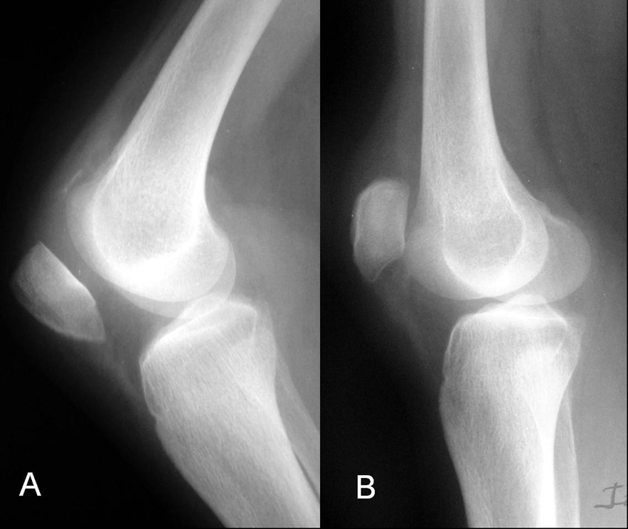
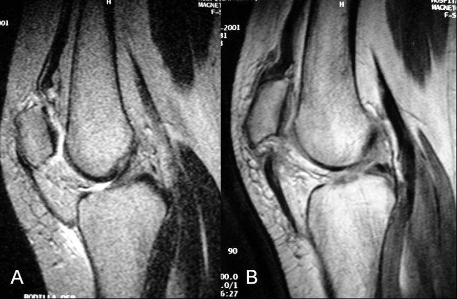
A 31-year-old female suffered sudden pain and loss of extension of both the knees after trying to get up from a sofa. No previous injuries of the knees or intensive sports activities were recorded. Her personal history consisted of chronic renal failure due to renal tubular acidosis diagnosed at the age of eight years and since then she was receiving maintenance hemodialysis until 1984, when a renal transplantation was performed. The transplantation failed six years later and a transplantectomy was performed. Since then, the patient underwent dialysis and recently, a tertiary hyperparathyroidism was detected, with high levels (2332 pg/mL) of parathyroid hormone (PTH), and a parathyroidectomy was proposed for its correction. Physical examination revealed local pain, swelling and lack of active extension. Laboratory examination was normal except for a normocytic anemia, creatinine of 1.6 mg/dL and serum calcium of 11.6 mEq/L. Radiographic examination revealed a high patella in the left knee and a low patella in the right knee (Figure 1). Insall-Salvati index was 0.65 for the right knee and 1.35 for the left knee. Magnetic resonance imaging (MRI) showed a complete rupture of the left patellar tendon at its proximal insertion and the avulsion of the right quadriceps tendon at its patellar insertion (Figure 2). The patient was operated and repair of both the tendons was performed. We observed that the rupture was present at the level of bone-tendon junction without an associated fracture. Tissue samples were obtained for pathological study. We performed the repair of the tendons using heavy nonabsorbable sutures, maintaining the original insertional point in order to avoid tilting of the patella. No additional reinforcing systems were used. At the end of the operation, the flexion grade of both knees was correct, without failure of the sutures during the passive mobilization. Pathological study revealed chronic degenerative changes, with no other specific alterations. Both legs were immobilized with plaster cast and walking with full weight-bearing was permitted at six weeks. One month after surgery, the patient underwent a surgical removal of the parathyroid glands, followed by a progressive normalization of the PTH levels. After four months follow-up, autonomous deambulation was possible without aids, and the range of movement was 0 to 70 degrees in the right knee and 0 to 90 degrees in the left knee. After ten months follow-up, a slight improvement in the range of movement was noticed for both knees (0 to 100 degrees) the patient was able to walk free of pain and was being satisfied with the results. | ||||||
| ||||||
| ||||||
|
Discussion
| ||||||
|
Rupture of quadriceps or patellar tendon is a relatively frequent injury in sport activities. However, simultaneous and spontaneous tendon rupture is a very rare condition. [1] [2] In the case of atraumatic rupture, it requires a low energy mechanism and a previous pathological disorder, such as primary, secondary and tertiary hyperparathyroidism, rheumatoid arthritis, diabetes mellitus, systemic lupus erythematosus, hyperuricemia and Ehlers-Danlos syndrome. [6] [7] The usual mechanism of patella tendon rupture is a sudden flexion of the knee coinciding with contraction of the quadriceps. Kellersmann et al. [8] found that the bilateral patellar tendon ruptures were described in literature as almost simultaneous, signifying that unilateral tendon rupture in one knee placed severe stress on the opposite knee that resulted in symmetric tendon rupture during the same incident. To our knowledge, only three previous cases of a patellar tendon and a contralateral quadriceps tendon rupture have been described, [3] [4] [5] and interestingly, one of them occurred due to hyperparathyroidism. [3] The mechanism to produce this rare pattern remains unclear.The diagnosis of bilateral rupture of the extensor mechanism of both the knees is made by clinical examination, helped by radiological study. After the rupture, the patient experiences pain and the clinical examination may reveal a palpable gap. The radiological study can determine if the patella is in a high or low position. Ultrasonography and MRI are complementary studies; they should be used when the clinical diagnosis is not certain. [9] MRI of the involved limb can confirm the clinical diagnosis. [10] The first case of bilateral simultaneous rupture was reported by Steiner et al. in 1949, affecting both quadriceps tendons. [1] In 1957, Wilson et al. [11] described a case of bilateral rupture of the quadriceps tendons in a patient with chronic nephritis and in 1962, Preston et al. [12] reported the first case of simultaneous extensor mechanism rupture in a patient with chronic renal failure undergoing haemodialysis. Previous reports have proposed a relationship between the duration of hemodialysis and the occurrence of spontaneous tendon ruptures, suggesting that tendinous weakness resulted in the patients from malnutrition, beta2-amyloidosis or accumulation of uremic toxins. Other studies have reported spontaneous tendon rupture related to secondary hyperparathyroidism. [2] [3] Chronic renal failure can cause osteodystrophy with muscular atrophy, debilitation of the extensor mechanism and a weakened tendon, due to degenerative changes in the collagen fibers. Moreover, the secondary hyperparathyroidism presents an increased resorbtion of the bone, debilitating the bone-tendon junction and facilitating the rupture-avulsion at this region. Our patient had chronic renal failure, was in a program of hemodialysis, and had a tertiary hyperparathyroidism with PTH levels of 2332 pg/mL. The diagnosis of tendon rupture should prompt an early surgical repair to prevent muscular retraction and local fibrosis. In our case, a satisfactory result was obtained due to prompt and correct diagnosis of the condition, probably aided by the young age of the patient, and correction of the hyperparathyroidism. | ||||||
|
Conclusion
| ||||||
|
Ruptures of the extensor mechanism of both knees must be managed adequately to restore the function with the aid of surgery and also to identify a possible second mechanism. A secondary or tertiary hyperparathyroidism must be ruled out in patients with chronic renal failure. On the other hand, patients with chronic renal failure and a known hyperparathyroidism need effective management to prevent spontaneous tendon rupture. Clinicians should be aware that careful management of hyperparathyroidism can prevent this severe complication. | ||||||
|
References
| ||||||
| ||||||

|
[HTML Abstract]
[PDF Full Text]
|
|
Author Contributions:
I López Zabala – Substantial contributions to conception and design, Acquisition of data, Analysis and interpretation of data, Drafting the article, Revising it critically for important intellectual content, Final approval of the version to be published MC Pulido – Substantial contributions to conception and design, Analysis and interpretation of data, Revising it critically for important intellectual content, Final approval of the version to be published D Popescu – Substantial contributions to conception and design, Analysis and interpretation of data, Revising it critically for important intellectual content, Final approval of the version to be published JA Fernández-Valencia – Substantial contributions to conception and design, Analysis and interpretation of data, Revising it critically for important intellectual content, Final approval of the version to be published |
|
Guarantor of submission:
The corresponding author is the guarantor of submission. |
|
Source of support:
None |
|
Conflict of interest:
Authors declare no conflict of interest. |
|
Copyright:
© I López Zabala et al. 2012; This article is distributed the terms of Creative Commons Attribution License which permits unrestricted use, distribution and reproduction in any means provided the original authors and original publisher are properly credited. (Please see Copyright Policy for more information.) |
|
|