| Table of Contents |  |
|
Case Report
|
| Anesthetic management of a newborn with giant occipital meningoencephalocele: Case report |
| Banu Cevik1, Arzum Orskiran1, Mesut Yilmaz2, Yasin Ekti1 |
|
1Dr. Lutfi Kirdar Kartal Education and Research Hospital, Anesthesiology and Intensive Care, Istanbul, Turkey.
2Dr. Lutfi Kirdar Kartal Education and Research Hospital, Clinic of Neurosurgery, Istanbul, Turkey. |
|
doi:10.5348/ijcri-2012-08-156-CR-3
|
|
Address correspondence to: Banu Cevik, MD Bagdat Cad. Noter Sok. Yazicioglu Apt., 10/12 Erenkoy-Istanbul Turkey Phone: +90 216 441 39 00 Fax: +90 216 3020523 Email: banueler@yahoo.com |
|
[HTML Abstract]
[PDF Full Text]
|
| How to cite this article: |
| Cevik B, Orskiran A, Yilmaz M, Ekti Y. Anesthetic management of a newborn with giant occipital meningoencephalocele: Case report. International Journal of Case Reports and Images 2012;3(8):10–12. |
|
Abstract
|
|
Introduction:
The anesthetic management of meningoencephalocele is a challange because of the positioning, handling of the airway and the difficulty in perioperative care.
Case Report: A 10-day-old neonate presented for surgical excision of giant occipital meningoencephalocele. Despite the difficulties, intubation and peroperative anesthetic management of the patient was successfully achieved. Conclusion: Previous similar case reports were reviewed and potential perioperative complications are highlighted. | |
|
Key Words:
Encephalocele, Occipital, Anesthesia
| |
|
Introduction
| ||||||
|
The term cephalocele refers to a defect in the skull and dura with extracranial extension of intracranial structures. Cephaloceles are divided into four types: meningoencephalocel, meningocele, atretic encephalocele and gliocele. Meningoencephalocele consists of a herniation of cerebrospinal fluid, brain tissue and meninges through the skull defect. [1] The cause of cephalocele has not been fully determined. Many differences in type and frequency of cephalocele among various ethnic groups have been observed. For example, in the Western hemisphere, the incidence is 1– 3 per 10,000 births. In Southeast Asia, the incidence is slightly higher, with approximately 1 in 5,000 live births. [2] The occipital bone is the most common location for cephalocele in the Caucasian populations of Europe and North America, accounting for about 80% of cases. [1] [2]In this case report, we have included a detailed discussion describing the special anesthetic considerations of these patients. | ||||||
|
Case Report
| ||||||
|
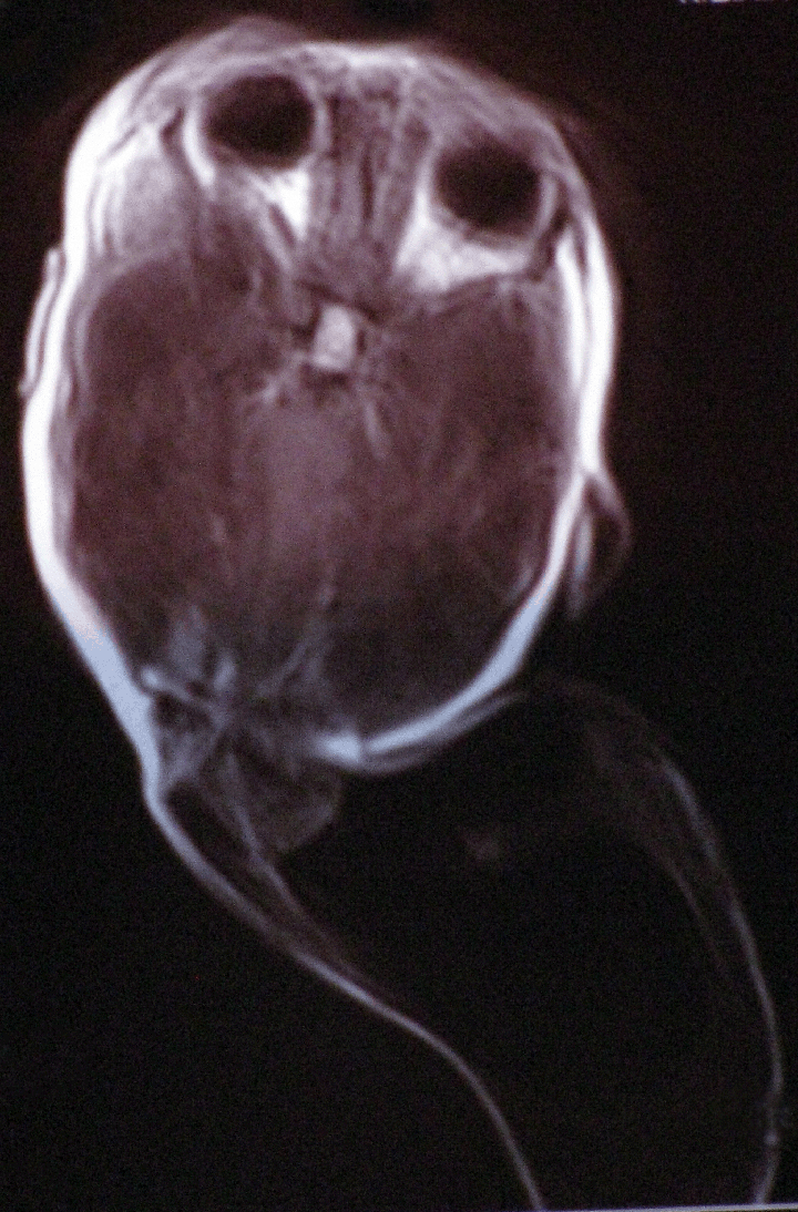
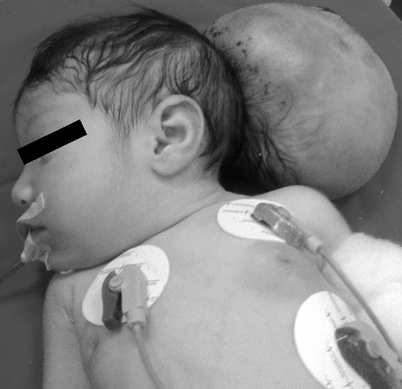
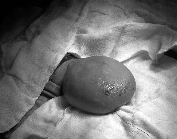
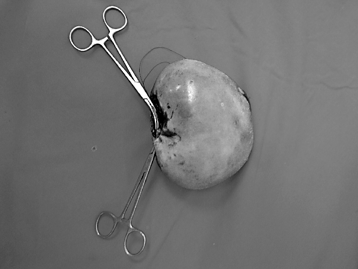
A 10-day-old female neonate presented with a giant cystic swelling in the occipital region and was scheduled for surgical excision. The full-term baby weighing 3710 g with Apgar scores of 5 and 8 at the first and fifth minutes, respectively. The baby was delivered by cesarean section in a municipal hospital. There was no evidence of family history and the mother had an irregular prenatal care period. The neonate was transferred to our hospital and admitted to the pediatric clinic for preoperative preparation. On preanesthetic evaluation, cardiovascular, respiratory and neurological system examination was normal. There was a swelling that measured 16x10 cm arising from posterior part of the head. No other congenital anomaly was detected. Magnetic resonance imaging (MRI) showed giant occipital meningoencephalocele with a minimal herniation of occipital lobe into the swelling (Figure 1). Laboratory evaluations were within normal limits. Following non-invasive arterial blood pressure, electrocardiogram, skin temperature and pulse oximetry monitoring, the neonate was induced with 6% sevoflurane in oxygen in the lateral position. After confirming adequate mask ventilation, muscle relaxation was achieved with 0.2 mg/kg vecuronium. The neonate was turned to supine position and bolsters were used to support the back of the neonate by another anesthesiologist. The sac was gently handled with a towel by a neurosurgeon. The intubation was successfully achieved with a 3.0 mm inner diameter endotracheal tube on the first attempt. After confirmation of appropriate placement and tube fixation, the patient was turned to head-lateral position (Figure 2). Anesthesia was maintained with 1–2% sevoflurane in an approximately 50% N2O 50% O2 mixture. The complete resection of the sac was achieved without any complication (Figures 3 and 4). In total, 250 mL fluid infusion and 50 mL blood transfusion was required and the patient was anesthetized for two hours. With the establishment of spontaneous respiration and gag reflex, the patient was extubated and transferred to the recovery room. For postoperative analgesia, 60 mg paracetamol was administered rectally. After an uneventful postoperative period the patient was discharged to neurosurgical clinic for further follow-up. | ||||||
| ||||||
| ||||||
| ||||||
| ||||||
|
Discussion
| ||||||
|
Encephalocele poses challenging operative approaches and related risks, thus the first question the neurosurgeon should consider is whether or not to repair the lesion. Factors such as the amount of the brain in the sac, the presence of other anomalies of the intracranial brain and the presence of other congenital anomalies should all be considered. Once the decision to operate has been made, a perioperative plan must be formulated by an anesthesiologist based on airway management, fluid balance and prevention of hypothermia. In our case, the occipital meningoencephalocele made supine position of head impossible due to the limited-neck extension and the likelihood of rupturing the membranes covering the spinal cord or brain. [3] Mask ventilation and intubation may be attempted in the lateral position [4] [5] or in alternative approaches including to place the child in supine position on a platform of rolled-up blankets while an assistant temporarily supports the head or placing the child' s head beyond the edge of the table with an assistant supporting it. [6] Before administering neuromuscular blocking agents and intubation, adequate mask ventilation must be verified. Another complication which may result is from derangement in the pontomedullary respiratory control center leading to inadequate spontaneous respiration. Aspiration complications may arise from lack of pharyngeal coordination, poor sucking reflex and absent gag reflex. [7] Anesthetic management of these children requires careful attention to positioning, airway management, monitoring body temperature and estimation of blood and fluid loss. Latex allergy precautions should be used with these children for their first anesthetic procedure. [8] | ||||||
|
Conclusion
| ||||||
|
Perioperative management of patients with giant meningoencephalocele may be challenging for both anesthesiologist and neurosurgeon. These patients must be managed closely with an interdisciplinary approach. | ||||||
|
References
| ||||||
| ||||||

|
[HTML Abstract]
[PDF Full Text]
|
|
Author Contributions:
Banu Cevik – Conception and design, Acquisition of data, Analysis and interpretation of data, Drafting the article, Critical revision of the article, Final approval of the version to be published Arzum Orskiran – Conception and design, Acquisition of data, Analysis and interpretation of data, Drafting the article, Critical revision of the article, Final approval of the version to be published Mesut Yilmaz – Conception and design, Acquisition of data, Analysis and interpretation of data, Drafting the article, Critical revision of the article, Final approval of the version to be published Yasin Ekti – Conception and design, Acquisition of data, Analysis and interpretation of data, Drafting the article, Critical revision of the article, Final approval of the version to be published |
|
Guarantor of submission:
The corresponding author is the guarantor of submission. |
|
Source of support:
None |
|
Conflict of interest:
Authors declare no conflict of interest. |
|
Copyright:
© Banu Cevik et al. 2012; This article is distributed the terms of Creative Commons Attribution License which permits unrestricted use, distribution and reproduction in any means provided the original authors and original publisher are properly credited. (Please see Copyright Policy for more information.) |
|
|