| Table of Contents |  |
|
Case Report
|
| Aneurysm of superior mesenteric vein |
| Mehrzad Lotfi1, Azadeh Hajati2, Seyedpouria Ostad3 |
|
1Shiraz University of Medical Sciences- Associate Professor, Department of Radiology, Namazi hospital, Shiraz, Fars, Iran, Islamic Republic.
2Shiraz University of Medical Sciences- Radiologist, Department of Radiology, Namazi hospital, Shiraz, Fars, Iran, Islamic Republic. 3Shiraz University of Medical Sciences- Resident of radiology, Department of Radiology, Namazi hospital, Shiraz, Fars, Iran, Islamic Republic . |
|
doi:10.5348/ijcri-2012-07-151-CR-12
|
|
Address correspondence to: Azadeh Hajati Mirzaye shirazi blvd Shabnam 50. No 150 Shiraz, Fars, Iran Islamic Republic Phone: 7188834776, (0098)9171123721; (0098)7116256852 Email: Azadeh.hajati@gmail.com |
|
[HTML Abstract]
[PDF Full Text]
|
| How to cite this article: |
| Lotfi M, Hajati A, Ostad S. Aneurysm of superior mesenteric vein. International Journal of Case Reports and Images 2012;3(7):49–51. |
|
Abstract
|
|
Introduction:
Venous aneurysms and pseudoaneurysms are rare entities; portal system aneurysms are shown as focal dilatations of the portal venous system. Superior mesenteric vein aneurysms are extremely rare.
Case Report: We describe a case of superior mesenteric vein aneurysm in an asymptomatic 73-year-old female. Although one cause of portal system aneurysm is portal hypertension, in this rare case no underlying cause was noted. Conclusion: Aneurysms of portal system are very rare entities. Although their cause is uncertain, portal hypertension should be suspected in the evaluation of portal aneurysms. Superior mesenteric vein aneurysms are rare. Most of them are asymptomatic and the diagnosis is confirmed by radiologic findings. Diagnosis in other cases is established after complications develop. According to the information in literature, portal system aneurysm is not as rare as previously presumed. As one of the important causes of portal aneurysm is portal hypertension and because portal system aneurysms can cause portal hypertension when thrombosis occur, evaluation of its presence has clinical significance in patients with portal hypertension. | |
|
Key Words:
Superior mesentric vein, Aneurysm, Portal hypertension, Ultrasound
| |
|
Introduction
|
|
Venous aneurysm and pseudoaneurysm are rare entities. [1] Aneurysms of portal system are rare entities and aneurysms of superior mesenteric vein are still rarer. [2] [3] They are shown as focal dilatations of the portal venous system. [2] Although their cause is uncertain, portal hypertension should be suspected in the evaluation of portal aneurysms. [4] Many aneurysms have no symptom and they are found through radiologic examinations. [5] Here, we reported a case of superior mesenteric vein aneurysm in asymptomatic person with no sign of portal hypertension. |
|
Case Report
|
|
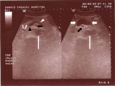
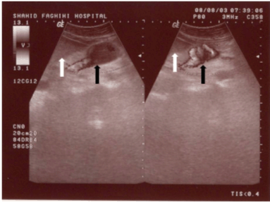
A 73-year-old female came to our sonogrphic clinic for abdomino-pelvic sonography as a routine work-up. In her medical history she had positive history of mild hyperlipidemia and mild essential hypertension and was on ß-blocker, nitroglycerine and anti-lipid therapy. Routine workup (CBC, LFT) was normal. She had no history of abdominal trauma or liver disease. Incidentally, on evaluation of para-aortic area, an anechoic structure was noted anterior to inferior vena cave (IVC) which was in continuation to portal confluence and was in the region of superior mesenteric vein (Figure 1). There was no sign of portal hypertension. Liver and spleen were normal (Figure 2). Color doppler flow mapping revealed flow inside the structure (Figure 3). Spectral analysis further confirmed venous type flow in this region (Figure 4). These findings confirmed the diagnosis of superior mesenteric vein aneurysm. This patient did not received any treatment because she was asymptomatic and there was no sign of portal hypertention. Follow-up sonography was done for her regularly but no increase in size of the aneurysm was noted and she has remained asymptomatic. |
|
|
|
|
|
|
|
|
|
Discussion
|
|
Arterial aneurysms are more common than venous aneurysms in clinical practice. The portal aneurysms are focal dilatations of the portal system. [2] Since portal vein aneurysms are rare conditions, only few cases of portal vein and superior mesenteric vein aneurysms have been reported in literature. According to one study about 60% of cases of portal vein aneurysm were congenital. [6] Portal vein aneurysms can be classified according to their location as intra or extrahapatic. [2] Age and sex has no influence on presence or type of aneurysm (extra or intrahepatic). [7] They can be congenital or acquired. [4] Finding their etiology is not always possible, however, because the most important cause is portal hypertension [2], it should be suspected in the evaluation of portal system aneurysms. Trauma can also be an etiologic factor for their development. Another factor is hepatocellular disease. Abdominal pain, jaundice, gastrointestinal hemorrhage or portal hypertension can be the accompanying symptoms, but sometimes patients are asymptomatic. [3] Thrombosis of these aneurysms can cause portal hypertension. [8] Superior mesenteric vein aneurysms are rare. Most of them are asymptomatic and the diagnosis is confirmed by radiologic findings but in other cases the diagnosis is established after complications develop. [5] Intra-hepatic portal vein aneurysms are seen as cystic lesions with internal flow but they are fusiform expansion in the main portal vein and its branches. [4] In sonography they are anechoic structures in gray scale and have spectral findings of portal venous system in color doppler study. [9] Evaluation of extra hepatic portal vein aneurysms is also possible by CT, magnetic resonance imaging (MRA) and angiography. [10] Due to rarity of superior mesentric vein aneurysms, their evolution is not known, however, the management of most patients is to follow-them regularly by imaging studies as the aneurysms are asymptomatic. The possibility of complications such as thrombosis and rupture should be explained to the patients. For few patients surgery such as aneurysmorrhaphy and simple resection of the aneurysm mesocaval shunts should be done due to complications or symptoms. [5] |
|
Conclusion
|
|
According to the published in the literature, portal system aneurysm is not as rare as previously presumed. As one of the important causes of these entities is portal hypertension and also it can cause portal hypertension when thrombosis occur in the aneurysm, evaluation of their presence has clinical significance in patients with portal hypertension. |
|
References
|
|


|
[HTML Abstract]
[PDF Full Text]
|
|
Author Contributions:
Mehrzad Lotfi - Conception and design, Acquisition of data, Analysis and interpretation of data, Drafting the article, Critical revision of the article, Final approval of the version to be published Azadeh Hajati - Conception and design, Acquisition of data, Analysis and interpretation of data, Drafting the article, Critical revision of the article, Final approval of the version to be published Seyedpouria Ostad - Conception and design, Acquisition of data, Analysis and interpretation of data, Drafting the article, Critical revision of the article, Final approval of the version to be published < |
|
Guarantor of submission:
The corresponding author is the guarantor of submission. |
|
Source of support:
None |
|
Conflict of interest:
Portal system aneurysms are unusual findings however should be sought in patients with portal hypertension because they can mimic mass lesions. Color Doppler ultrasound is an appropriate method for their investigation. |
|
Copyright:
© Mehrzad Lotfi et al. 2012; This article is distributed the terms of Creative Commons Attribution License which permits unrestricted use, distribution and reproduction in any means provided the original authors and original publisher are properly credited. (Please see Copyright Policy for more information.) |
|
|