| Table of Contents |  |
|
Case Report
|
| Solitary fibrous tumor of the larynx |
| Yehuda Schwarz1, Sharon Tamir1, Constantin Reinus1, Jean Yves Sichel1, Uri Peleg1 |
|
1Institutional affiliations of all authors: Shaare Zedek Medical Center, Jerusalem, Israel.
|
|
doi:10.5348/ijcri-2012-07-150-CR-11
|
|
Address correspondence to: Yehuda Schwarz Netzach Jerusalem 6/4, Efrat Israel Phone: 972-2-9938416 Fax: 972-2-9938416 Email: Inbalzim@yahoo.com |
|
[HTML Abstract]
[PDF Full Text]
|
| How to cite this article: |
| Schwarz Y, Tamir S, Reinus C, Sichel JY, Peleg U. Solitary fibrous tumor of the larynx. International Journal of Case Reports and Images 2012;3(7):45–48. |
|
Abstract
|
|
Introduction:
Solitary fibrous tumors of the larynx are extremely rare. To date only occasional cases have been reported in the literature. We present this case report as an additional case to existing literature.
Case Report: This report presents a patient with progressive dysphonia for two years. Examination revealed a bulging mass in the left paraglottic space. A CT scan was performed which demonstrated a well delineated, submucosal mass. The mass was firm and non-mobile and due to this fact obtaining material for biopsy was technically impossible. A surgical excision of the mass via a cervical approach was performed. The mass was completely removed and there was an immediate improvement in the patient's voice quality. Histological examination and immunohistochemical studies revealed a solitary fibrous tumor. Conclusion: This is a rare presentation of a solitary fibrous tumor in the larynx. Management options are discussed and the literature on this uncommon entity is reviewed. | |
|
Key Words:
Solitary fibrous tumor, Larynx
| |
|
Introduction
| ||||||
|
Solitary fibrous tumors of the larynx are extremely rare. To date only occasional cases have been reported in the literature. [1] Solitary fibrous tumors are uncommon benign spindle cell neoplasms of mesenchymal origin. [2] Majority of these tumors (more than 50%) occur in the chest cavity, however, they can be found everywhere in the body. In the head and neck region, the most commonly affected site is the oral cavity. [2] [3] There are few series that documented the clinical presentation, surgical management and outcome of these tumors in the head and neck region. Generaly it is thought that they present as asymptomatic slow-growing masses that can be very difficult to distinguish from other soft tissue tumors. The diagnosis is therefore strongly dependent on the microscopic appearance and characteristic immunohistochemical staining. These tumors are commonly treated by local excision. We present this case report as an additional case to existing literature. | ||||||
|
Case Report
| ||||||
|
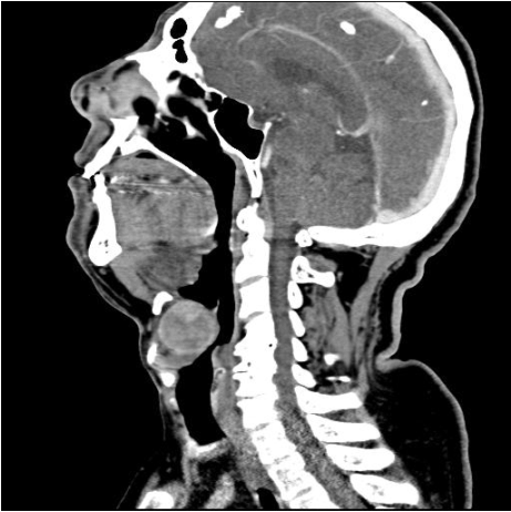
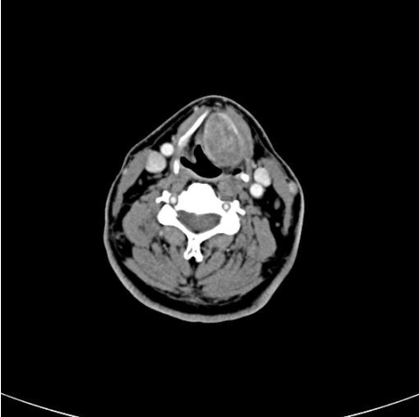
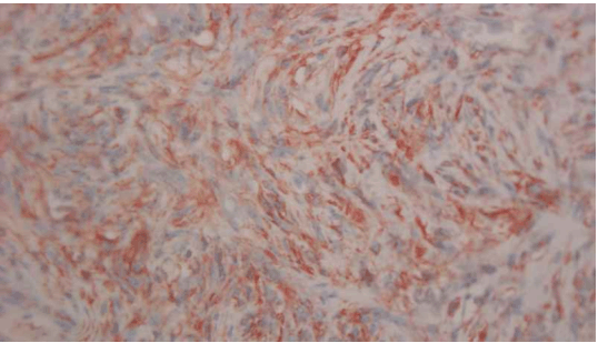
A 66-year-old male presented to our clinic with a two year history of progressively disabling dysphonia which had disrupted his career as a cantor. His physical examination was normal. Fiberoptic laryngoscopy revealed a bulging mass in the left paraglottic space extending to the vallecula and displacing the left vocal cord. A computed tomography (CT) scan was performed which demonstrated a 3.5 cm well delineated submucosal mass, located in the left paraglottic space and extending to the pre-epiglottic space and left vallecular region (Figures 1, 2). There was no evidence of cartilaginous destruction. Direct laryngoscopy revealed a firm and non-mobile mass. Due to this fact obtaining sufficient material with a punch biopsy forceps was technically impossible. Therefore, a surgical excision of the mass via a cervical approach was undertaken, with removal of the left thyroid cartilage lamina. Using this approach excellent exposure of the left paraglottic space containing the tumor was achieved. The mass was completely removed without any violation of the airway. Post-operative recovery was uneventful and an immediate improvement in the patient's voice quality was noted. Histological examination showed a well circumscribed mass composed of spindle cells with vesicular nuclei (Figure 3). The cells were arranged in short fascicles. Foci of storiform pattern, areas of hyalization and myxoid stromal changes were present. Branching vascular channels were also present. Immunohistochemical studies showed strong cytoplasmatic positivity of the cells for vimentin, CD 34 and bcl-2 (Figure 4), while staining for cytokeratin, desmin, actin and S100 were negative. A final diagnosis of solitary fibrous tumor of the larynx was given. | ||||||
| ||||||
|
| ||||||
| ||||||
| ||||||
|
Discussion
| ||||||
|
Solitary fibrous tumor was initially described by Klemperer et al. in 1931. [1] They described a tumor arising from the pleural alveolar tissue. The tumor grew slowly and only caused symptoms after it had reached a substantial size. Solitary fibrous tumors are uncommon benign spindle cell neoplasms of mesenchymal origin. [2] The majority of these tumors (over 50%) occur in the chest cavity, however, they can be found everywhere in the body. In the head and neck region, the most commonly affected site is the oral cavity. [2] [3] Thompson et al. reported a case of solitary fibrous tumor of the larynx and reviewed seven other cases. [4] The authors noted the extreme rarity of this entity. Tumor presentation depends on the exact location of the mass. There are no specific clinical signs, and symptoms are usually progressive. When the origin is in the larynx, the most common symptoms are hoarseness, breathing difficulties, foreign body sensation, cough and throat clearing. [4] CT scan shows a submucosal dense homogeneous enhancement of the mass on post contrast studies. CT imaging is essential to rule out adjacent bony or cartilaginous involvement, which usually does not occur with solitary fibrous tumors. [2] Magnetic resonance imaging (MRI) features of laryngeal Solitary fibrous tumors are nonspecific, and are similar to those of other more common tumors such as hemangiopericytomas, neurogenic tumors, and soft tissue sarcomas. [2] On MRI images, solitary fibrous tumor is isointense relative to muscle on T1 images and it enhances with gadolinium. The intensity on T2 images is variable. [5] Due to the non-specific physical and imaging findings of this entity, the differential diagnosis may include squamous cell carcinoma, sarcoma, fibrous histiocytoma, schwannoma, neurofibroma and fibroma. [2] Histology usually reveals a well circumscribed mass characterized by ovoid and spindle cells with a majority of cells appearing fibroblastic. These tumors often show a rich vasculature. Necrosis is not a usual feature of these lesions. [3] The use of immunohistochemical staining is important in confirming the diagnosis. The tumor cells show cytoplasmic reactivity for CD34, bcl-2, and vimentin. When trying to further confirm the diagnosis of solitary fibrous tumor, immunohistochemical staining for S-100, cytokeratin, muscle-specific antigen and desmin should be negative thus excluding the possibility of other tumors. [3] In our case, obtaining material for histological diagnosis via direct laryngoscopy proved to be difficult due to the firm nature of the mass. This fact and the tumor's size led us to perform open surgery through an external approach. Because of the limited number of cases reviewed in the literature, a "gold standard" surgical approach is not agreed upon. There are several surgical approaches possible. Thomson et al reported partial laryngectomy and pharyngotomy. [4] Endoscopic CO2 laser surgery has gained popularity in the last few years. [4] In our opinion, an external surgical approach in large tumors seems optimal. It can be achieved through a relatively small skin incision, which provides an excellent exposure with easier access to the larynx. This enables complete removal of the tumor without penetrating the airway and better control over bleeding. Endoscopic removal may be considered in selected cases. | ||||||
|
Conclusion
| ||||||
|
Solitary fibrous tumors of the larynx are very rare. Imaging features of CT scan and MRI are not specific. Histology is mandatory for the diagnosis. Whenever possible complete surgical excision should be regarded as the treatment of choice. Although transoral approach has become more popular we feel that the external approach is preferred due to its greater exposure and easier access to the larynx. | ||||||
|
Acknowledgment
| ||||||
|
Schwarz Henry, M.D. – Provided guidance and general support during the writing of this paper. | ||||||
|
References
| ||||||
| ||||||


|
[HTML Abstract]
[PDF Full Text]
|
|
Author Contributions:
Yehuda Schwarz - Substantial contributions to conception and design, Acquisition of data, Analysis and interpretation of data, Drafting the article, Revising it critically for important intellectual content, Final approval of the version to be published Sharon Tamir - Substantial contributions to conception and design, Acquisition of data, Analysis and interpretation of data, Drafting the article, Revising it critically for important intellectual content, Final approval of the version to be published Constantin Reinus - Substantial contributions to conception and design, Acquisition of data, Analysis and interpretation of data, Drafting the article, Revising it critically for important intellectual content, Final approval of the version to be published Jean Yves Sichel - Substantial contributions to conception and design, Acquisition of data, Analysis and interpretation of data, Drafting the article, Revising it critically for important intellectual content, Final approval of the version to be published Uri Peleg - Substantial contributions to conception and design, Acquisition of data, Analysis and interpretation of data, Drafting the article, Revising it critically for important intellectual content, Final approval of the version to be published |
|
Guarantor of submission:
The corresponding author is the guarantor of submission. |
|
Source of support:
None |
|
Conflict of interest:
Authors declare no conflict of interest. |
|
Copyright:
© Yehuda Schwarz et al. 2012; This article is distributed the terms of Creative Commons Attribution License which permits unrestricted use, distribution and reproduction in any means provided the original authors and original publisher are properly credited. (Please see Copyright Policy for more information.) |
|
|