| Table of Contents |  |
|
Case Report
|
| Split optic nerve penetrated by a carotid-ophthalmic aneurysm another instance of a rare presentation: A case report |
| Caleb E Feliciano1, Duke Samson2 |
|
1Department of Surgery-Section of Neurourgery, University of Puerto Rico-Medical Sciences Campus, PO Box 365067, San Juan, PR 00936-5067.
2Department of Neurosurgery, University of Texas-Southwestern at Dallas 2353 Harry Hines Blvd, Dallas, TX 75390-8855. |
|
doi:10.5348/ijcri-2012-07-149-CR-10
|
|
Address correspondence to: Caleb E. Feliciano, MD Department of Surgery-Section of Neurourgery University of Puerto Rico-Medical Sciences Campus, PO Box - 365067 San Juan, PR 00936-5067 USA Phone: 787-765-8276 Fax: 787-765-8276 Email: caleb.feliciano@upr.edu |
|
[HTML Abstract]
[PDF Full Text]
|
| How to cite this article: |
| Feliciano CE, Samson D. Split optic nerve penetrated by a carotid-ophthalmic aneurysm another instance of a rare presentation: A case report. International Journal of Case Reports and Images 2012;3(7):39–44. |
|
Abstract
|
|
Introduction:
Penetration of the optic nerve by aneurysms of the paraclinoid carotid artery is rare. We present the seventh aneurysm-related split optic nerve case reported in the literature, review the pathophysiologic mechanisms and discuss the technical aspects for successful clip ligation and complication avoidance.
Case Report: A 64-year-old male with history of hypertension, chronic obstructive pulmonary disease and polycystic kidneys presented with headache and dizziness. Magnetic resonance imaging and angiographic work-up confirmed the presence of a right-sided partially thrombosed carotid-ophthalmic aneurysm. There was no evidence of significant visual dysfunction. A right pterional craniotomy with frontal extension was performed. The aneurysm dome was noted to extend subfrontally after passing through a split optic nerve. Aneurysm neck was clipped using a combination of standard ophthalmic and fenestrated clips. During immediate post-operative period the patient had worsening of visual acuity, yet showed progressive visual improvement on clinical follow-up. Conclusion: The pathophysiologic mechanisms for aneurysm development range from congenital to mechanical/hemodynamic. Our patient's clinical presentation suggests a slowly growing lesion that may have penetrated through a weak spot in the overlying optic nerve. This may have been caused by either congenital fenestration of the optic nerve, persistent vestigial artery or an old unrecognized hemorrhage. Optic nerve penetration by carotid-ophthalmic aneurysms occurs rarely, yet is being increasingly reported and recognized. Knowledge of this potential configuration can help avoid inadvertent damage and improve results by novel application of fenestrated and standard clips. | |
|
Key Words:
Carotid-ophthalmic aneurysm, Optic nerve fenestration, Paraclinoid aneurysm, Split optic nerve, Surgical technique
| |
|
Introduction
| ||||||
|
Carotid-ophthalmic (CO) aneurysms constitute 0.3–1% of intracranial aneurysms and 0.9–6.5% of aneurysms of the internal corotid artery (ICA). [1] Although they commonly present with subarachnoid hemorrhage, presentation with visual dysfunction due to mass effect on the optic pathways can also occur, [2] [3] usually after reaching a size of more than 1 cm. [4] [5] In Yasargil series, [6] 18% of patients presented with visual symptoms. Day [4] reported that 30% cases with CO aneurysms can present with visual dysfunction. CO aneurysms usually displace the optic nerve (ON) supero-medially against the falciform ligament as they grow. Penetration of the optic nerve by aneurysms of this region occurs rarely. Only six case reports were found in literature showing this occurrence (seven after including a case of de novo ON splitting without associated aneurysm [7], one of them by a ruptured anterior communicating artery aneurysm, and the rest by paraclinoidal ICA aneurysms. [1][5] [7] [8] [9] [10] [13] To this instance, we present the seventh case reported in literature and review the proposed pathophysiologic mechanisms and the technical aspects for successful clip ligation and complication avoidance. | ||||||
|
Case Report
| ||||||
|
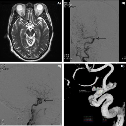
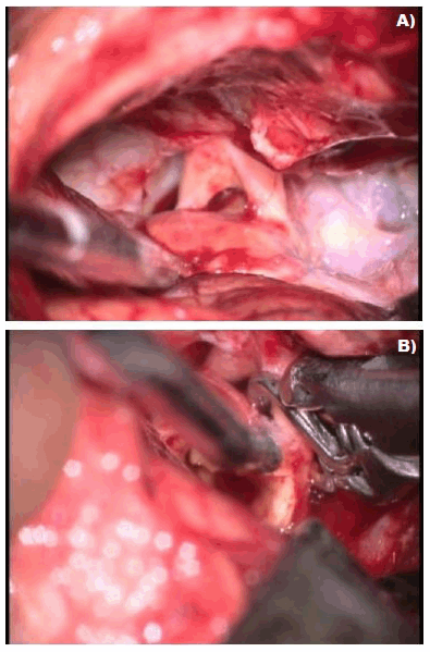
The patient was a 64-year-old male with history of hypertension, chronic obstructive pulmonary disease (COPD) and polycystic kidney disease (PKD) who presented with an episode of headache and dizziness. On physical examination the patient was fully alert and oriented, as well as able to provide his own history. Cranial nerve evaluation, motor, sensory and cerebellar examinations revealed no gross neurologic deficits. There was no evidence of significant visual dysfunction, except for the fact that the patient reported using glasses. No formal visual field testing (perimetry) was done. Due to a significant history of protracted smoking and family history of subarachnoid hemorrhage, magnetic resonance imaging/magnetic resonance angiography (MRI/MRA) work-up were done which confirmed the presence of a right-sided partially thrombosed carotid-ophthalmic aneurysm measuring approximately one cm in its largest diameter (Figure 1). In view of the aneurysm findings, the patient was offered endovascular treatment with stent-assisted coiling in the referring hospital, however he had significant tortuosity in the access vessels and the procedure was deemed unsafe. At our clinic evaluation, he was offered surgical exploration for clip ligation due to the high estimated risk of hemorrhage and possibility for development of significant mass effect symptoms. Operative Technique: Under general endotracheal anesthesia, the patient was positioned supine and his head turned and fixed 45° to the left using the Mayfield-Kees head holder. After prepping and draping, a curvilinear skin incision was made beginning anterior to the tragus and ending midline behind the hairline. Separate skin and temporalis muscle flaps were elevated and secured with fish-hook retractors. A rectangular bone flap centered at the pterion with frontal extension was performed. Subsequently, the sphenoid wing was drilled until visualization of the lateral superior orbital fissure. The dura was opened and the optico-carotid cistern reached and opened for CSF drainage. A dense arachnoiditis was noted between the optic nerve and the gyrus rectus. This was sharply dissected, and the aneurysm dome was noted to extend subfrontally below the gyrus rectus. It protruded through a split optic nerve (Figure 2A). Attention was then directed to the neck of the aneurysm and after adequate proximal and distal control, it was clipped with two large Yasargil ophthalmic clips that spanned the entire atherosclerotic portion of the neck. The aneurysm dome was subsequently opened and decompressed by removing the atherosclerotic debris and old thrombus. On reaching the proximal neck of the aneurysm brisk arterial bleeding was noted. A thin part of the medial aneurysm neck was not adequately sealed by the clips due to significant luminal irregularity. A third large straight fenestrated clip was then placed, including the lateral half of the optic nerve in the fenestration and effectively occluding the neck of the aneurysm (Figure 2B). Patency of the surrounding vessels was confirmed by Doppler insonation. Postoperative visual examination revealed worsening of visual acuity on the right eye to finger counting and mild right inferior visual field loss. The patient was seen one month after surgery and visual acquity was noted to be steadily improving. | ||||||
|
| ||||||
|
| ||||||
|
Discussion
| ||||||
|
Optic nerve penetration by carotid-ophthalmic aneurysms occurs rarely, yet is being increasingly reported and recognized. The pathophysiologic mechanisms proposed in the literature range from congenital to mechanical/hemodynamic causes. However, there is no clear evidence in favor of a specific mechanism. One report by House et al. [9] proposed the de novo fenestration of an ON. This could predate the penetration by an enlarging aneurysm. Congenital splitting has been described by Snead in 1915, [11] making this a plausible explanation. Other reports have pointed out the existence of vestigial remnants of optic nerve/chiasmal perforation or anastomotic arteries that may actually be the origin of these aneurysms or provide areas of weakness along the optic apparatus through which an aneurysm may expand. [1] [5] [7] [10] [12] [13] Other reports propose that at the moment of rupture of a CO aneurysm the optic nerve may split causing sudden visual deterioration and provide a pathway for further aneurismal growth. [8] Jea et al. [5] proposed that a slow growing aneurysm may expand through the optic nerve along the pathway of least resistance, if hemodynamic and anatomical factors favor growth in that direction, splitting the nerve longitudinally along the nerve fibers. A similar mechanism was proposed by Fujita et al. for a patient with rapidly progressive visual loss accounted for by a rapidly growing CO aneurysm. [14] Table 1 illustrates the important aspects of the cases that have been reported so far, as well as the pathophysiologic mechanisms proposed. Other similar cases have been commented on, yet not reported. Therefore, this condition may be more common than expected. In our case, there was no obvious intraoperative evidence of hemorrhage. In addition, a vestigial remnant of an arterial perforator or congenital anastomotic artery was not found. Taking into account that visual deterioration was not the chief medical complaint of the patient, it suggests a slowly growing lesion that may have penetrated through a weak spot in the overlying ON. The weakened spot may have been caused by any of the prior mentioned mechanisms, whether congenital fenestration of the ON, persistent vestigial artery or even an old unrecognized hemorrhage. The exact pathophysiologic mechanism is difficult to point out, and every mechanism discussed may in fact play a role in different cases. Increasing knowledge of this potential configuration can help avoid inadvertent damage and improve results through careful surgical technique and novel application of fenestrated and standard aneurysm clips. | ||||||
| ||||||
|
Conclusion
| ||||||
|
In published literature the pathophysiologic mechanisms proposed for development of carotid- ophthalmic aneurysms range from congenital to mechanical/hemodynamic. Optic nerve penetration by carotid-ophthalmic aneurysms rarely occurs, yet is increasingly reported and recognized. Knowledge of this potential configuration can help avoid inadvertent damage and improve results by novel application of fenestrated and standard clips. | ||||||
|
References
| ||||||
| ||||||


|
[HTML Abstract]
[PDF Full Text]
|
|
Author Contributions:
Caleb E Feliciano - Substantial contributions to conception and design, Acquisition of data, Drafting the article, Revising it critically for important intellectual content, Final approval of the version to be published Duke Samson - Substantial contributions to conception and design, Acquisition of data, Drafting the article, Revising it critically for important intellectual content, Final approval of the version to be published |
|
Guarantor of submission:
The corresponding author is the guarantor of submission. |
|
Source of support:
None |
|
Conflict of interest:
Authors declare no conflict of interest. |
|
Copyright:
© Caleb E Feliciano et al. 2012; This article is distributed the terms of Creative Commons Attribution License which permits unrestricted use, distribution and reproduction in any means provided the original authors and original publisher are properly credited. (Please see Copyright Policy for more information.) |
|
|