| Table of Contents |  |
|
Case Report
|
| Pituitary apoplexy resembling acute meningitis without visual defect and ophthalmoplegia |
| Shu-Yi Wang1, Chieh-Sen Chuan2, 3 |
|
1Departments of Endocrinology and Metabolism, Changhua Christian Hospital, Changhua, Taiwan.
2Departments of Neurology, Changhua Christian Hospital, Changhua, Taiwan. 3Department of Life Sciences, National Chung-Hsing University, Taichung City, Taiwan. |
|
doi:10.5348/ijcri-2012-07-146-CR-7
|
|
Address correspondence to: Chieh-Sen Chuang, MD Department of Neurology, Changhua Christian Hospital Changhua, Taiwan. No. 135, Nanxiao St., Changhua City Changhua County 500 Taiwan Email: Email: 83954@cch.org.tw |
|
[HTML Abstract]
[PDF Full Text]
|
| How to cite this article: |
| Wang SY, Chuang CS. Pituitary apoplexy resembling acute meningitis without visual defect and ophthalmoplegia. International Journal of Case Reports and Images 2012;3(7):26–29. |
|
Abstract
|
|
Introduction:
Pituitary apoplexy is a rare but life-threatening disorder. The clinical presentation includes severe headache, impaired consciousness, fever, visual disturbances, variable ocular paresis and hypoadrenalism. It usually results from sudden hemorrhage or infarction-induced swelling in a pituitary adenoma. Signs of meningeal irritation are very rare. The presentation of headache, fever, pleocytosis and meningism might lead to a misdiagnosis of septic meningitis.
Case Report: We described a 71-year-old man who suffered from acute headache, fever, chills, dizziness and photophobia. Stiffness in the neck was notable and the cerebrospinal fluid study revealed neutrophilic pleocytosis. Empiric antibiotic therapy was administered for suspected septic meningitis but the symptoms persisted. Further brain imaging study showed pituitary adenoma with recent hemorrhage and the endocrine survey revealed a low cortisol level. Corticosteroid was added for pituitary apoplexy, and the patient recovered without surgical management. Conclusion: When a patient presents with fever, headache and meningeal irritation, it is important to include pituitary apoplexy in the differential diagnosis of infectious meningitis. Early treatment of pituitary apoplexy allows for aggressive endocrine management or neurosurgical decompression when required. | |
|
Key Words:
Pituitary Apoplexy, Meningitis, Hypoadrenalism, Pituitary adenoma
| |
|
Introduction
| ||||||
|
Pituitary apoplexy is a rare but potentially life-threatening condition with an incidence of about 0.6–10.5% among cases of pituitary adenoma. [1] It is caused by a hemorrhage or infarction in patients with pituitary adenoma. The main clinical features are headache, cranial nerve palsy, decreased visual acuity and visual field defects, nausea or vomiting, an altered level of consciousness and some degree of pituitary insufficiency. Because of the overlap of clinical symptoms with other common medical conditions, misdiagnosis of pituitary apoplexy and delay in management are not uncommon. Signs of meningeal irritation are rarely reported in the presentation of pituitary apoplexy. [2] However, the presence of neck stiffness, headache, fever and alteration of consciousness may lead to misdiagnosis as a case of infectious meningoencephalitis and delay the correct management. The following is a report of a patient with pituitary apoplexy with a rare initial presentation resembling bacterial meningoencephalitis. | ||||||
|
Case Report
| ||||||
|
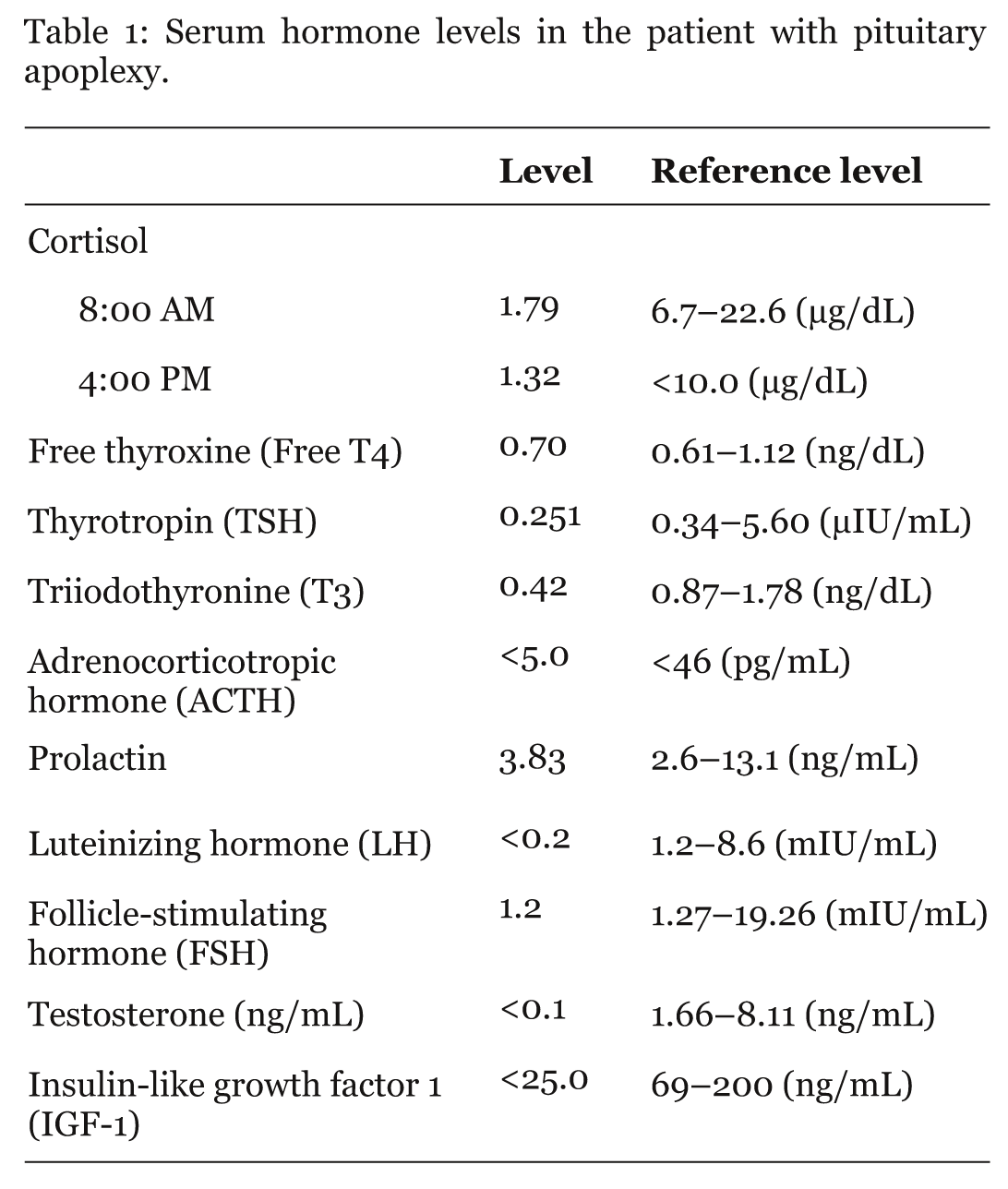
A 71-year-old man presented with an acute onset of headache, fever, chills, dizziness and photophobia for one day. He was transferred to our hospital due to the preliminary diagnosis of bacterial meningitis. Stiffness in the neck was remarkable. Cerebrospinal fluid (CSF) analysis revealed an increased leukocyte count of 520/mm3 with neutrophilic granulocytes predominating (95%) and elevated total protein content (78.8 mg/dL). CSF glucose was 44 mg/dL, the serum glucose level was 85 mg/dL and the CSF red blood cell count was 180/mm3. Emergency computed tomography (CT) of the brain showed an isodense lesion in the sellar and suprasellar region (Figure 1A, B). A tumor, such as an adenoma or aneurismal vascular lesion, was suspected. Thus, the endocrine function was checked at the same time. After admission, a fourth-generation cephalosporin antibiotic (cefepime) and vancomycin were administrated to treat the suspected bacterial meningitis. However, the symptoms of fever, dizziness and headache persisted and consciousness deteriorated. Magnetic resonance imaging (MRI) of the brain showed a pituitary mass lesion with recent hemorrhage (Figure 2A, B). The endocrinologic profile (Table 1) revealed a lower cortisol level in the morning (cortisol (AM): 1.79 ug/dl (reference range: 6.7~22.6 ug/dl)). Corticosteroid (dexamethasone) substitutions were immediately started. Afterward, consciousness and body temperature normalized within one day. The blood and CSF culture findings for the detection of bacteria were negative during the period of admission. The patient refused an operation to remove the pituitary mass. Three months after discharge, the brain MRI revealed that the size of the pituitary and hematoma had decreased (Figure 2C, D). | ||||||
| ||||||
|
| ||||||
| ||||||
|
Discussion
| ||||||
|
Pituitary apoplexy is an uncommon syndrome resulting from hemorrhage or infarction of a pre-existing pituitary adenoma. In about 60–80% of cases, pituitary apoplexy occurs spontaneously in previously asymptomatic patients and in patients who are not previously diagnosed with pituitary adenomas. [3] However, several precipitating or predisposing factors have been reported in patients with pituitary apoplexy, including postpartum hemorrhage, closed head trauma, hypotension, hypertension, diabetes, history of irradiation, cardiac surgery, anticoagulant therapy or bleeding disorder and treatment with dopamine agonists. [4] [5] Precipitating factors are reported in 25–30% of pituitary apoplexy patients. This 71-year-old man had no remarkable predisposing factors such as hypertension, diabetes mellitus, coagulation disorder or recent head trauma that would make him prone to pituitary apoplexy. In a retrospective study of clinical features and surgical outcomes of pituitary apoplexy, the most common symptom was visual deficit and the second most common symptom was headache. Nausea or vomiting, cranial nerve palsy, impairment of consciousness and endocrine dysfunction were also the main features. [6] After hemorrhagic infarction of the pituitary adenoma the sudden and rapid expansion of the contents of the sella turcica and rapid increase in intrasellar pressure resulted in compression of perisellar structures or meningeal irritation. This would lead to the symptoms of severe headache, visual disturbances and impairment of pituitary function. [4] The symptoms of headache, fever, alteration of consciousness and meningeal irritation in patients with pituitary apoplexy would resemble the characteristics of acute meningoencephalitis, subarachnoid hemorrhage, or meningitis. This misdiagnosis may lead to a delay in immediate management. Our patient presented with acute headache, associated with fever, chills, dizziness, and photophobia without significant initial defects of visual fields and eye movement. In addition, CSF laboratory findings revealed neutrophilic pleocytosis and an increased protein count. All the above findings led to the preliminary diagnosis of acute bacterial meningoencephalitis. The variability of clinical presentations in pituitary apoplexy poses great challenges for the initial diagnosis. Leakage of blood or necrotic tissue into the subarachnoid space may cause headache, an altered level of consciousness, visual disturbance, fever or signs of meningism. The incidence of symptom of headache in patients with clinical pituitary apoplexy was about 72%. [6] The possible mechanisms of headache were enlargement of the walls of sella turcica, dura mater compression, meningeal irritation or involvement of the trigeminal nerve. [7] Even though the high temperature was suggestive of infection, it can also be a typical feature in pituitary apoplexy associated with secondary adrenal insufficiency. [8] Ophthalmoplegia was also a classic finding in about 12% of those with clinical pituitary apoplexy and might be due to an enlarged pituitary or hemorrhage extending laterally into the cavernous sinus. [6] The most misleading results were the CSF analysis findings. Several case reports have shown the presence of neutrophilic pleocytosis in cases of pituitary apoplexy that might be misinterpreted as infectious meningoencephalitis. The abnormal CSF findings could be attributed to the leakage of blood and necrotic debris from the pituitary gland into the subarachnoid space. [5] [9] It is important to stabilize patients with pituitary apoplexy early on as it is a life-threatening condition. However, the definite diagnosis of a pituitary mass lesion using brain image studies is difficult, even if hemorrhage is present. [10] Bills et al. reported a 46% rate of detection of apoplexy using CT scans in 37 patients with pituitary tumor apoplexy. [11] Whole-brain CT might obtain limited views of the pituitary fossa, thus restricting diagnostic sensitivity. In addition, CT may not be able to precisely differentiate cystic or degenerative changes from previous hemorrhage. [12] A sellar MRI study is more sensitive in evaluating the pituitary gland and may visualize hemorrhage not seen on the CT scan. [13] In our case, initial brain CT without contrast showed an isodense lesion in the sellar area but no significant hemorrhagic signal. However, brain MRI with contrast done for a more thorough examination revealed a pituitary adenoma with recent hemorrhage. The initial management for pituitary apoplexy includes conservative or surgical interventions. Patients presenting with clinical symptoms require immediate medical attention, thorough clinical evaluation, and continuous monitoring. Many patients need intravenous fluids and blood transfusions. If necessary, high-dose corticosteroid is administered to restore endogenous hormone function and to prevent edema on parasellar structures. The proper time for surgical decompression is still controversial. If the altered mental status or impaired visual acuity does not recover after supportive clinical management the patients would then require urgent surgical decompression as definitive treatment. [3] [10] Several studies have illustrated that a better visual outcome was found in patients undergoing surgery within eight days of onset of symptoms. [1] Our patient did not have the symptoms of visual deficit or ophthalmoplegia. He refused surgical intervention; however, administering steroid ameliorated the clinical features and the adenoma showed no further growth in the follow-up brain MRI. | ||||||
|
Conclusion
| ||||||
|
Pituitary apoplexy cannot be excluded clinically in a patient who has a headache, fever and meningism, even when ophthalmological symptoms are absent in the acute phase. Further examination of the endocrinological profile and neuroimaging are necessary for timely treatment. | ||||||
|
References
| ||||||
| ||||||


|
[HTML Abstract]
[PDF Full Text]
|
|
Author Contributions:
Shu-Yi Wang - Substantial contributions to conception and design, Acquisition of data, Analysis and interpretation of data, Drafting the article, Revising it critically for important intellectual content, Final approval of the version to be published Chieh-Sen Chuang - Substantial contributions to conception and design, Acquisition of data, Analysis and interpretation of data, Drafting the article, Revising it critically for important intellectual content, Final approval of the version to be published |
|
Guarantor of submission:
The corresponding author is the guarantor of submission. |
|
Source of support:
None |
|
Conflict of interest:
Authors declare no conflict of interest. |
|
Copyright:
© Shu-Yi Wang et al. 2012; This article is distributed the terms of Creative Commons Attribution License which permits unrestricted use, distribution and reproduction in any means provided the original authors and original publisher are properly credited. (Please see Copyright Policy for more information.) |
|
|