Volume 3; Number 6 (June 2012)
|
Cover Figure |
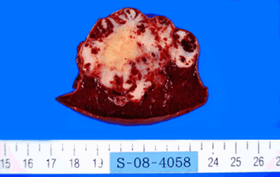 |
Figure 1: The cut surface of the tumor showed 0.3–0.5 cm red-brown multinodular lesions interspersed with a 5 cm white lobular tumor at the periphery. (Splenic sclerosing angiomatoid nodular transformation) (Page 35)
|
Go Back to Table of Contents |
|