| Table of Contents |  |
|
Case Report
|
| Free floating, intraspinal, neuroepithelial cyst: A case report |
| Mihir Ravindra Bapat1, Mehandi Hassan Shaukat Ali Ansari2, Kiran Prakash Paknikar2, Prasanna Chandrakant Rathi2, Chirag Santosh Patel2 |
|
1Director of Spine Surgery, Orthopedics Department, Kokilaben Dhirubhai Ambani Hospital, Mumbai, India.
2Clinical Associate, Orthopedics Department, Kokilaben Dhirubhai Ambani Hospital, Mumbai, India. |
|
doi:10.5348/ijcri-2012-06-136-CR-9
|
|
Address correspondence to: Dr. Mehandi Hassan Ansari Center for Bone and Joints, Kokilaben Dhirubhai Ambani Hospital Four Bungalows, Andheri West Mumbai - 400053 India Phone: +91- 9699872286 Fax: +91 - 22 - 30972030 Email: mehandi0120@gmail.com |
|
[HTML Abstract]
[PDF Full Text]
|
| How to cite this article: |
| Bapat MR, Ansari MHSA, Paknikar KP, Rathi PC, Patel CS. Free floating, intraspinal, neuroepithelial cyst: A case report. International Journal of Case Reports and Images 2012;3(6):38–41. |
|
Abstract
|
|
Introduction:
Neuroepithelial (NE) cysts are congenital tumours of the central nervous system. Intraspinal NE cysts are rare. We report a case of intradural, extramedullary NE cyst with concomitant lumbar disc herniation with significant overlap of symptoms. Interesting is to note the free floating nature of the cyst and the change in the symptoms of the patient with the change in position of the cyst.
Case Report: A 41-year-old lady presented with acute onset low back pain with bilateral, radiating leg pain. MRI showed lumbar, intradural NE cyst with significant L5-S1 prolapse disc. The patient refused surgery and managed herself with intermittent analgesics for 12 months. However, with change in position of cyst from the posterior to the anterior aspect of the cauda, the patient had intractable leg pain with perineal pain and hypoaesthesia and urinary symptoms. Surgery was done to excise the cyst with L5-S1 microdiscectomy. Conclusion: NE cysts are rare, benign cysts of the central nervous system that have the capacity to expand by accumulating CSF like fluid, and require excision or decompression before any damage is caused by the pressure effect. Interesting in the case was to note the free floating nature of the cyst and the change in the symptoms of the patient with change in position of the cyst. | |
|
Key Words:
Intradural neuroepithelial cyst
| |
|
Introduction
| ||||||
|
Neuroepithelial cysts are non neoplastic lesions of the central nervous system, lined by a single layer of cuboidal or columnar epithelium.These are rare, congenital lesions usually confined to the central nervous system, intracranially in the paraventricular white matter of frontal and parietal lobe. Intraspinal neuroepithelial cysts are very rare. [1] Herein the authors report a case of lumbar intradural, neuroepithelial cyst in a 41-year-old woman with concomitant disc herniation at L5-S1 level. | ||||||
|
Case Report
| ||||||
|
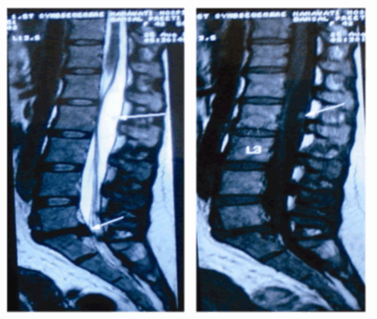
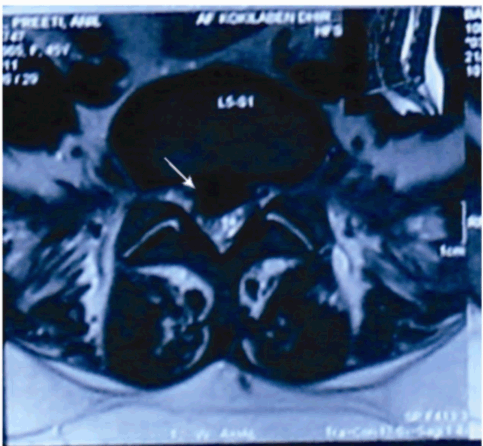
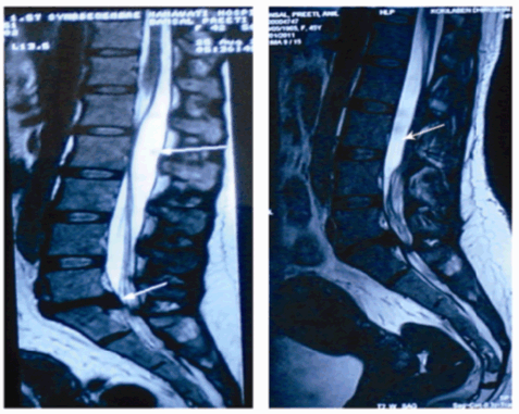
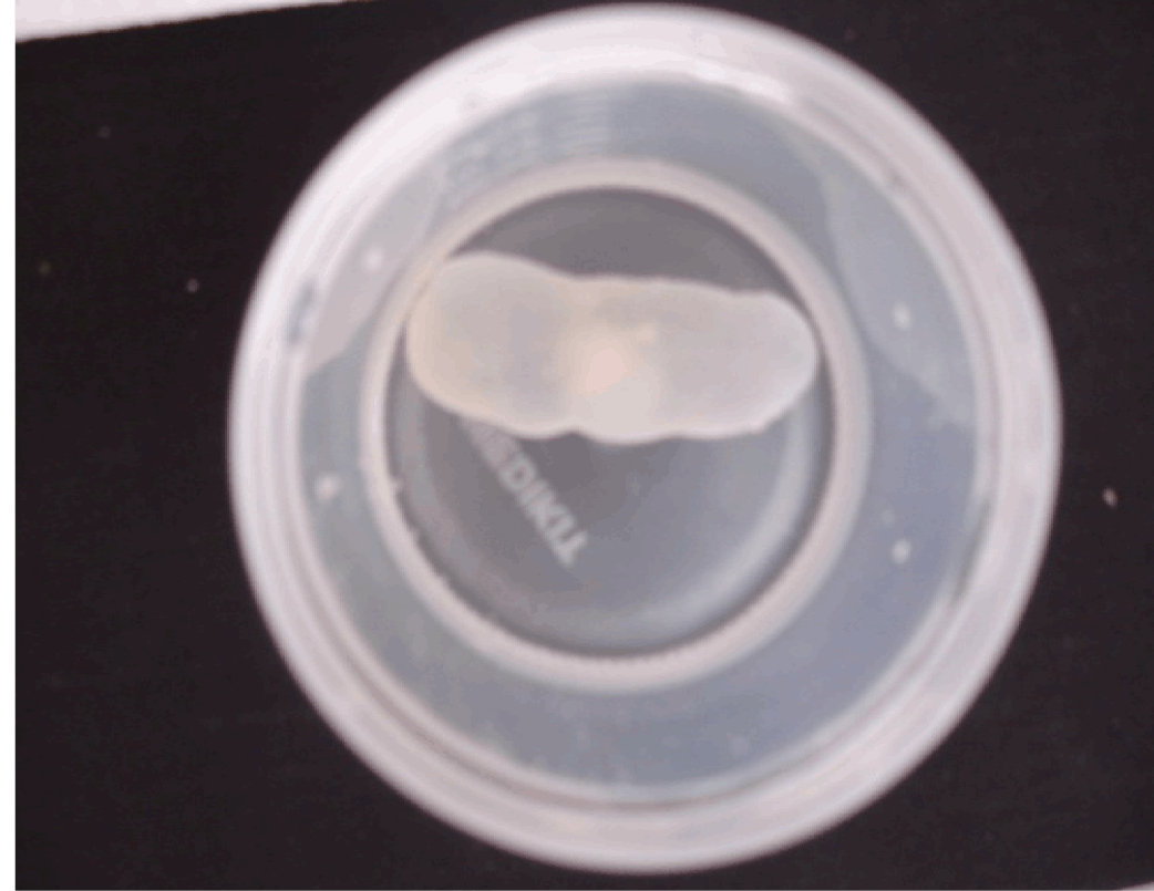
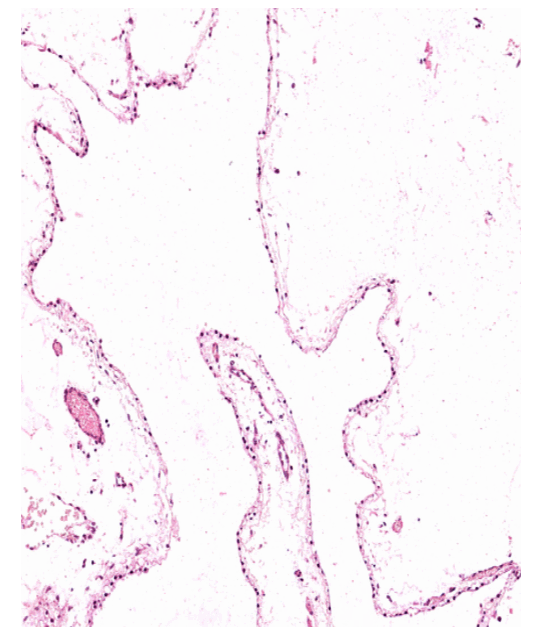
A 41-year-old lady presented with complaints of gradual, progressive deterioration of lumbar axial pain since six months, with aggravation since seven days. The pain was radiating to both lower limbs along the posterior aspect of thigh and calf. There was no obvious history of trauma. Examination revealed positive straight leg raising test in both lower limbs with no motor or sensory deficit. An MRI was done, that showed diffuse disc bulge at L5-S1 level with an inferiorly migrated central extrusion causing severe compression of the thecal sac (Figure 1, 2). It also revealed an intradural, extramedullary, cystic space occupying lesion with an internal solid-cystic nodule, extending from L1 to L2-3 causing compression and anterior displacement of filum and cauda equina (Figure 1). The lesion showed signal intensities similar to those of CSF on T1, T2 and FLAIR images. There was no enhancement noted after administration of contrast in the MR scans. The imaging features were suggestive of a benign cyst with possibilities of a cysticercosis or an arachnoid cyst. The patient was advised surgery for the removal of cyst with L5-S1 microdiscectomy, but the patient refused surgery and continued her activities, managing well with oral analgesics intermittently for a period of 12 to 14 months. However, later she presented with acute aggravation of lumbar axial pain and spasms with poor response to analgesics. The pain was continuous, radiating to both lower limbs, not relieved by rest, along with perineal pain and hypoesthesia. Motor power was normal in both lower limbs with urinary urgency and frequency without difficulty in initiation and incontinence. Repeat MRI showed no interval change in the degree of disc extrusion, however the cyst that was initially on the posterior aspect of the filum and cauda equina, had shifted anteriorly with posterior displacement of cauda (Figure 3). The patient was posted for surgery for L5-S1 discectomy with excision of cyst. At surgery a L1-L3 laminectomy was done. Careful durotomy of the bulging dura revealed a thin walled, oval shaped cyst of size 4.5x2x1 cm (Figure 4). Externally the surface was smooth. The cyst was removed enbloc. On opening, it was unilocular, fluctuant, transilluminant cyst containing clear fluid with a papery thin, transparent wall. Also, L5-S1 microscopic discectomy was done. Histological study of the operative specimen revealed a thin walled cyst lined by single layer of benign ciliated columnar cells. The wall showed loose connective tissue with no neural tissue (Figure 5). Analysis of the cyst fluid was similar with CSF. Patient was relieved of her symptoms in the immediate post operative period and was made ambulatory on the second post operative day. Two years post surgery, the patient is comfortable and completely relieved of her pain. | ||||||
|
| ||||||
| ||||||
| ||||||
| ||||||
| ||||||
|
Discussion
| ||||||
|
Neuroepithelial cysts (Ependymal cyst, Glioependymal cyst) are rare, congenital, ependymal lined, benign cysts. They are typically located in the lateral ventricle but can be seen in the brain parenchyma of temperopareital and frontal lobes and less commonly in the subarachnoid space. Intraspinal neuroepithelial cysts are very rare comprising 0.4% 0f the primary spinal tumors. [1] Many theories exist regarding the origin of neuroepithelial cyst, however more commonly they are believed to origin from the extruded ependymal cells which are located close to the anterior cord substance at the time of closure of neural tube and these cells have lost continuity and become isolated from the floor plate of the neural tube. [2] The future intramedullary or extramedullary location depends on the site of isolated ependymal lesion. They are usually separated from the central canal and more often, located on the anterolateral aspect of the cord. Intradural extramedullary ependymal cysts constitute 0.2–0.4% of the total primary spinal tumors with an overall 2:1 female predominance with a predilection for the fourth decade. [3] There is no age or gender predominance for intramedullary spinal ependymal cysts. [4] Among the intramedullary neuroepithelial cysts the thoracolumbar region is the most common. [4] Intradural extramedullary lesions are more common in the region of conus medullaris and cauda equina. [3] These are thin walled cysts that are detached from the normal ependyma, ranging from 2–3 mm in diameter to up to 9 cm. The best diagnostic clue is there imaging features; they are thin walled with CSF like signal intensities in all pulse sequences. [5] These lesions may be indistinguishable from arachnoid cyst or choroid plexus cyst. On histological studies, neuroepithelial cysts are lined with simple columnar or cuboidal epithelium and absence of basement membrane. [1] [6] Electron microscopy can characterize ependymal cysts by presence of intercellular junction complexes, the absence of a basement membrane, membranebound granules in noncilliated cells, and absence of a coating on the luminal surface of the cells. [6] No mucinous production or glycoprotein can be detected by periodic acid-Schiff, alcian blue, mucicarmine staining. [7] Immunocytochemical stain can be useful in differential diagnosis of cystic lesion. [8] Wackym, et al. reported a spinal ependymal cyst which showed positivity for cytokeratin antibody reaction and negativity for glialfibrillary acidic protein and S-100. [7] They are usually clinically asymptomatic. The symptoms can be related to obstruction to CSF outflow or mass effect in accordance to their localization and ability to expand due to accumulation of CSF like cyst fluid. Depending on the location and size of the cyst symptoms include slowly progressive myelopathy with or without radiculopathy (i.e., intermittent paraparesis or tetraparesis, paraesthesia and radicular pain) or radiculopathy alone. Whenever spinal neuroepithelial cysts are symptomatic, surgery with total excision of the cyst is the treatment of choice as early as possible before any damage to the cord. However, total excision of the cyst may not be possible always due to absence of cleavage line between the cord and the cyst, especially in the intramedullary lesions. [9] The aim of surgery should be to create communication between the cystic cavity and the subarachnoid space and to undertake a biopsy of cystic wall. [9] Although Rhee, et al. report recurrence of cyst, fenestration or marsupialization is the most safe surgical method. [10] Some authors have also reported cystosubarachnoid shunt. [11] | ||||||
|
Conclusion
| ||||||
|
Neuroepithelial cysts are rare, benign cysts of the central nervous system that have the capacity to expand by accumulating CSF like fluid, and require excision or decompression before any damage is caused by the pressure effect. In this case, the patient presented with intradural, lumbar spinal tumor with L5-S1 disc prolapse with significant overlap of symptoms, which required both the etiologies to be dealt at the same sitting. Also, interesting was to note the free floating nature of the cyst and the change in the symptoms of the patient with change in position of the cyst. | ||||||
|
References
| ||||||
| ||||||

|
[HTML Abstract]
[PDF Full Text]
|
|
Author Contributions:
Mihir Ravindra Bapat - Conception and design, analysis and interpretation of data, Drafting the article, Critical revision of the article, Final approval of the version to be published Mehandi Hassan Shaukat Ali Ansari - Conception and design, acquisition of data, analysis and interpretation of data, Drafting the article, Critical revision of the article, Final approval of the version to be published Kiran Prakash Paknikar - Significant contributions in acquisition of data, analysis and interpretation of data, Critical revision of the article, Final approval of the version to be published Prasanna Chandrakant Rathi - Significant contributions in acquisition of data, analysis and interpretation of data, Critical revision of the article, Final approval of the version to be published Chirag Santosh Patel - Analysis and interpretation of data, Critical revision of the article, Final approval of the version to be published |
|
Guarantor of submission:
The corresponding author is the guarantor of submission. |
|
Source of support:
None |
|
Conflict of interest:
Authors declare no conflict of interest. |
|
Copyright:
© Mihir Ravindra Bapat et al. 2012; This article is distributed the terms of Creative Commons Attribution License which permits unrestricted use, distribution and reproduction in any means provided the original authors and original publisher are properly credited. (Please see Copyright Policy for more information.) |
|
|