| Table of Contents |  |
|
Case Report
|
| Laparoscopic splenectomy for splenic sclerosing angiomatoid nodular transformation: A case report |
| Kunihiro Shinjo1, Koichi Sato1, Hiroshi Maekawa1, Mutsumi Sakurada1, Hajime Orita1, Tomoaki Ito1, Masayuki Saita1, Sei Matsumori1, Yoshihiro Komatsu1, Miki Yamano2 |
|
1Department of Surgery, Juntendo Shizuoka Hospital, Juntendo University School of medicine, Izunokuni, Shizuoka, Japan.
2Department of Pathology, Juntendo Shizuoka Hospital, Juntendo University School of medicine, Izunokuni, Shizuoka, Japan. |
|
doi:10.5348/ijcri-2012-06-135-CR-8
|
|
Address correspondence to: Kunihiro Shinjo, MD, Ph.D. Department of Surgery, Juntendo Shizuoka Hospital Juntendo University School of medicine 1129 Nagaoka, Izunokuni-shi, Shizuoka Japan - 410-2295 Phone: 81-55-948-3111 Fax: 81 -55-946-0514 Email: old113@yahoo.co.jp |
|
[HTML Abstract]
[PDF Full Text]
|
| How to cite this article: |
| Shinjo K, Sato K, Maekawa H, Sakurada M, Orita H, Ito T, Saita M, Matsumori S, Komatsu Y, Yamano M. Laparoscopic splenectomy for splenic sclerosing angiomatoid nodular transformation: A case report. International Journal of Case Reports and Images 2012;3(6):34–37. |
|
Abstract
|
|
Introduction:
Sclerosing angiomatoid nodular transformation (SANT) is a rare benign and proliferative vascular lesion arising within the red pulp of the spleen. The lesion is often identified incidentally on imaging and the diagnosis is confirmed on pathologic assessment of the resected spleen. The lesion can be cured by splenectomy. Until now, little has been published on the laparoscopic approach for treatment of SANT.
Case Report: A 5-cm splenic tumor was discovered during a routine examination of an asymptomatic 38-year-old man. We were initially suspicious of fibrous hamartoma, but could not rule out malignancy. Consequently, laparoscopic splenectomy was performed, and histopathological findings showed that the tumor was a sclerosing angiomatoid nodular transformation. Conclusion: We report a rare case of sclerosing angiomatoid nodular transformation, completely resected by laparoscopy. In such cases, laparoscopic splenectomy seems to be a feasible, safe, and effective treatment. | |
|
Key Words:
Sclerosing angiomatoid nodular transformation, SANT, Laparoscopic splenectomy
| |
|
Introduction
| ||||||
|
Sclerosing angiomatoid nodular transformation (SANT) is a relatively new, rare, and unique lesion of the spleen that was initially reported by Martel et al. in 2004. [1] SANT is a benign and non-neoplastic vascular lesion; however, its pathogenesis is not fully understood. Splenectomy is curative. To date, about 50 cases have been reviewed in the English literature, [1] [2] [3] [4] [5] [6] few of which describe a laparoscopic approach to surgical treatment. [6] In this case, we demonstrate the utility of laparoscopic splenectomy for SANT. | ||||||
|
Case Report
| ||||||
|
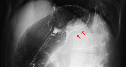
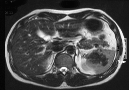
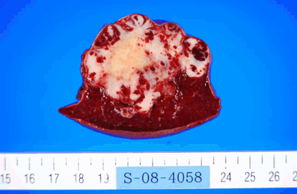
A 38-year-old man without any symptoms was found to have a splenic mass on routine medical examination. Physical examination found nothing remarkable. Laboratory test results were within normal limits. Tumor marker levels were not elevated (CEA: 1 ng/ml, AFP: 2 ng/ml, CA19-9: 2 ng/ml). Upper gastrointestinal (GI) series revealed extrinsic compression of the upper body of the stomach (Figure 1). An abdominal computed tomography (CT) scan revealed a 5-cm tumor and progressive delayed enhancement in the hilus of the spleen (Figure 2). Abdominal magnetic resonance imaging (MRI) revealed that the tumor was isointense on a T1-weighted image and hypointense on a T2-weighted image (Figure 3). The findings were confirmed by abdominal ultrasonography and we subsequently followed the patient by clinical and radiological examination. CT scan showed that the tumor was not enlarged four months after the initial presentation. It was thought that there was little possibility of malignancy, but on patient's request laparoscopic splenectomy was performed for diagnosis. Surgery was performed under general anesthesia in the right semilateral decubitus position. Four ports were used: the first 10-mm trocar was on the median line, two cm above the umbilicus. Two additional 10-mm trocars and a 5-mm trocar were on the left subcostal line between the median to the axillary line. An endoscopic linear cutter device (Endo GIA, Ethicon Endosurgery, Cincinnati, OH, USA) was used to dissect the splenic artery and vein. The excised spleen was placed into a specimen retrieval bag and extracted after enlarging the site of the telescopic trocar. A tube drain was placed in the residual cavity. The total operative time was 100 min and blood loss was 10 ml. The removed spleen was 11×7×5.5 cm in size and 150 g in weight. Macroscopic examination showed 0.3–0.5 cm red-brown multinodular lesions interspersed with a 5-cm white lobular tumor at the periphery (Figure 4). Histopathologically, the main component of the white portion consisted of hyperplastic collagen fibers. The red-brown lesions were multiple angiomatoid nodules in a fibrotic stroma. Each angiomatoid nodule was made up of slit-like, round irregular-shaped vascular spaces lined by endothelial cells and interspersed ovoid cells (Figure 5). Immunohistochemical studies revealed three different types of blood vessels within the angiomatoid nodules: i) cord capillaries (CD34+/CD8-/CD31+), ii) sinusoid vessels (CD34-/CD8+/CD31+), and iii) small veins (CD34-/CD8-/CD31+). Thus, a diagnosis of SANT was made. | ||||||
| ||||||
| ||||||
| ||||||
| ||||||
| ||||||
|
Discussion
| ||||||
|
Sclerosing angiomatoid nodular transformation (SANT) is a distinct pathological vascular lesion of the spleen newly described by Martel et al. in 2004. [1] Fewer than 50 cases have been described in the English literature. The most common presentation was incidental findings of an asymptomatic splenic mass. No cases had evidence of recurrent disease after splenectomy. [1] [2] [3] [4] [5] [6] The report by Martel et al. [1] described the splenic lesion as solitary and sharply demarcated from the surrounding parenchyma. In our case, the cut surface revealed a mass of coalescent red-brown nodules embedded in a dense fibrous stroma. The individual nodules had an angiomatoid appearance, in the sense that they were composed of slit-like, round or irregular-shaped vascular spaces lined by plump endothelial cells and interspersed by a population of spindly or ovoid cells. Nuclear atypia was minimal, mitotic figures were extremely rare, and necrosis was consistently absent. The internodular stroma consisted of variably myxoid to dense fibrous tissue with scattered plump myofibroblasts, plasma cells, lymphocytes, and siderophages. Immunostaining revealed 3 distinct types of vessels in the angiomatoid nodules: a cord capillary-like type that co-expressed CD34 and CD31 but not CD8, a sinusoid-like type that expressed CD8 and CD31 but not CD34, and small veins that expressed only CD31. These features are therefore distinct from those of littoral cell angioma, conventional hemangioma, and hemangioendothelioma of the spleen. The characteristic morphological appearance, immunophenotype, and benign clinical course suggest that SANT is a distinctive non-neoplastic vascular lesion. [3] [4] In the present case, histopathological and immunostaining findings coincided with the peculiarities of SANT. The advantages of laparoscopic splenectomy have been well described in previous studies. Shorter length of hospital stay, less need for narcotics and earlier return to activity are only a few advantages of laparoscopic splenectomy over open splenectomy. [7] [8] Laparoscopic splenectomy has become a standard surgical procedure for the management of some splenic tumors, especially benign ones. [9] [10] [11] [12] [13] In our patient, we demonstrated that the laparoscopic approach was safe and effective in a comparatively short time. | ||||||
|
Conclusion
| ||||||
|
We report the experience of a rare case of SANT, completely resected by laparoscopic surgery. Laparoscopic splenectomy seems to be a feasible, safe, and effective method for the treatment of SANT. | ||||||
|
References
| ||||||
| ||||||


|
[HTML Abstract]
[PDF Full Text]
|
|
Author Contributions:
Kunihiro Shinjo - Conception and design, Acquisition of data, Analysis and interpretation of data, Drafting the article, Critical revision of the article, Final approval of the version to be published Koichi Sato - Conception and design, Acquisition of data, Analysis and interpretation of data, Final approval of the version to be published Hiroshi Maekawa - Conception and design, Acquisition of data, Analysis and interpretation of data, Final approval of the version to be published Mutsumi Sakurada - Conception and design, Acquisition of data, Analysis and interpretation of data, Drafting the article, Critical revision of the article, Final approval of the version to be published Hajime Orita - Conception and design, Acquisition of data, Analysis and interpretation of data, Drafting the article, Critical revision of the article, Final approval of the version to be published Tomoaki ito - Conception and design, Acquisition of data, Analysis and interpretation of data, Drafting the article, Critical revision of the article, Final approval of the version to be published Masayuki Saita - Conception and design, Acquisition of data, Analysis and interpretation of data, Drafting the article, Critical revision of the article, Final approval of the version to be published Sei Matsumori - Conception and design, Acquisition of data, Analysis and interpretation of data, Drafting the article, Critical revision of the article, Final approval of the version to be published Yoshihiro Komatsu - Conception and design, Acquisition of data, Analysis and interpretation of data, Drafting the article, Critical revision of the article, Final approval of the version to be published Miki Yamano - Conception and design, Acquisition of data, Analysis and interpretation of data, Drafting the article, Critical revision of the article, Final approval of the version to be published |
|
Guarantor of submission:
The corresponding author is the guarantor of submission. |
|
Source of support:
None |
|
Conflict of interest:
Authors declare no conflict of interest. |
|
Copyright:
© Kunihiro Shinjo et al. 2012; This article is distributed the terms of Creative Commons Attribution License which permits unrestricted use, distribution and reproduction in any means provided the original authors and original publisher are properly credited. (Please see Copyright Policy for more information.) |
|
|