| Table of Contents |  |
|
Case Report
|
| Synovial sarcoma of ethmoid sinus masquadering as chronic sinusitis: A case report |
| Krishnangshu Choudhury Bhanja1, Shatarupa Dutta2, Priyanjit Kumar Kayal2, Suman Ghorai3, Anjali Majumder4 |
|
1Senior Resident, Department of Radiotherapy, Institute of Post graduate Medical Education and Research, Kolkata, West Bengal, India.
2Junior Resident, Department of Radiotherapy, Institute of Post graduate Medical Education and Research, Kolkata, West Bengal, India. 3Assistant Professor, Department of Radiotherapy, Institute of Post graduate Medical Education and Research, Kolkata West Bengal, India. 4Professor, Department of Radiotherapy, Institute of Post graduate Medical Education and Research, Kolkata, West Bengal, India. |
|
doi:10.5348/ijcri-2012-04-111-CR-8
|
|
Address correspondence to: Krishnangshu Bhanja Choudhury C-11/4, Green Tower Golf Green Kolkata West Bengal India Phone: 91-9830932869 Email: krishnangshuchoudhury@gmail.com |
|
[HTML Abstract]
[PDF Full Text]
|
| How to cite this article: |
| Bhanja KC, Dutta S, Kayal PK, Ghorai S, Majumder A. Synovial sarcoma of ethmoid sinus masquadering as chronic sinusitis: A case report. International Journal of Case Reports and Images 2012;3(4):30–33. |
|
Abstract
|
|
Introduction:
Synovial sarcomas are malignant soft tissue tumors usually occurring in adolescent and young adults of 15–40 years of age. It usually originates from the synovium but sometimes have a tendency to arise from unknown parasynovial tissues as parapharyngeal, paranasal, retroperitoneal and mediastinal regions. Paranasal sinuses are rarely involved subsites of head and neck region.
Case Report: We report case of a 55-year-old hypertensive patient who presented with intermittent nasal bleeding. Computed tomography (CT) scan suggests left anterior ethmoidal mass with anterior cranial fossa extension. Biopsy was suggestive of synovial sarcoma. She had history of polyp extraction two times but biopsy was suggestive of inflammatory origin. This patient received EBRT 50Gy in 25 fractions. No recurrence was noticed in one year follow up. Conclusion: We report unusual ethmoid sinus synovial sarcoma masquerading as chronic sinusitis in an elderly female patient, unlikely age presentation. | |
|
Key Words:
Synovial sarcoma, Ethmoid sinus
| |
|
Introduction
| ||||||
|
Synovial sarcomas are malignant soft tissue tumors comprising 5–10% of all soft tissue sarcomas. It usually occurs in adolescent and young adults of 15–40 years of age. Synovial sarcomas usually originate from the synovium but sometimes have a tendency to arise from unknown parasynovial tissues such as parapharyngeal, paranasal, retroperitoneal and mediastinal regions. [1] [2] With majority of cases seen in the extremities, less than 10% cases are reported in the head and neck, most common site being hypopharynx. [3] Paranasal sinuses are rarely involved. We report an unusual case of synovial sarcoma of the ethmoid sinus initially masquerading as chronic sinusitis. | ||||||
|
Case Report
| ||||||
|
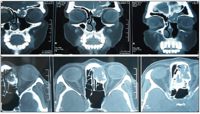
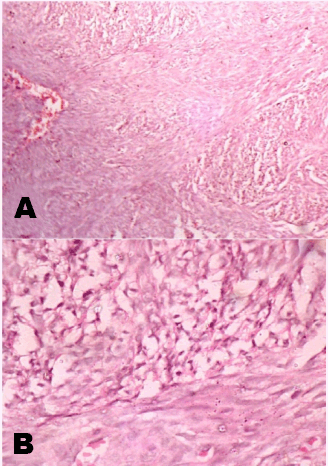
One 55-year-old, hypertensive female patient was admitted in the emergency department with history of intermittent nasal bleeding and headache of one week duration. She was being treated for chronic sinusitis for last eight months. She had undergone left rhinotomy with removal of frontonasal polyp and then removal of ethmoidal polyp after three months interval. The biopsies were diagnostic of inflammatory non-malignant lesions. With onset of new symptoms of nasal bleeding, endoscopy examination was repeated to show recurrence of polyp. Computed tomography scan of the face showed a mass in left anterior ethmoid sinus with extension into the floor of anterior cranial fossa. Medially the lesion eroded the lamina papyracea with no obvious intraorbital extension. Localised bone erosion was noted (Figure 1). Left ethmoid sinus was explored and mass was excised. The biopsy report was suggestive of epithelial cell aggregates with densely cellular fascicles of spindle cells surrounding the epithelial cells, suggestive of biphasic synovial cell sarcoma (Figure 2A, B). Diagnosis was confirmed with immunohistochemical positivity for EMA (focal), Mic-2, bcl-2, calponin, S-100 and CD-56 (focal) but negative for CD-34 and cytokeratin. CT Scan of thorax and ultrasonography of abdomen were negative for metastatic disease. Postoperatively she received only radiation, 50 Gray (Gy) in five weeks, in 25 fractions (200 centiGy dose per fraction). One year into follow up there is no evidence of recurrence of disease. | ||||||
| ||||||
| ||||||
|
Discussion
| ||||||
|
Synovial sarcomas can arise from any portion of the body containing pluripotent mesenchymal cells near or even remote from articular surfaces, tendons, tendon sheaths, juxta-articular membranes and facial aponeuroses or anywhere unassociated with joint cavities including head and neck, lower back, chest and abdominal wall. [2] [4] [5] [6] [7] Uncommonly, these tumors may arise in the head and neck (approximately 9% of all synovial sarcomas). In the head and neck region, hypopharynx is the most common site probably due to having abundant synovial tissues. [8] Synovial sarcomas in the paranasal sinuses are very uncommon. The first description of paranasal sinus synovial sarcoma was reported in 1970 by Trible et al. [9] of a metastatizing synovial sarcoma to the sphenoid sinus. Since that time, there have been only few reported cases of synovial sarcomas involving the paranasal sinuses. [4] [5] [6] Patients were predominant males with age range from 12–54 years (mean 31 years). Synovial cell sarcoma of the head and neck is seen most commonly in patients who are more than a decade younger than patients who have the same cancer in the trunk and extremities; and after onset of symptoms, patients with synovial cell sarcoma of the head or neck are treated at an age nearly two decades younger than patients with the same disease in the trunk and extremities. [6] To our knowledge, we report the first case of synovial cell sarcoma involving the ethmoid sinuses in an elderly woman masquerading as chronic sinusitis. Synovial cell sarcoma is composed of two morphologically distinct types of cells that form a characteristic biphasic pattern. The biphasic synovial cell sarcoma includes epithelial cells and spindle cells. Histologically, the cuboidal or columnar epithelial cells form glands or papillary structures surrounded by broad sheets of spindle cells. Because the spindle cells have sparse cytoplasm and indistinct borders, the spindle cells appear to have overlapping nuclei. In the monophasic form of synovial cell sarcoma (which became recognized more recently), the spindle cell component dominates, whereas detection of the epithelial component requires the use of special immunohistochemical markers. [3] [4] [5] Ninety percent of synovial sarcomas are characterized by a specific balanced translocation (X; 18). Patients with biphasic histological tumours demonstrated SYT-SSX1 gene fusions. [11] These patients fared worse than patients with SYT-SSX2- positive tumours that were associated with the monophasic phenotype. Cytogenetic analysis and molecular techniques to detect translocation have aided in diagnosing synovial sarcomas, especially monophasic fibrous types and poorly differentiated forms that can be difficult to differentiate from other types of sarcomas. [5] [6] Spindle cells stain positive for keratin and epithelial membrane antigen. Vimentin is demonstrable in spindle cells but absent in epithelial cells. S-100 staining may give positive results. Rhabdomyosarcoma, liposarcoma, leiomyosarcoma, angiosarcoma, minor salivary gland pleomorphic adenomas, osteosarcoma should be included in the differential diagnosis. [6] The reported five years survival rates in patients of synovial sarcomas were from 36 to 76% and 10 years survival rates were from 20 to 63%. As the treatment guidelines are limited because of very few reported cases of paranasal sinuses involvement, treatment is based on experiences with tumours developing in the extremities. [4] [5] [6] [7] [8] [9] Primary therapy in most patients with synovial sarcomas is operative excision followed by adjuvant radiotherapy or chemotherapy. [4] Some authors advocated that postoperative radiation therapy has not been found to increase survival rate, although it has improved local control rates and chemotherapy may prevent or delay the occurrence of metastasis. [8] The beneficial effect of chemotherapy as the treatment modality of synovial sarcomas is difficult to assess. Post treatment recurrence rates for synovial cell sarcoma arising from all body sites is 50%, with most cases recurring within the first two years after treatment. Wide excision with negative margins and the use of radiation therapy yield the best results. Kartha et al. emphasized that outcome was not related to the extent of tumour but with timing of radiation and local recurrence. [10] In the present case, the patient underwent surgical excision first, followed by adjuvant radiotherapy with no sign of recurrence at the time of reporting. Thus, regular follow up evaluation and close observation will be needed for possibility of local recurrence. | ||||||
|
Conclusion
| ||||||
|
We report unusual ethmoid sinus synovial sarcoma masquerading as chronic sinusitis in an elderly female patient of unlikely age presentation. | ||||||
|
References
| ||||||
| ||||||

|
[HTML Abstract]
[PDF Full Text]
|
|
Author Contributions:
Krishnangshu Choudhury Bhanja - Substantial contributions to conception and design, Acquisition of data, Drafting the article, revising it critically for important intellectual content, Final approval of the version to be published Shatarupa Dutta - Substantial contributions to conception and design, Analysis and interpretation of data, Drafting the article, Final approval of the version to be published Priyanjit Kumar Kayal - Substantial contributions to conception and design, Drafting the article, revising it critically for important intellectual content, Final approval of the version to be published Suman Ghorai - Drafting the article, revising it critically for important intellectual content, Substantial contributions to conception and design, Final approval of the version to be published Anjali Majumder - Substantial contributions to conception and design, Revising it critically for important intellectual content, Final approval of the version to be published |
|
Guarantor of submission:
The corresponding author is the guarantor of submission. |
|
Source of support:
None |
|
Conflict of interest:
Authors declare no conflict of interest. |
|
Copyright:
© Krishnangshu Choudhury Bhanja et. al. 2012; This article is distributed the terms of Creative Commons Attribution License which permits unrestricted use, distribution and reproduction in any means provided the original authors and original publisher are properly credited. (Please see Copyright Policy for more information.) |
|
|