| Table of Contents |  |
|
Case Report
|
| A case report on abnormal origin of right recurrent laryngeal nerve from right vagus in thorax |
| Aniruddha Sarkar1 |
|
1Assistant Professor, Department of Anatomy, Medical College, City-Kolkata, State-West Bengal, Country-India.
|
|
doi:10.5348/ijcri-2012-03-97-CR-2
|
|
Address correspondence to: Dr. Aniruddha Sarkar S/o- Mr. Arun Kumar Sarkar (Advocate) Post- Raghunathganj, Dist.-Murshidabad Locality- Pakurtala, State- West Bengal - 742225 India Phone: (+91) 9474474468 Email: drani77@gmail.com |
|
[HTML Abstract]
[PDF Full Text]
|
| How to cite this article: |
| Sarkar A. A case report on abnormal origin of right recurrent laryngeal nerve from right vagus in thorax. International Journal of Case Reports and Images 2012;3(3):4-7. |
|
Abstract
|
|
Introduction:
Variations in the course of right recurrent laryngeal nerve are very common. But the right recurrent laryngeal nerve taking origin in the thorax instead of the root of the neck is a very rare anomaly. There are only few reports available regarding such anomaly of right recurrent laryngeal nerve.
Case Report: During a routine gross anatomy dissection of front of the neck in an adult female cadaver right recurrent laryngeal nerve was found to take origin in the thoracic cavity instead of origin in the root of the neck. The right recurrent laryngeal nerve had a recurrent course passing behind the right subclavian artery forming a loop in the thorax. Conclusion: The right recurrent laryngeal nerve in thorax may be damaged during surgical procedures like pacemaker placement in the heart, during the mediastinoscopy etc. So, during any thoracic surgery the surgeons should be very cautious about the abnormal origin of the right recurrent laryngeal nerve in thorax. We are trying to give focus by presenting this case on the abnormal origin of the right recurrent laryngeal nerve so that it can be prevented from injury during the thoracic surgeries or maneuvers, if an abnormal origin is present. | |
|
Key Words:
Thoracic, Right recurrent laryngeal nerve
| |
|
Introduction
| ||||||
|
The occurrence of non-recurrent laryngeal nerve is more common in right side (0.6%) than in left side (0.04%). [1] They are associated with the vascular anomalies such as an aberrant origin of the right subclavian artery from the descending thoracic aorta or the right sided aortic arch. [1] Recurrent laryngeal nerve displacements may occur by cervical and substernal goiters. Such nerves are at risk during lobectomy unless the surgeon anticipates the unusual location and is very careful. Rarely, the nerves are so stretched that spontaneous palsy may results. After careful dissection and preservation, functional recovery may occur postoperatively. [2] When there is a vascular anomaly of the right subclavian artery, the recurrent laryngeal nerve no longer "recurs" around this artery but proceeds from the vagus nerve in a more transverse direction to the larynx. In such a situation, the nerve is much more likely to be damaged during operation unless care is taken to visualize its course in the neck. Two cases of a right non-recurrent nerve were encountered in 203 thyroidectomies. [3] In its abnormal, non-recurrent course the nerve passes transversely from under the carotid sheath and takes a position which is at right-angles to the normal recurrent laryngeal nerve. [3] The abnormal origin of right recurrent laryngeal nerve can create problem during thoracic surgeries if the nerve is not detected during the surgical procedures. A very rare presentation of right recurrent laryngeal nerve was seen during a routine dissection of neck. The nerve was seen to take origin from the right vagus in the thoracic cavity but not in the root of the neck as usual. The case was diagnosed incidentally during a routine dissection for medical students. The present case not only aims at explaining the possible embryological basis of this rare congenital malformation, but also the risk factors in the surgical interventions associated with this anomaly where the right recurrent laryngeal nerve taken origin in thorax instead of the root of the neck. Hence, the knowledge of uncertain anatomy of such atypical origin of the right recurrent laryngeal nerve might be very helpful both for the anatomists as well as the surgeons. | ||||||
|
Case Report
| ||||||
|
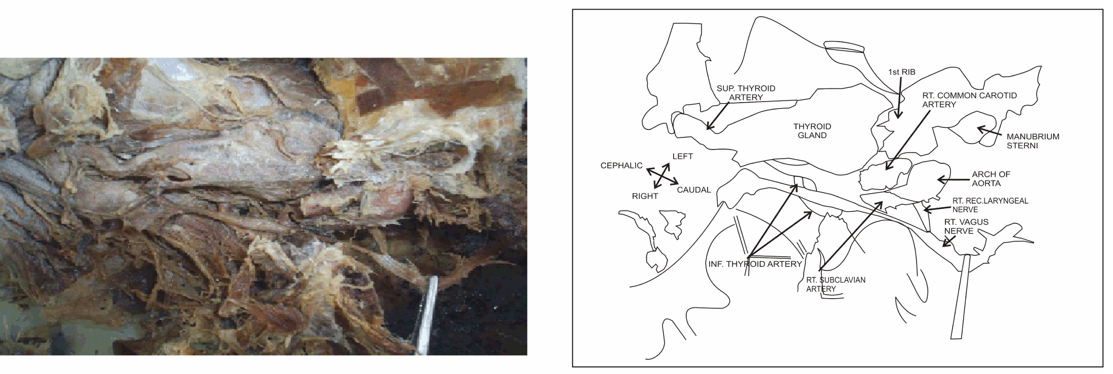
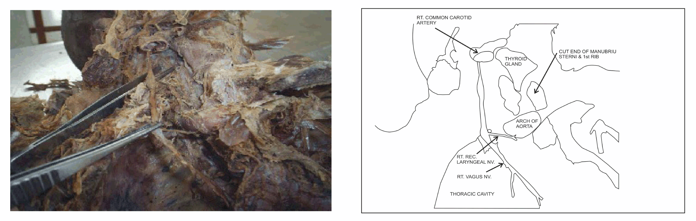
During a routine dissection of neck of a female cadaver the right recurrent laryngeal nerve was found that it was not hooking the inferior border of the right subclavian artery at the root of the neck as usual but the nerve was present in the right tracheo-oesophageal groove. During tracing the course of the nerve we saw that the right recurrent laryngeal nerve was taking origin from the right vagus below the first rib and the clavicle of right side in the thoracic cavity (figure 1). The first rib and the clavicle had been cut to show the upward course of the right recurrent laryngeal nerve. After origin the right recurrent laryngeal nerve had a recurrent course and it was going behind the right subclavian artery ultimately to enter the right tracheo-oesophageal groove as like the normal course of the right recurrent laryngeal nerve. The distance in between the origin of the right recurrent laryngeal nerve from the right vagus and the inferior border of the right subclavian artery measured about two cm (figure 2, figure 3D). The right subclavian artery, the arch of the aorta and the descending thoracic aorta were in normal position. No fibrous band or any cord like structure was seen even on minute dissection on right side around the sixth arterial arch. No other abnormalities were detected. | ||||||
| ||||||
| ||||||
|
| ||||||
|
Discussion
| ||||||
|
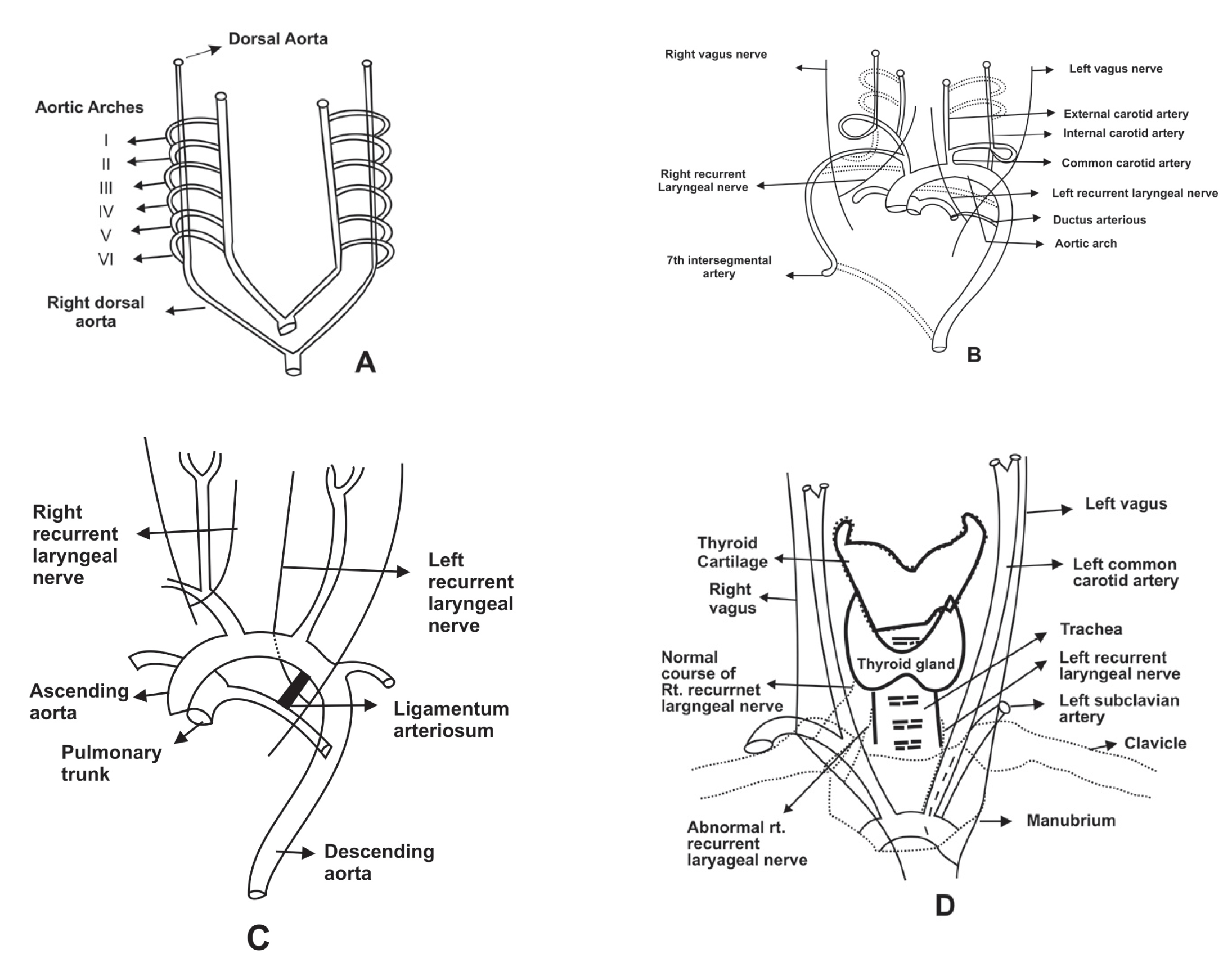
The courses of the recurrent laryngeal nerves become different on both sides as a result of the caudal shift of the heart and the disappearance of various portions of the aortic arches. Initially both right and left recurrent laryngeal nerves are at the level of sixth pharyngeal arches. As the heart descends caudally along with the lengthening of neck, recurrent laryngeal nerves after arising from the vagus have an ascending course (recurrent) by hooking the sixth aortic arch (figure 3 A). As the development progress, on the right side the distal portion of the sixth aortic arch and the fifth aortic arch disappears so that the right recurrent laryngeal nerve moves up and is ultimately hooked by the right subclavian artery (developing from fourth aortic arch). On the left side the distal portion of the sixth aortic arch will persist as the ligamentum arteriosum which will obstruct the left recurrent laryngeal nerve to ascend up in neck (figure 3B,C). [4] [5] In the presented case the right recurrent laryngeal nerve failed to ascend up and it was at the level of sixth aortic arch in the thoracic cavity. The right recurrent laryngeal nerve was most probably obstructed in ascending up. The obstructions might be due to the persistence of some fibrous bands which are the remnant of distal portion of the right fifth and sixth aortic arches. There are few reports available regarding the non-recurrent course of the recurrent laryngeal nerve but such anomaly as the case presented here is not reported fill date. This very rare incidental finding is important from surgical point of view. If surgeon is unaware of such rare anomaly then the right recurrent laryngeal nerve might be injured during neck or thoracic surgeries of right side. | ||||||
|
Conclusion
| ||||||
|
The present case report showing the abnormal origin of the right recurrent laryngeal nerve, aims at highlighting the unusual anatomy and possible embryological explanation of this very rare congenital abnormality. Though the abnormality was detected during routine dissection but the anomaly should be remembered not only by the anatomists but also by surgeons by which perioperative and postoperative complications due to the damage of such a nerve having an abnormal course can be avoided. | ||||||
|
References
| ||||||
| ||||||

|
[HTML Abstract]
[PDF Full Text]
|
|
Author Contributions:
Aniruddha Sarkar - substantial contributions to conception and design, acquisition of data, or analysis and interpretation of data, drafting the article or revising it critically for important intellectual content, final approval of the version to be published |
|
Guarantor of submission:
The corresponding author is the guarantor of submission. |
|
Source of support:
None |
|
Conflict of interest:
Authors declare no conflict of interest. |
|
Copyright:
© Aniruddha Sarkar 2012; This article is distributed under the terms of Creative Commons Attribution License which permits unrestricted use, distribution and reproduction in any means provided the original authors and original publisher are properly credited. (Please see http://www.ijcasereportsandimages.com/copyright-policy.php for more information.) |
|
|