
| Table of Contents |  |
|
Case Report
|
| Compound palmar ganglion: A tubercular manifestation of flexor tenosynovitis of the wrist |
| K Arun Kumar1, B Kanthimathi2, CS Krishnamurthy3, S Sujai4 |
|
1Post Graduate, Division of Orthopaedics, Rajah Muthiah Medical College, Annamalai University, Chidambaram, Tamilnadu, India.
2Professor of Orthopaedics, Division of Orthopaedics, Rajah Muthiah Medical College, Annamalai University, Chidambaram, Tamilnadu, India. 3Professor of Orthopaedics, Division of Orthopaedics, Rajah Muthiah Medical College, Annamalai University, Chidambaram, Tamilnadu, India. 4Post Graduate, Division of Orthopaedics, Rajah Muthiah Medical College, Annamalai University, Chidambaram, Tamilnadu, India. |
|
doi:10.5348/ijcri-2012-02-93-CR-7
|
|
Address correspondence to: Dr Arun Kumar K Sakthi Medicare center, 112 Bazaar Street, Chidambaram Tamil Nadu India, Pin - 608002 Phone: +919840538029 Email: dr.arunkumar.orth@gmail.com |
|
[HTML Abstract]
[PDF Full Text]
|
| How to cite this article: |
| Arun Kumar K, Kanthimathi B, Krishnamurthy CS, Sujai S. Compound palmar ganglion: A tubercular manifestation of flexor tenosynovitis of the wrist. International Journal of Case Reports and Images 2012;3(2):28-31. |
|
Abstract
|
|
Introduction:
Compound palmar ganglion of tuberculous origin is uncommon. The clinical picture is very typical and is always confirmed by histopathology. The condition is best managed in its early stages before it spreads to the underlying bones causing destruction.
Case Report: Here, we report a 55-year-old male who presented with pain and progressive swelling over the left wrist and hand. Examination revealed positive cross fluctuation, restriction of movements and islands of numbness over median nerve territory. He was diagnosed to have chronic flexor tenosynovitis of left wrist and was treated with debulking tenosynovectomy along with anti tubercular therapy. He responded to treatment achieving full functional recovery. Conclusion: Compound palmar ganglion is considered a severe form of extra-pulmonary musculoskeletal tuberculosis. Intra-operative finding of melon seed bodies or rice bodies as seen in our case is pathognomonic of tuberculous tenosynovitis. According to literature, extensive debridement and full course chemotherapy brings about a better prognosis. Early diagnosis, complete debulking and appropriate anti-tubercular therapy is the recommended treatment. It can improve the patient functionally by preventing a subsequent arthrodesis which is a major concern for both the surgeon and the patient. | |
|
Key Words:
Compound palmar ganglion, Chronic flexor tenosynovitis, Melon seed bodies
| |
|
Introduction
| ||||||
|
Chronic flexor tenosynovitis of the wrist, commonly of tuberculous origin is also called compound palmar ganglion. Though the incidence is very less, it is not uncommon in developing countries. The clinical picture is very typical and is always confirmed by histopathology. The disease can progress and result in a gross destruction of structures around the wrist and hence requires excision without delay. The palmar synovial bursa, covered by a dense palmar fascia, is not a common site for tuberculosis. But once infected, it can cause inflammation of all tendon sheaths about the hand and wrist resulting in median nerve compression. [1] It can also lead to destruction of underlying bones if left untreated. Early recognition and complete surgical excision of the diseased tissue along with appropriate anti-tubercular therapy gives a better prognosis. | ||||||
|
Case Report
| ||||||
|
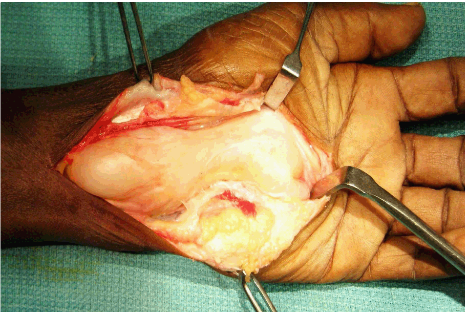
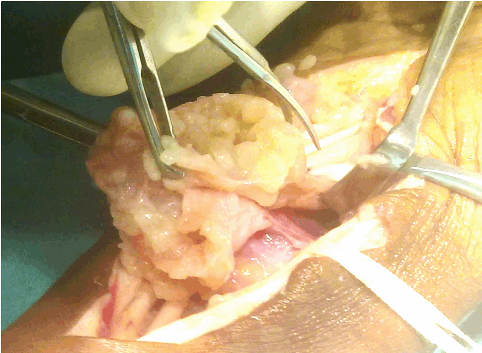
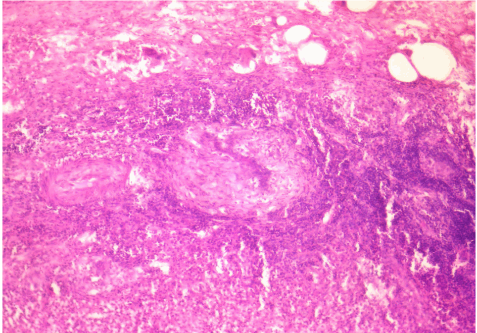
A 55-year-old male butcher, presented with complaints of an increasing swelling over the volar aspect of his left hand and wrist. The condition was associated with pain which was worse at night, disturbing sleep. He had loss of appetite and loss of weight for six months prior to admission. Evening rise of temperature was more marked for the past one month. There was no similar history in the family and no history of contact with any tuberculous patient. Examination revealed two swellings proximal and distal to the flexor retinaculum with a positive cross fluctuation (figure 1). Entire sensory territory of the median nerve was numb. Movements of wrist and fingers were limited along with loss of power in the fingers. Radiographs of the wrist and hand were normal without any involvement of the underlying bones. The chest was normal clinically and radiographically. Blood parameters were within normal limits except an elevated ESR which was 40 and 84 at 1/2 and one hour respectively. There was no evidence of immunodeficiency. There were no other detectable foci of infection. Excision and biopsy was planned and carried out without delay. With a usual approach to the volar wrist and hand, the skin and fascia was incised and retracted. Careful dissection and release of the flexor retinaculum revealed a single continuous fluctuant mass which was filled with fibrinous material and straw colored fluid (figure 2). Fluid sample was taken for PCR. The ganglion also contained melon seed bodies (figure 3), all of which were evacuated completely. Inflamed tendon sheaths were excised after protection of the median nerve. A thorough wash was given and the wound was closed. Histopathology of the specimen revealed inflammatory lesions with large granulomas of epitheloid cells and multiple giant cells with central caseous necrosis (figure 4). PCR was positive for mycobacterium tuberculosis. According to WHO guidelines, patient was started with two months of isoniazid (H), rifampicin (R), pyrazinamide (Z), ethambutol (E) in the intensive phase followed by 4 months continuation phase of HR thrice a week. [2] Finger mobilization was started in the first week postoperatively which was well tolerated by the patient. Later, mobilization of the wrist was started. Subsequent review of the patient showed a complete recovery of numbness and regaining of power in the operated hand. By eight weeks, the patient had returned to normal activity with full function of the affected side. There were no contractures and the scar remained healthy. One year later, having completed anti tubercular therapy, patient is comfortable with using his left hand. There are no signs of recurrence and no other foci of infection. | ||||||
| ||||||
| ||||||
| ||||||
| ||||||
|
Discussion
| ||||||
|
Tuberculosis is still widely present in many developing countries, especially more so in immunocompromised individuals. Though tuberculosis affects various organ systems in the body, involvement of hand and wrist is quite rare. Hence the diagnosis and confirmation of this clinical entity is delayed because of the rarity of the condition. Mycobacterium tuberculosis is the most common causative organism for such an extensive lesion over the wrist and hand and is always confirmed by culture. Operative finding of melon seed bodies are highly suggestive of tuberculous tenosynovitis. [3] Early diagnosis, complete debulking and appropriate anti-tubercular therapy is needed to overcome this condition. With lack of pulmonary symptoms, diagnosing this condition is quite difficult. Patients usually present after six months to one year of being diseased when the condition had already progressed to an extent. In that situation, a debulking tenosynovectomy and chemotherapy is a must for a better prognosis. Moreover, it is the recommended treatment of choice favoured in various reports in the literature. [4] [5] [6] [7] Interfering with the disease before it involves the underlying bones is the main goal of treatment. Early surgery can improve the patient functionally by preventing a subsequent arthrodesis which is a major concern for both the surgeon and the patient. Early post operative mobilization helps to regain a powerful hand grip and also prevents stiffness and adhesions. The one year follow up of our case reveals no signs of recurrence and the patient had returned to his best functional status. | ||||||
|
Conclusion
| ||||||
|
Thus, we conclude insisting that the possibility of tuberculosis in a chronic flexor tenosynovitis of the wrist should always be kept in mind. The delay between the onset of symptoms and the diagnosis should be minimized so that a timely interference could be planned which would do much good for the patient. | ||||||
|
References
| ||||||
| ||||||


|
[HTML Abstract]
[PDF Full Text]
|
|
Author Contributions:
K Arun Kumar - Substantial contributions to conception and design, Acquisition of data, Analysis and interpretation of data, Drafting the article, Revising it critically for important intellectual content, Final approval of the version to be published B Kanthimathi - Substantial contributions to conception and design, Acquisition of data, Analysis and interpretation of data, Drafting the article, Revising it critically for important intellectual content, Final approval of the version to be published CS Krishnamurthy - Substantial contributions to conception and design, Acquisition of data, Analysis and interpretation of data, Drafting the article, Revising it critically for important intellectual content, Final approval of the version to be published S Sujai - Substantial contributions to conception and design, Acquisition of data, Analysis and interpretation of data, Drafting the article, Revising it critically for important intellectual content, Final approval of the version to be published |
|
Guarantor of submission:
The corresponding author is the guarantor of submission. |
|
Source of support:
None |
|
Conflict of interest:
Authors declare no conflict of interest. |
|
Copyright:
© K Arun Kumar et al. 2012; This article is distributed under the terms of Creative Commons Attribution License which permits unrestricted use, distribution and reproduction in any means provided the original authors and original publisher are properly credited. (Please see http://www.ijcasereportsandimages.com/copyright-policy.php for more information.) |
|
|