
| Table of Contents |  |
|
Case Report
|
| Perforated pyometra presenting as a pelvic abscess: A case report |
| Afrasyab Khan1, Jagdish Prasad2 |
|
1House Surgeon, Whakatane Hospital. Whakatane, New Zealand.
2Consultant General Surgery, Whakatane Hospital, Whakatane, New Zealand. |
|
doi:10.5348/ijcri-2012-02-89-CR-3
|
|
Address correspondence to: Dr. Afrasyab Khan House Surgeon Whakatane Hospital Whakatane 3158 New Zealand Fax: 073060969 Email: afrasyabkhan@gmail.com |
|
[HTML Abstract]
[PDF Full Text]
|
| How to cite this article: |
| Khan A, Prasad J. Perforated pyometra presenting as a pelvic abscess: A case report. International Journal of Case Reports and Images 2012;3(2):10-14. |
|
Abstract
|
|
Introduction:
Pyometra - the accumulation of purulent material inside the uterus - can be asymptomatic in 50% of cases. A perforated pyometra usually presents with an acute abdomen at the outset and it is unusual for it to present as a pelvic abscess.
Case Report: We present the case of a 57-years-old lady, who presented with increasing epigastric pain. She was hypotensive with epigastric tenderness. She had an elevated white count and elevated liver enzymes. Imaging did not reveal the etiology of her condition. She started to have peritoneal signs on hospital day nine and was taken to the operating theatre for exploratory laparotomy. A pelvic abscess was found that was communicating with a perforated pyometra. Hysterectomy and bilateral salpingo-oophorectomy was performed. No evidence of malignancy was found on histo pathologic examination. The patient had post operative ileus and was on parenteral nutrition. She soon recovered and was discharged home. Conclusion: A perforated pyometra is usually seen in post menopausal women and presents as an acute abdomen. It is associated with blockage of the endocervical canal. The patient in this case had a pelvic abscess that had walled off the perforation. The patient had symptoms for a while as overt peritonitis had not developed. No malignancy was identified. A high index of suspicion is needed to diagnose a perforated pyometra in postmenopausal patients presenting as an acute abdomen or a pelvic abscess. It may not be associated with malignancy and presentation may be overtly acute or insidious. | |
|
Key Words:
Pyometra, Abscess, Acute abdomen, Peritonitis
| |
|
Introduction
| ||||||
|
The accumulation of pus or purulent material in the uterine cavity is known as a pyometra. This condition can be asymptomatic in up to 50% of patients. [1] Spontaneous rupture of a pyometra can occur and the common presentation is with an acute abdomen. [1] [2] [3] We present an interesting case of a perforated pyometra that presented as a pelvic abscess. | ||||||
|
Case Report
| ||||||
|
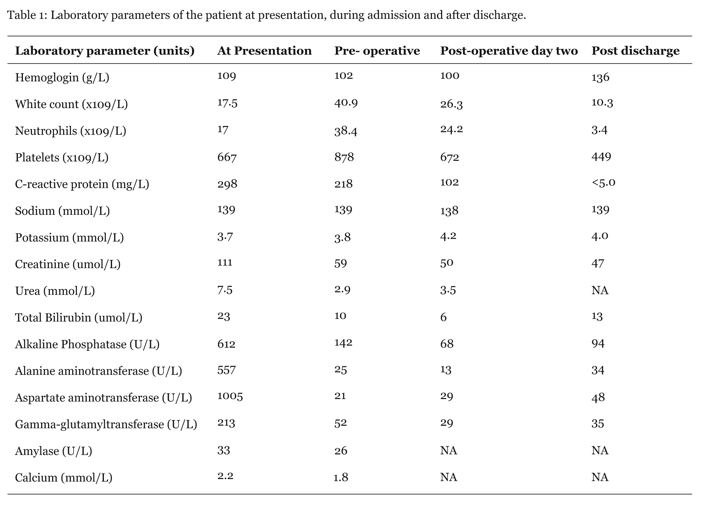
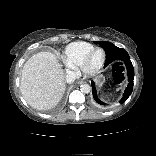
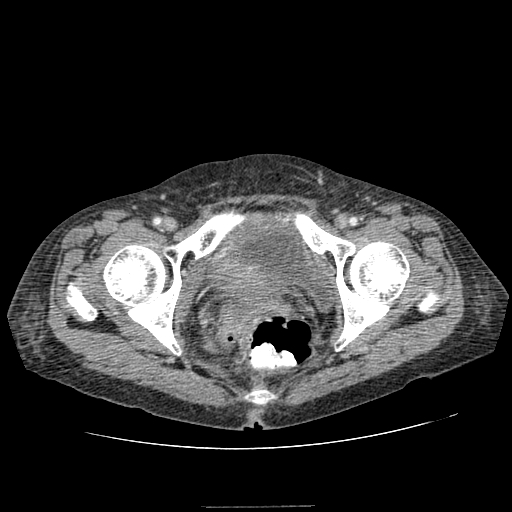
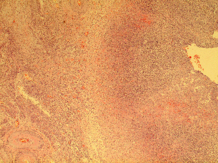
A 57-years-old lady presented to the Emergency Department(ED) with complaints of epigastric pain and vomiting. She had these symptoms for a month along with dysuria but increasing severity and her generally feeling unwell prompted presentation to the Emergency Department. A urine dipstick and culture did not show any abnormality. She did not have any per-vaginal discharge or bleeding on initial presentation. Significant findings on examination were hypotension (70/32 mmHg) and epigasric tenderness without any peritoneal signs. On initial investigations she had a raised white blood cell (WBC) count and raised liver enzymes (table 1). Blood samples were sent for culture and sensitivity testing. The initial chest and abdominal X-rays did not reveal any abnormality. After resuscitation in the ED and administration of empiric antibiotics, she was admitted to the hospital. Initial antibiotics were intravenous amoxicillin 500 mg every eight hours, intravenous metronidazole 500 mg every eight hours and intravenous gentamicin 240 mg once daily. Hepatitis serology was requested. A CT scan of the abdomen and pelvis was done which revealed that she had some free intra-abdominal fluid and isolated non-specific mesenteric nodes (figure 1, 2). Broad spectrum antibiotics were continued. Gentamicin was given for a total of three days and then stopped. Metronidizole was continued. Amoxicillin was changed after three days to co-amoxiclav intravenous 625 mg three times a day for broader coverage. She remained stable in the hospital. Operative intervention was not advocated initially as her condition remained stable and there was no definite diagnosis yet. The patient had her first spike of fever on hospital day five. Blood cultures, urine cultures and hepatitis serology were all negative. An endovaginal ultrasound and ascitic tap did not help in finding the etiology of her condition. Ascitic fluid sample showed no growth in culture. On hospital day nine, she had a sudden rise in temperature and an increase in WBC count. On examination she now had increased tenderness and started to develop peritoneal signs. After discussion with the patient, it was decided to go ahead with exploratory laparotomy. Significant findings in the exploratory laparotomy included a pelvic abscess that was walled off by matted small bowel, ceacum and sigmoid colon. A 10 cm pyometra that had ruptured and was communicating with the pelvic abscess was identified. Approximately 500 ml of pus was drained from the abscess cavity and the pelvis. Samples were sent for testing. The pelvis was washed out. A hysterectomy and bilateral salpingo-oophorectomy was performed. Further washout was done with saline and a drain was placed. Histology showed acute and chronic inflammation of the uterus and fallopian tubes with necrosis and abscess formation (figure 3-6). There was no evidence of malignancy on gross and microscopic examination of the specimens. The pus sample showed no aerobic or anaerobic growth. Post operatively the patient was shifted to the intensive care unit (ICU) and was hemodynamically stable. She was tolerating sips of water. She was soon transferred to the surgical ward. Antibiotics were stopped on post operative day nine. Return of bowel function was delayed and she was started on total parental nutrition on post operative day four. She had prolonged post operative ileus and was on tota parentral nutrition. However, her condition soon improved and she started tolerating oral intake. She was discharged home on post operative day thirty three. | ||||||
| ||||||
| ||||||
| ||||||
| ||||||
| ||||||
| ||||||
| ||||||
|
Discussion
| ||||||
|
A perforated or ruptured pyometra is mainly seen in postmenopausal women. [1] It is associated with endocervical malignancy and stenosis of the cervical canal. [4] [5] [6] Pyometra has a reported incidence of 0.38- 0.5% in gynecologic patients. [7] [8] Patients present with abdominal pain and there may be fever and vaginal discharge. [9] It may present as a case of peritonitis or acute abdomen. [1] [2] [3] However, in this case a pelvic abscess was found and had walled off the site of perforation. Etiologies include malignant growths of the uterus and pelvis, benign growths in the uterus and pelvis, atrophic cervicitis, pelvic radiation and puerperal infections. [10] [11] Diagnosis is by ultrasonography; or by computed tomography which is usually done suspecting some other cause of presenting signs and symptoms. In an emergency, broad spectrum antibiotics, supportive treatment and total hysterectomy with bilateral salpingo-oophorectomy and irrigation of abdominal cavity is the treatment of choice. In non-emergent cases drainage of the uterus via transcervical approach with antibiotics has been described. [12] The diagnosis of a ruptured pyometra is usually not apparent until exploration of the abdomen is done for the presenting peritonitis and a perforated pyometra is found. A very high index of suspicion for the diagnosis of a perforated pyometra is needed in post menopausal women with acute abdomen or a pelvic abscess. There have been case reports of a ruptured pyometra with no malignancy found in the uterus or cervix. [2] [4] [13] [14] Similarly in this case, no malignancy was found on gross and microscopic examination of the uterus and adnexa after surgery. The ascitic fluid sample in this case did not grow any organism in culture probably because the perforation was well walled off in the form of a pelvic abscess and she had also been on broad spectrum antibiotic therapy. Imaging with CT can be repeated if the index of suspicion for an infective focus or an abscess is high and the initial imaging failed to reveal any abscess. | ||||||
|
Conclusion
| ||||||
|
While a perforated pyometra is a rare condition, a high index of suspicion is necessary in post menopausal women presenting with acute abdomen or with ascitic fluid showing acute inflammation and uncharacterized pelvic or uterine abnormality. In cases of acute inflammatory conditions of the abdomen or pelvis in which a cause is not identified and there is no appropriate response to broad spectrum antibiotic treatment, an abscess must always be considered as a possible etiology even if initial imaging failed to identify an abscess. A pyometra may not be associated with malignancy and presentation due to perforation may be overtly acute or insidious. | ||||||
|
References
| ||||||
| ||||||


|
[HTML Abstract]
[PDF Full Text]
|
|
Author Contributions:
Afrasyab Khan - Substantial contributions to conception and design, acquisition, analysis and interpretation of data, Drafting the article, revising it critically for important intellectual content, Final approval of the version to be published Jagdish Prasad - Substantial contributions to conception and design; and interpretation of data, Drafting the article, revising it critically for important intellectual content, Final approval of the version to be published |
|
Guarantor of submission:
The corresponding author is the guarantor of Submission. |
|
Source of support:
None |
|
Conflict of interest:
The authors declare no conflict of interest. |
|
Copyright:
© Afrasyab Khan et al. 2012; This article is distributed under the terms of Creative Commons Attribution License which permits unrestricted use, distribution and reproduction in any means provided the original authors and original publisher are properly credited. (Please see http://www.ijcasereportsandimages.com/copyright-policy.php for more information.) |
|
|