| Table of Contents |  |
|
Case Report
|
| Langerhans cell histiocytosis in the right scapula in a young man |
| Georgios Tsirakis1, Maria Kaparou2, Peggy Kanellou2, Georgios Kontakis3, Michael Alexandrakis4 |
|
1Consultant Haematologist, Department of Haematology, University Hospital of Heraklion, Crete, Greece.
2Resident in Haematology, Department of Haematology, University Hospital of Heraklion, Crete, Greece. 3Associate Professor of Orthopedics, Department of Orthopedics and Traumatology, University Hospital of Heraklion, Crete, Greece. 4Associate Professor of Haematology, Department of Haematology, University Hospital of Heraklion, Crete, Greece. |
|
doi:10.5348/ijcri-2012-01-83-CR-5
|
|
Address correspondence to: Michael Alexandrakis University Hospital of Heraklion Department of Haematology P.O. box 1352, Heraklion, Crete Greece, P.C. 71110 Phone: 00302810392425 Fax: 00302810392426 Email: alexandm@med.uoc.gr |
|
[HTML Abstract]
[PDF Full Text]
|
| How to cite this article: |
| Tsirakis G, Kaparou M, Kanellou P, Kontakis G, Alexandrakis M. Langerhans cell histiocytosis in the right scapula in a young man. International Journal of Case Reports and Images 2012;3(1):16-19. |
|
Abstract
|
|
Introduction:
Langerhans cell histiocytosis (LCH) is a proliferative histiocytic disorder with a variable clinical manifestations. It may be localized or disseminated. The aetiology and pathogenesis of the disease are unknown. Various mechanisms regarding the aberrant expression of chemokines' receptors or dysregulation of chemokine production in the lesions, probably take part in the pathophysiology of the disease.
Case Report: We report a rare case of Langerhans cell histiocytosis in the right scapula. A 28-year-old man presented with pain in right shoulder. A localized osteolysis of glenoid with disruption of the cortex, accompanied by extensive bone edema was detected with CT and MRI, whereas Tc-99m bone scan revealed increased concentration of the radionuclide in the affected area. The bone lesion biopsy revealed Langerhans cell histiocytosis (CD1a+, S-100+, CD68+), and a palpable spleen was also revealed. After the removal of the lesion, he was treated with systematic chemotherapy and the patient remains with no active disease for four years. Conclusion: Langerhans cell histiocytosis is a disease of histiocytes with multiple pathophysiologic aetiologies. The diagnosis is based on morphological and immunohistochemical characteristics. The clinical manifestations vary depending on the organs or systems affected. It most frequently affects bone, skin and pituitary and less commonly haematopoietic system, spleen, lungs and central nervous system. In the bones, the lesions are usually localized to the skull, particularly in the jaw, long tubular bones, vertebrae, pelvis and ribs. Scapula is a very rare site for LCH. This is one of very few case reports of LCH in the scapula. | |
|
Key Words:
Langerhans cell histiocytosis, Scapula, Osteolysis
| |
|
Introduction
| ||||||
|
Langerhans cell histiocytosis (LCH) is a rare group of disorders with a common characteristic of the proliferation of CD1a+ dendritic cells. [1] The aetiology remains poorly defined. Infection with Epstein-Barr virus in certain cases has been described probably as a triggering mechanism. [2] Another important risk factor for pulmonary involvement from LCH is smoking. In previous studies it was reported that more than 90% of cases of pulmonary LCH occur in adults who smoke. [3] The incidence of LCH in adults may reach 1-2 cases per million and is significantly lower than the children. The mean age at diagnosis of LCH is 35±14 years. [4] Clinical manifestations of LCH in adult patients vary depending of the organs or systems affected. Bones are major organs involved with more usual locations in the skull, particularly in the jaw, long tubular bones, vertebrae, pelvis and ribs. We report a very rare case of a 28-year-old man with LCH of the right scapula. | ||||||
|
Case Report
| ||||||
|
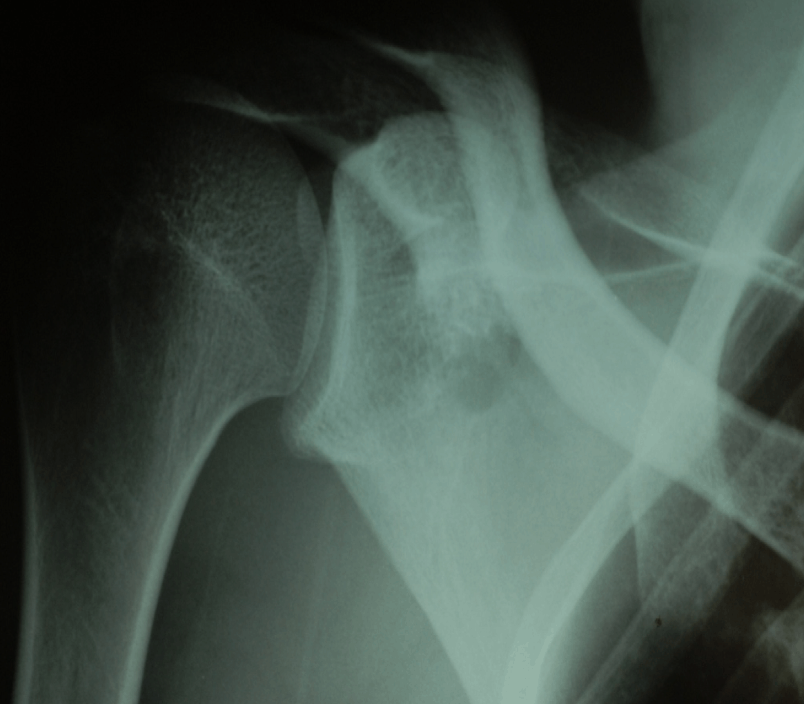
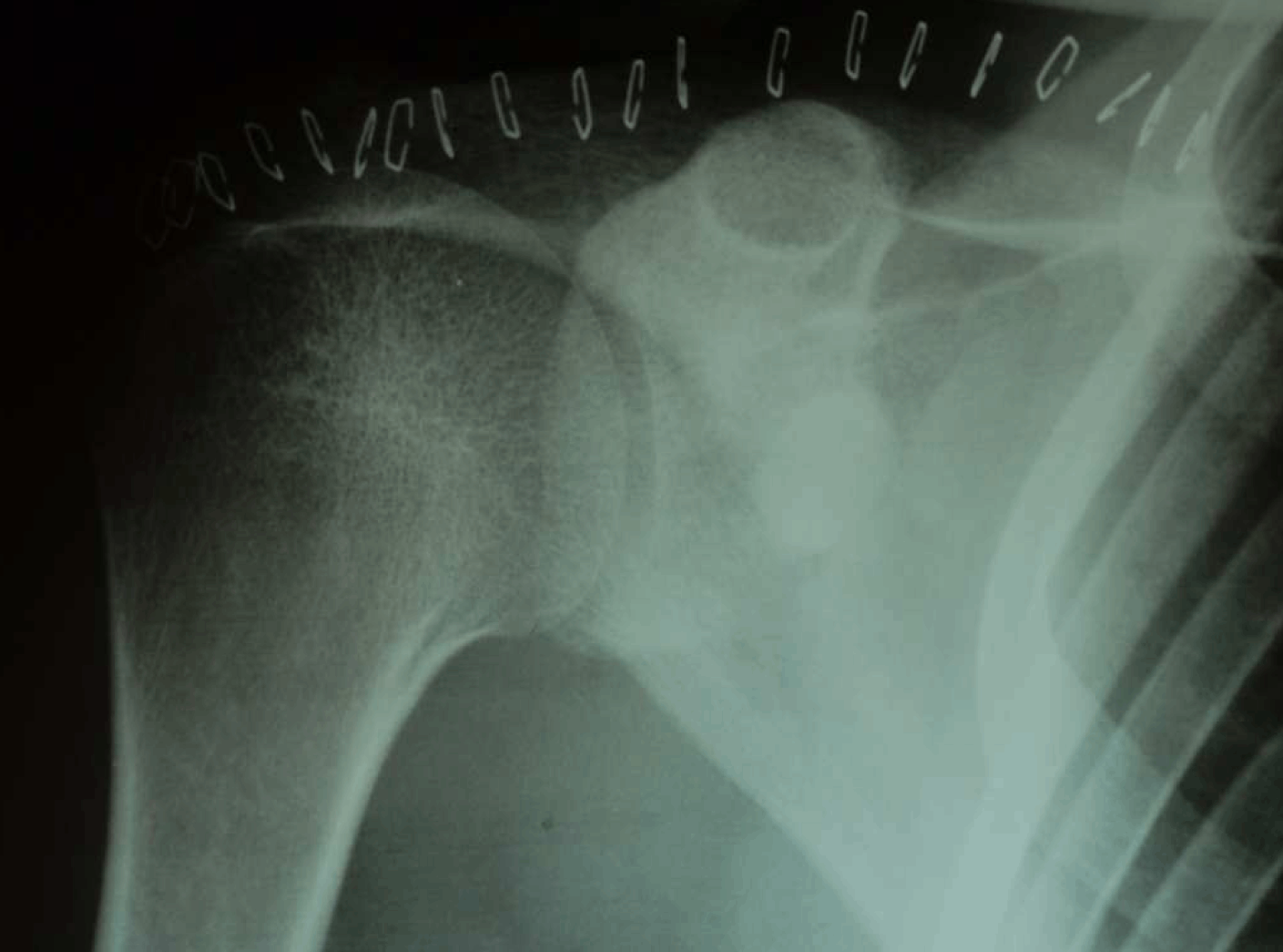
A 28-year-old male presented with a one month history of pain in the region of the right shoulder and posterior scapular area. He described it as a constant aching, which decreased with rest and analgesic drugs and increased when he raised his arm overhead and lifted objects. Pain was initially intermittent and then became constant. Although he had already started painkillers, his clinical condition gradually deteriorated. He did not complain of fever, weight loss, night sweats. There was no other severe co-morbidity. Clinical examination revealed a palpable spleen (enlarged three cm below costal margin in the midclavicular line), without evidence of lymphadenopathy or any other sign of disease on CT of thorax, abdomen and pelvis. Laboratory tests revealed WBC - 6.6x10³/µl, (neutrophils - 4.2x10³/µl, lymphocytes 2x10³/µl), haemoblobin - 15.6 g/dl, platelets - 234x103/µl. Plasma electrolytes, calcium, total protein, albumin, globulins, lactate dehydrogenase, immunoglobulins and erythrocyte sedimentation rate were normal. Studies of serum protein electrophoresis, coagulation, urine and biochemical studies of renal and liver function were also normal. The X-ray showed a lytic lesion in the area of the right scapula (figure 1) and the findings of the CT and MRI showed a localized osteolysis of glenoid with disruption of the cortex, accompanied by extensive bone edema, compatible with malignancy. The skeletal radiograph survey did not reveal other bone lesions. Tc-99m bone scan revealed increased concentration of the radionuclide in the affected area of the right scapula. An open biopsy was performed, with intralesional removal of the pathologic tissue from the bone and muscle. After the curettage, the defect was filled by PMMA (Poly-Methyl-Methacrylate) cement which produces local exothermic reaction [5] (figure 2). The bone lesion biopsy showed extensive necrosis with histiocytic cells that had elongated, grooved nuclei with higher nuclear to cytoplasmic ratio than typical macrophages (figure 3). The features of cytoplasm and nucleus were suggestive for Langerhans cells. Immunohistochemical staining for CD1a complex, S-100 protein and CD68 markers specific for Langerhans cells were positive. He was considered to have multisystem LCH (MS-LCH) (unifocal bone involvement and involvement of one risk organ, such as the spleen) which was an indication for systemic therapy. He started an initial 6-week course therapy with vinblastine (6 mg/m2 weekly) and prednisone (40 mg/m2 daily) and continued a maintenance therapy with pulses of vinblastine (6 mg/m2) and prednisone (40 mg/m2 daily for five days) every three weeks and daily continuous 6-mercaptopurine (50 mg/m2) for total treatment duration of 12 months, according to Histiocyte Society Evaluation and Treatment Guidelines. After the end of therapy the spleen was unpalpable. Four years post end of therapy, the patient remains with no active disease. | ||||||
| ||||||
| ||||||
|
| ||||||
|
Discussion
| ||||||
|
LCH is characterized by the infiltration of one or more organs by a clonal population of CD1a+ dendritic Langerhans cells, with various proportions of macrophages, T-lymphocytes, eosinophils and multinucleated giant cells. [6] The CD1a+ cells are retained in an immature state, due to dysregulation of chemokine production and/or chemokine receptor expression, which enables their recruitment to inflammation sites. Furthermore, CD1a+ cells overexpress inflammatory chemokines, such as CCL20/MIP-3a, important for T-cell recruitment, and CCL5/RANTES and CXCL11/I-TAC that recruit other inflammatory cell types. T - cells, surrounding the reactive "rim" of the lesions, are CD4+ CD45RO+, most of which express CXCR3, relating to Th1-type immune response. [7] LCH cells have a high expression of CD40 and interact with CD40L+ T-cells, causing their activation, via an uncontrolled production of several cytokines. The following cytokine storm includes interleukins (IL-1a, IL-2, IL-3, IL-4, IL-5, IL-7), tumor necrosis factor-a, interferon-? and colony stimulating factor (GM-CSF) and causes the recruitment of Langerhans cells progenitors, their maturation and their rescue from apoptosis. They also play a role in bone resorption, fibrosis and necrosis. [6] Recently it was suggested that a regulatory T-cells expansion fails to eliminate LCH cells. [8] The most frequently affected organs are the bone, skin and pituitary. [4] Other tissues often involved are liver, spleen, haematopoietic system, lungs, lymph nodes and central nervous system. [9] The clinical course of the disease is characterized from the organs or systems affected. It varies from one single lesion in the involved organs or involvement of multiple sites, with rapid and progressive disease affecting the quality of life. [10] LCH can be divided in single system (uni- or multifocal) and multisystem disease according to number of affecting organs or systems. There are some organs and tissues with increased risk of involvement such as lungs, spleen, liver and blood, where the evidence of involvement could be only indirect, based on imaging techniques, clinical evaluation or on the cytopenias, without histological confirmation of infiltration. Indications for treatment are multisystem disease and single system disease with multifocal bone lesions or with CNS-risk lesions (craniofacial bones, eye, ear or oral involvement and vertebral lesions with intraspinal soft tissue extension). Bone lesions are usually localized to the skull (51%), particularly in the jaw (30%), long tubular bones (17%), vertebrae (13%), pelvis (13%) and ribs (6%). [4] [6] [10] An unusual skeletal site in LCH is scapula with clinical manifestations from the upper limb, such as pain and limited movement. Our patient reported symptoms from this lesion; meanwhile he had a palpable spleen which is a risk organ for systematic involvement. Patients without involvement of risk organs, as well as those with involvement of risk organs who respond to standard initial therapy, have an excellent chance of long-term survival. We conclude that combination of local treatment, with removal of the lesion and filling of the defect, after the curettage, by PMMA (Poly-Methyl-Methacrylate) cement which produces local exothermic reaction and systematic chemotherapy, in our patient with unifocal bone lesion and involvement of one risk organ, was the appropriate therapy. | ||||||
|
Conclusion
| ||||||
|
LCH is a rare disease of children and young adults. Scapula is a very rare location of LCH and we present here a case of this unusual involvement. | ||||||
|
References
| ||||||
| ||||||


|
[HTML Abstract]
[PDF Full Text]
|
|
Author Contributions:
Georgios Tsirakis - Acquisition of data, Analysis and interpretation of data, Drafting the article, Critical revision of the article, Final approval of the version to be publishe. Maria Kaparou - Analysis and interpretation of data, Critical revision of the article, Final approval of the version to be published Peggy Kanellou - Analysis and interpretation of data, Critical revision of the article, Final approval of the version to be published Georgios Kontakis - Acquisition of data, Drafting the article, Critical revision of the article, Final approval of the version to be published Michael G Alexandrakis - Conception and design, Drafting the article, Critical revision of the article, Final approval of the version to be published |
|
Guarantor of submission:
The corresponding author is the guarantor of submission. |
|
Source of support:
None |
|
Conflict of interest:
Authors declare no conflict of interest. |
|
Copyright:
© Georgios Tsirakis et al. 2012; This article is distributed the terms of Creative Commons Attribution License which permits unrestricted use, distribution and reproduction in any means provided the original authors and original publisher are properly credited. (Please see Copyright Policy for more information.) |
|
|