Volume 2; Number 12 (December 2011)
|
Cover Figure |
 |
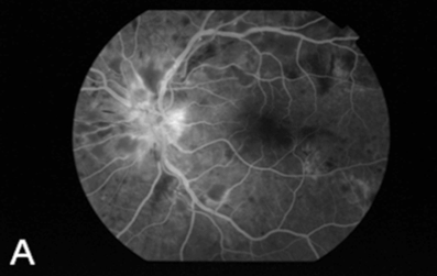
Figure 2: Fluorescein angiography shows the marked hyperfluorescence from the deep layers of the retinal pigment epithelium in the peripapillary area and peripheral fundus. (Page 17)
|
Go Back to Table of Contents |
|