| Table of Contents |  |
|
Clinical Image
|
| Abdominal compartment syndrome: A lethal disease |
| Ravish Shah1 |
|
1Assistant Professor, Division of Nephrology, The Ohio state university, 395 West 12th avenue, Ground floor, Columbus, OH-43210, USA.
|
|
doi:10.5348/ijcri-2011-12-78-CI-8
|
|
Address correspondence to: Ravish Shah Assistant Professor, Division of Nephrology The Ohio state university 395 West 12th avenue Ground floor, Columbus OH-43210, USA Phone: 614-293-4055 Fax: 614-293-3073 Email: ravish.shah@osumc.edu |
|
[HTML Abstract]
[PDF Full Text]
|
| How to cite this article: |
| Shah R. Abdominal compartment syndrome: A lethal disease. International Journal of Case Reports and Images 2011;2(12):34-36. |
|
Case Report
| ||||||
|
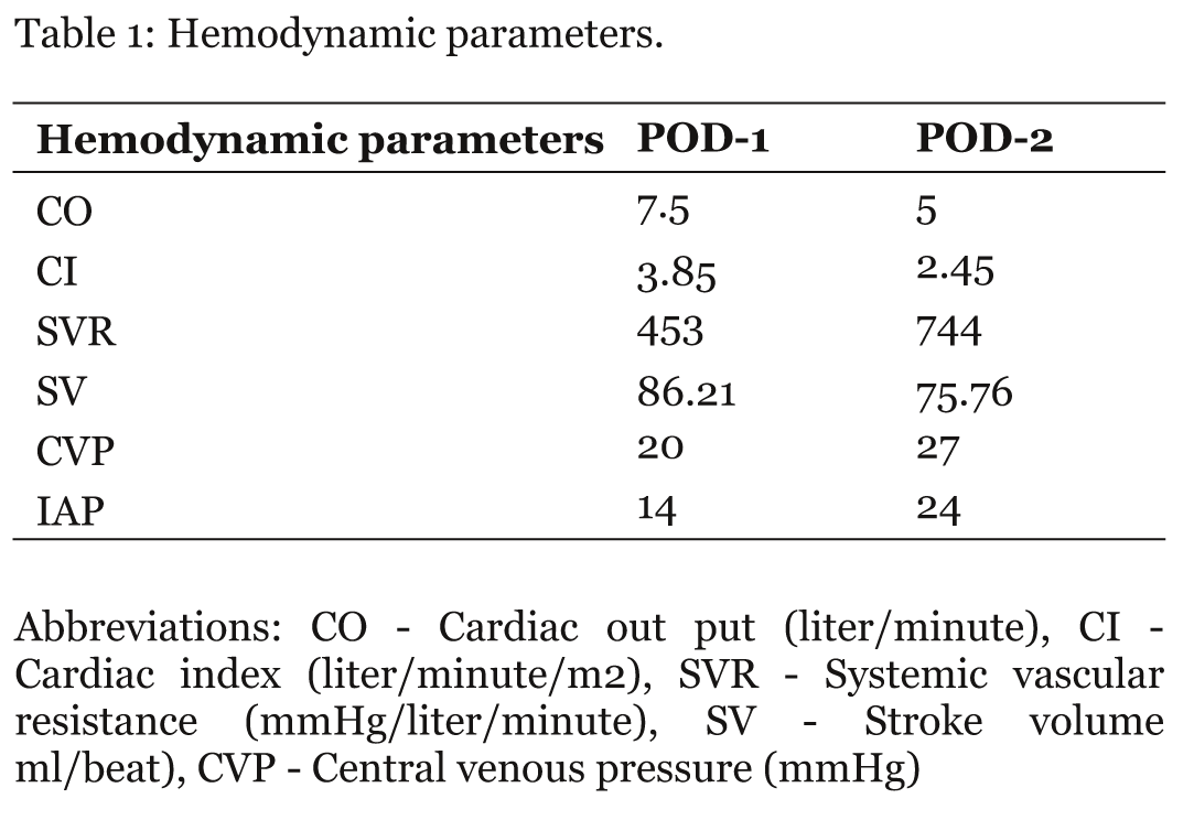
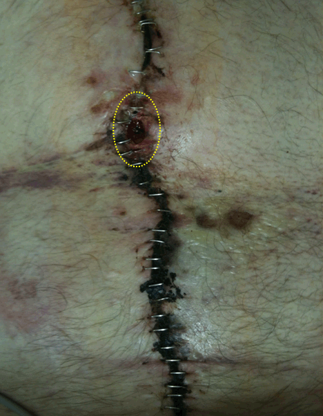
A 63-year-old Caucasian male with a history of ventral hernia secondary to multiple motor vehicular accidents and prior abdominal surgeries was admitted for an elective ventral hernia repair. A primary closure of ventral hernia with a skin mesh was performed. Post-operatively he remained intubated and was transferred to surgical intensive care unit. At baseline, his laboratory data included serum creatinine - 0.9 mg/dl, hemoglobin - 10 gm/dl, white cell count - 9x10³/mm3 and lactic acid - 1 mmol/L. Peri-operatively, he was 10 liters net positive with a urine output (VOP) of 1200 cc in 24 hours. His serum creatinine was 1.3 mg/dl with one liter urine output on post-operative day (POD)-1. A two millimeter (mm) gap was noted in between the surgical staples. Intra-abdominal pressure (IAP) measured by transvesical route was noted to be elevated at 14 cm of H2O. He was started on the intensive regimen for management of intra-abdominal hypertension (IAH) including nasogastric decompression, elevation of head of the bed > 30°, discontinuation of intravenous fluids and a trial of neuromuscular blockage. On POD-2, he was noted to have increasing abdominal distension, with worsening hemodynamic parameters (table 1). The gap in the surgical wound increased to five mm (figure 1) and the IAP was noted to be 24 cm of H2O. His condition significantly deteriorated during next few hours with development of oliguric acute kidney injury (AKI) (UOP 200 cc/24hours), acute respiratory distress syndrome, high anion gap metabolic acidosis and refractory hypotension requiring escalating doses of multiple vasopressors (norepinephrine 3 µgm/kg/min, epinephrine 1 mcg/kg/min, vasopressin 0.04 units/minute), to maintain mean arterial pressure of about 60 mmHg. His laboratory data revealed serum creatinine - 5 mg/dL, hemoglobin - 7.8 gm/dL, white cell count - 20x103/mm3 and an elevated lactic acid - 18 mmol/liter. He was diagnosed with the refractory abdominal compartment syndrome (ACS). Given his clinical deterioration and hemodynamic instability, the treatment goals were addressed with the family. The patient was made comfortable care per his former wishes, at family's request. | ||||||
| ||||||
| ||||||
|
Discussion
| ||||||
|
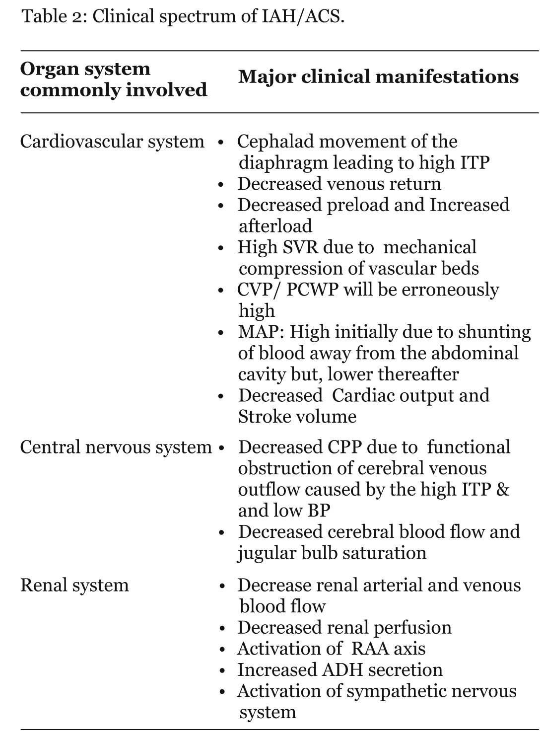
Abnormally elevated IAP has been increasingly recognized in the critically ill as a causes of significant morbidity and mortality. [1] Depending on the severity of increased IAP and organ function, the World Society on Abdominal Compartment Syndrome have published consensus definition of IAH and ACS. [1] IAH is defined by a sustained or repeated pathologic increase in IAP = 12 mmHg. IAH is graded as follows: grade I, IAP 12 - 15 mmHg; grade II, IAP 16 - 20 mmHg; grade III, IAP 21 - 25 mmHg; grade IV, IAP > 25 mmHg. ACS is defined as a sustained IAP = 20 mmHg (with or without abdominal perforation pressure <60 mmHg) that is associated with new organ dysfunction/failure. [1] Causes can be divided based on the mechanism of IAP elevation: i) condition associated with increase in intra-abdominal volume: gastric distention, ileus, volvulus, intra-abdominal or retroperitoneal masses, ascites; ii) conditions leading to reduced abdominal wall compliance: abdominal surgery, especially with tight abdominal closures, rectus sheath hematomas, surgical correction of large abdominal hernias; iii) combination of decreased abdominal wall compliance and increased intra-abdominal volume: obesity, sepsis, severe sepsis, and septic shock, severe acute pancreatitis, massive fluid resuscitation, major burns. [1] Increased IAP within the closed abdominal cavity can not only lead to decreased perfusion and ischemia of intra-abdominal organs but can also cause organ dysfunction beyond the abdominal cavity due to the close anatomic relationships with contiguous cavities. [2] Major clinical manifestations of the commonly involved organ systems are described in table 2. The key to recognizing ACS in a critically ill patient is the demonstration of elevated IAP. Clinical examination is an inaccurate indicator of IAP estimation with a sensitivity and positive predictive value of around 40- 60%. [3] [4] Moreover the radiologic investigations with plain radiography of the abdomen, abdominal ultrasound or CT scan are also insensitive. [2] Although different direct or indirect methods for estimating IAP can be used, the transvesical measurement has achieved the most widespread adoption worldwide because of its simplicity and minimal cost. [2] Different medical/minimally invasive options, based on following principles have been recommended to decrease IAP: i) stratagies applied to improve abdominal wall compliance, ii) evacuation of intraluminal contents or abdominal fluid collections, iii) correction of capillary leak and positive fluid balance. [1] [2] A decompressive laparotomy should be considered if medical treatment is not effective or in the presence of refractory ACS. Although invasive, decompressive laparotomy is effective in decreasing IAP and improving organ function. [4] [5] | ||||||
| ||||||
|
| ||||||
|
Conclusion
| ||||||
|
IAH and ACS are an important cause of AKI in critically ill patients. It is crucial for the clinicians to be aware of the existence of IAH, its pathologic implications, and available methods for IAP measurement. If medical management fails and the patient progresses to ACS, an immediate surgical decompression is warranted. | ||||||
|
Acknowledgement
| ||||||
|
I thank Dr. Daniel Allendorf for critical revisions of the manuscript. | ||||||
|
References
| ||||||
| ||||||

|
[HTML Abstract]
[PDF Full Text]
|
|
Author Contributions:
Ravish Shah - Substantial contributions to conception and design, Acquisition of data, Analysis and interpretation of data, Drafting the article, Revising it critically for important intellectual content, Final approval of the version to be published |
|
Guarantor of submission:
The corresponding author is the guarantor of submission. |
|
Source of support:
None |
|
Conflict of interest:
Authors declare no conflict of interest. |
|
Copyright:
© Ravish Shah et al. 2011; This article is distributed under the terms of Creative Commons Attribution License which permits unrestricted use, distribution and reproduction in any means provided the original authors and original publisher are properly credited. (Please see copyright policy for more information.) |
|
|