| Table of Contents |  |
|
Case Report
|
| Diverticulitis as a cause of septic thrombophlebitis: A literature review |
| Jessica Rose1, Rostam Khoubyari1, James McClenathan1 |
|
1University Physicians Hospital at Kino, University of Arizona.
|
|
doi:10.5348/ijcri-2011-12-77-CR-7
|
|
Address correspondence to: Jessica Rose Department of Surgery The University of Arizona, 1501 N. Campbell Avenue, Rm. 4334 P.O. Box 245058, Tucson, AZ 85724 Phone: 607-725-4619 Fax: 520-626-2247 Email: jessfrose@gmail.com |
|
[HTML Abstract]
[PDF Full Text]
|
| How to cite this article: |
| Rose J, Khoubyari R, McClenathan J. Diverticulitis as a cause of septic thrombophlebitis: A literature review. International Journal of Case Reports and Images 2012;2(12):28-33. |
|
Abstract
|
|
Introduction:
Perforated diverticulitis, with fistula formation between the colon and inferior mesenteric vein, is a cause of portal venous gas. In contrast to more common causes of portal venous air, such as mesenteric ischemia and gangrene, the prognosis is much better for patients with perforated diverticulitis. However, the diagnosis can be difficult to make, given the wide range of nonspecific symptoms. Delays in diagnosis increase the morbidity and mortality rates.
Case Report: In this report, we describe a case of perforated diverticulitis with portal venous gas in a 46-year-old male from our institution, and review the literature on this topic. Conclusion: With modern imaging technology and early diagnosis, septic phlebitis can be managed surgically, with good outcomes. | |
|
Key Words:
Diverticulitis, Perforated diverticulitis, Septic phlebitis, Portal venous gas, Portal venous fistula.
| |
|
Introduction
| ||||||
|
Diverticulitis is a common diagnosis with many known complications. About 8% of patients experience a perforation and 20% a fistula. [1] [2] In only 1-2% of patients the perforation extends into the mesentery. [1] Fistulas can be colocolic, colocutaneous, colocoxal, coloureteral, coloenteral, colosalpingeal, colouterine, colovesical, cologastric, colovenous, and some other less common forms. [3] Septic phlebitis (also known as pylephlebitis or portal venous gas) is an uncommon but potentially morbid complication of diverticulitis in which the perforation extends into the mesentery and erodes into a vein. We recently saw such a patient at our institution. | ||||||
|
Case Report
| ||||||
|
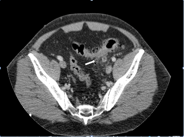
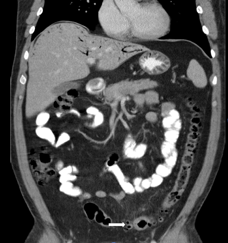
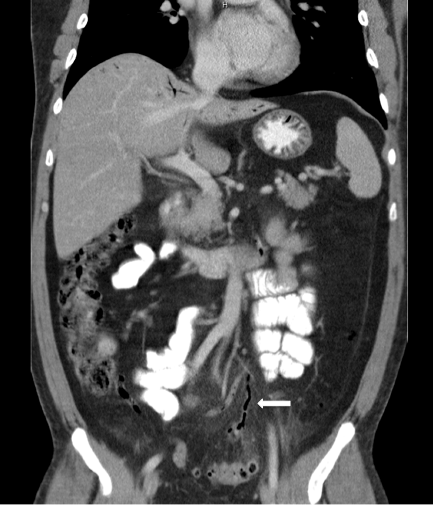
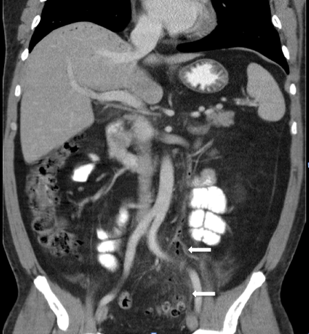
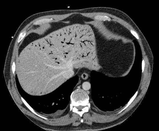
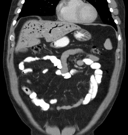
Our patient, a 46-year-old male, had a past medical history of infectious colitis, hypertension, and hyperlipidemia. He came to our emergency department after two days of fever (as high as 39.4°C), chills, headache and dull left lower quadrant abdominal pain. Previously, he had been admitted to another institution with rectal bleeding and had undergone a colonoscopy which revealed resolving infectious colitis. When he arrived in our emergency department, he was in no acute distress, was afebrile and had stable vital signs. His physical examination was significant for mild left lower quadrant tenderness. His initial workup included laboratory studies: WBC counts - 4.5x10³/mm3 with band cells, blood glucose - 232 mg/dl, total bilirubin - 1.8 mg/dl, AST - 96 IU/L, ALT - 110 IU/L and INR - 1.5. Secondary to his elevated liver function test findings, he underwent an abdominal ultrasound examination, which revealed innumerable echogenic foci in the liver. Those foci were thought to reflect calcification but other causes were not excluded. His ultrasound examination also revealed nonspecific gallbladder wall thickening, without cholelithiasis. To better elucidate the ultrasound findings, we ordered computed tomography (CT) scan of the abdomen and pelvis. It showed portal venous gas, as well as gas within branches of the inferior mesenteric vein (IMV) extending from the sigmoid colon, with sigmoid diverticulitis (figures 1-6). | ||||||
| ||||||
|
| ||||||
| ||||||
|
| ||||||
| ||||||
|
| ||||||
| ||||||
|
| ||||||
| ||||||
|
| ||||||
| ||||||
|
The patient underwent an exploratory laparotomy. During the operation, we found an inflamed sigmoid diverticulum with erosion into the mesentery. The liver appeared grossly normal. A sigmoid resection and a Hartmann procedure with a left colostomy was performed. His postoperative recovery was uneventful. The pathology report showed diverticulitis with rupture and acute serositis, diverticulosis and vascular congestion. | ||||||
|
Discussion
| ||||||
|
To more fully understand the case history of our patient, we performed a literature search, using PubMed. We identified and read all English-language articles on septic phlebitis, pylephlebitis, portal venous gas and colovenous fistulas that resulted from perforated diverticulitis. In light of the variety of clinical presentations, perforated diverticulitis needs to be considered in the differential diagnosis of septic patients. Without proper management of this disease, it can be fatal. In fact, one series reported a mortality rate of 65%, with the majority of diagnoses occurring at autopsy. [4] Diverticulitis with fistula formation to mesenteric vessels was the only diagnosis that we included in our literature review; however, pylephlebitis has other causes. Any condition involving infection or inflammation of the region drained by the IMV and portal vein can cause pylephlebitis. [5] The most common causes of portal venous gas include ulcerative colitis, Crohn's disease, bowel ischemia with necrosis, peroxide enemas, hemorrhagic pancreatitis, bowel obstruction, gastric ulcers, appendicitis, abscesses, toxic ingestions and trauma; it can also be iatrogenic, after certain procedures. [6] [7] [8] [9] [10] [11] Gas in the portal venous system is due either to gas under pressure in the bowel lumen or to transmigration of gas-forming bacteria; transmigration patients have a poorer prognosis. [7] Clinical Presentation:
Patients with pylephlebitis can have laboratory values that are consistent with infection with leukocytosis and left shift. They also can harbor bacteremia with blood cultures that grow fecal bacteria such as Escherichia coli, Bacteroides, Proteus, group B streptococci, Enterococcus, Pseudomonas, and others. [2] [4] [6] [8] [14] Secondary to the involvement of the portal veins and subsequent inflammation of the liver, such patients can also have hyperbilirubinemia, transaminitis, and elevated alkalkine phosphatase and gamma-glutamyltransferase levels. [8] Imaging Studies:
In addition, CT can reveal intrahepatic abnormalities such as transient hepatic attenuation differences and early abscess formation. [17] Magnetic resonance imaging (MRI) and magnetic resonance angiography (MRA) can also be used to similarly image that same area. [18] Contrast enemas, although not usually the initial study of choice, can also show evidence of colovenous fistulas from perforated diverticulitis. If a fistula is present, a contrast enema will show intravasation of the contrast material into the IMV. This study has been used in patients whose CT findings are non-diagnostic and who are unable to undergo intravenous (IV) injection of a contrast material. In one patient, however, a contrast enema almost led to the mistaken finding of a coloureteral fistula, because of the pattern of contrast intravasation. [4] Thus, contrast enemas should be performed with water-soluble contrast material. Sometimes barium is used, [19] but if it begins to go into the patient's circulation, the contrast enema must be immediately terminated; moreover, the patient must be placed in the reverse Trendelenburg position, to prevent the barium from going into the cardiopulmonary circulation. Although barium intravasation is rare, it is quite dangerous. One study reported a 67% mortality rate. [1] If delayed images are obtained from contrast enemas, contrast material is frequently appreciated in the IMV, portal vein splenic vein, and liver. [12] Ultrasonography, especially with Doppler, has been used to diagnose colovenous fistulas. The images will show portovenous air and thrombus in the portal vein and in the inferior vena cava (IVC). [18] In our own patient, this was the study that prompted the CT scan and final diagnosis. Celiac angiography, although not a first-line study, will also show septic thrombophlebitis. [1] The affected mesenteric vein will have a filling defect within the lumen, illustrating the thrombus. [20] Treatment:
Most of the literature supports combination antibiotic therapy. Drugs most frequently used are third-generation cephalosporins, beta-lactams, and aminoglycosides, [2] but any group of antibiotics that cover fecal bacteria would be appropriate. Our own institution typically uses ciprofloxacin and metronidazole. In patients with significant splenomesenteric varicosities, preoperative embolization is occasionally performed to prevent variceal hemorrhage when the sigmoid colon is mobilized during resection. [9] Antibiotics and bowel rest with a delayed operation, have also been described, but we do not recommend such a strategy. [18] In patients with extensive thrombosis or progression of thrombus, anticoagulation may be indicated. [14] Advocates of heparinization state that it will prevent thrombus progression that could eventually lead to bowel infarction, an extraordinarily rare event. [5] Prognosis:
| ||||||
|
Conclusion
| ||||||
|
Septic pylephlebitis secondary to a colovenous fistula is an uncommon sequelae of perforated diverticulitis. Although it is difficult to diagnose, a good prognosis is possible with proper surgical care. Current medical imaging has allowed for diagnosing this condition more readily. We urge early surgical treatment with a sigmoid resection and Hartmann pouch, plus a course of IV antibiotics covering common fecal bacteria. Despite the severity of the usual patient's clinical condition, the diverticulitis typically appears to be mild per both the CT scan and the surgical specimen. [7] | ||||||
|
References
| ||||||
| ||||||

|
[HTML Abstract]
[PDF Full Text]
|
|
Author Contributions:
Jessica Rose - Substantial contributions to conception and design, Analysis and interpretation of data, Drafting the article, Final approval of the version to be published Rostam Khoubyari - Substantial contributions to conception and design, Analysis and interpretation of data, Drafting the article, Final approval of the version to be published James McClenathan - Substantial contributions to conception and design, Analysis and interpretation of data, Drafting the article, Final approval of the version to be published |
|
Guarantor of submission:
The corresponding author is the guarantor of Submission. |
|
Source of support:
None |
|
Conflict of interest:
The authors declare no conflict of interest. |
|
Copyright:
© Jessica Rose et al. 2011; This article is distributed under the terms of Creative Commons Attribution License which permits unrestricted use, distribution and reproduction in any means provided the original authors and original publisher are properly credited. (Please see www.ijcasereportsandimages.com /copyright-policy.php for more information.) |
|
|