| Table of Contents |  |
|
Case Report
|
| Bilateral exudative multifocal retinal detachment: An unusual presentation of accelerated hypertension with obstructive uropathy |
| G Nageswar Rao1, Suresh Chandra Dash2, Gayatri Kanungo1, Arttatrana Pal3 |
|
1Department of Ophthalmology, Kalinga Institute of Medical Sciences, KIIT University, Bhubaneswar, India.
2Department of Nephrology, Kalinga Institute of Medical Sciences, KIIT University, Bhubaneswar, India. 3School of Biotechnology, KIIT University, Bhubaneswar, India. |
|
doi:10.5348/ijcri-2011-12-74-CR-4
|
|
Address correspondence to: Arttatrana Pal Assistant Professor, Molecular biology Lab School of Biotechnology, KIIT University Bhubaneswar 751024 India Phone: +91-674-2725349 Fax: +91-674-2725732 Email: arttatrana@yahoo.com |
|
[HTML Abstract]
[PDF Full Text]
|
| How to cite this article: |
| Rao GN, Dash SC, Kanungo G, Pal A. Bilateral exudative multifocal retinal detachment: An unusual presentation of accelerated hypertension with obstructive uropathy. International Journal of Case Reports and Images 2011;2(12):15-18. |
|
Abstract
|
|
Introduction:
We report a case of accelerated hypertension with obstructive uropathy presenting with four days history of headache and profound loss of vision due to exudative retinal detachment, macular ischemia and bilateral hypertensive choroidopathy in young adult.
Case Report: A 27-yr-old male presented with bilateral vision loss. A detailed ocular examination, fundus fluorescein angiography, abdominal ultra sound, cystoscopy, brain CT scan, and laboratory examinations were carried out. Physical examination was notable for a high blood pressure and the visual acuity was present in both eyes. Visual acuity was noted by hand movement and counting fingers at one meter distance in right eye and left eye respectively. Further examination revealed signs of intense headache, vomiting, irritability, and blood pressure of 220/130 mm of Hg. Fundoscopy showed bilateral arteriolar narrowing, retinal hemorrhages, cotton wool exudates, and bullous exudative retinal detachments. Fundus fluorescein angiography revealed macular ischemia and pinpoint leaks. His urinary bladder was distended and Cystoscopy discovered high bladder neck and bladder neck stenosis causing obstruction. Ultrasonography showed dilated ureters suggestive of obstructive uropathy causing chronic interstitial nephropathy and chronic renal failure stage III leading to hypertension. Treatment with antihypertensive therapy over the next four months resulted in improvement in systemic blood pressure and subsided retinal detachment. Subsequently, the high bladder neck obstruction was relieved by appropriate urologic intervention. Conclusion: This case report highlights the clinical presentation of accelerated hypertension with obstructive uropathy which is rare and the importance for ophthalmologists in first detecting the accelerated hypertension which led to successful recovery with treatment of antihypertensives. | |
|
Key Words:
Accelerated hypertension, Exudative retinal detachment, Macular ischemia, Obstructive uropathy.
| |
|
Introduction
| ||||||
|
Ocular damage due to accelerated hypertension or malignant hypertension is a well-established phenomenon. Many factors are known to influence the incidence of accelerated hypertension. The symptoms are due to force on the inside walls of the arteries resulting in end organ damage. The patients usually present with headache, fatigue, dizziness, vomiting, shortness of breath and blurred vision. Common ocular manifestations include retinal arteriolar narrowing, flame shaped hemorrhages, soft exudates and papilledema in case of malignant hypertension. [1] However, bilateral exudative multifocal retinal detachment is a rare presentation of accelerated hypertension. We present a patient with accelerated hypertension with obstructive uropathy presenting with progressive loss of vision of four days duration. | ||||||
|
Case Report
| ||||||
|
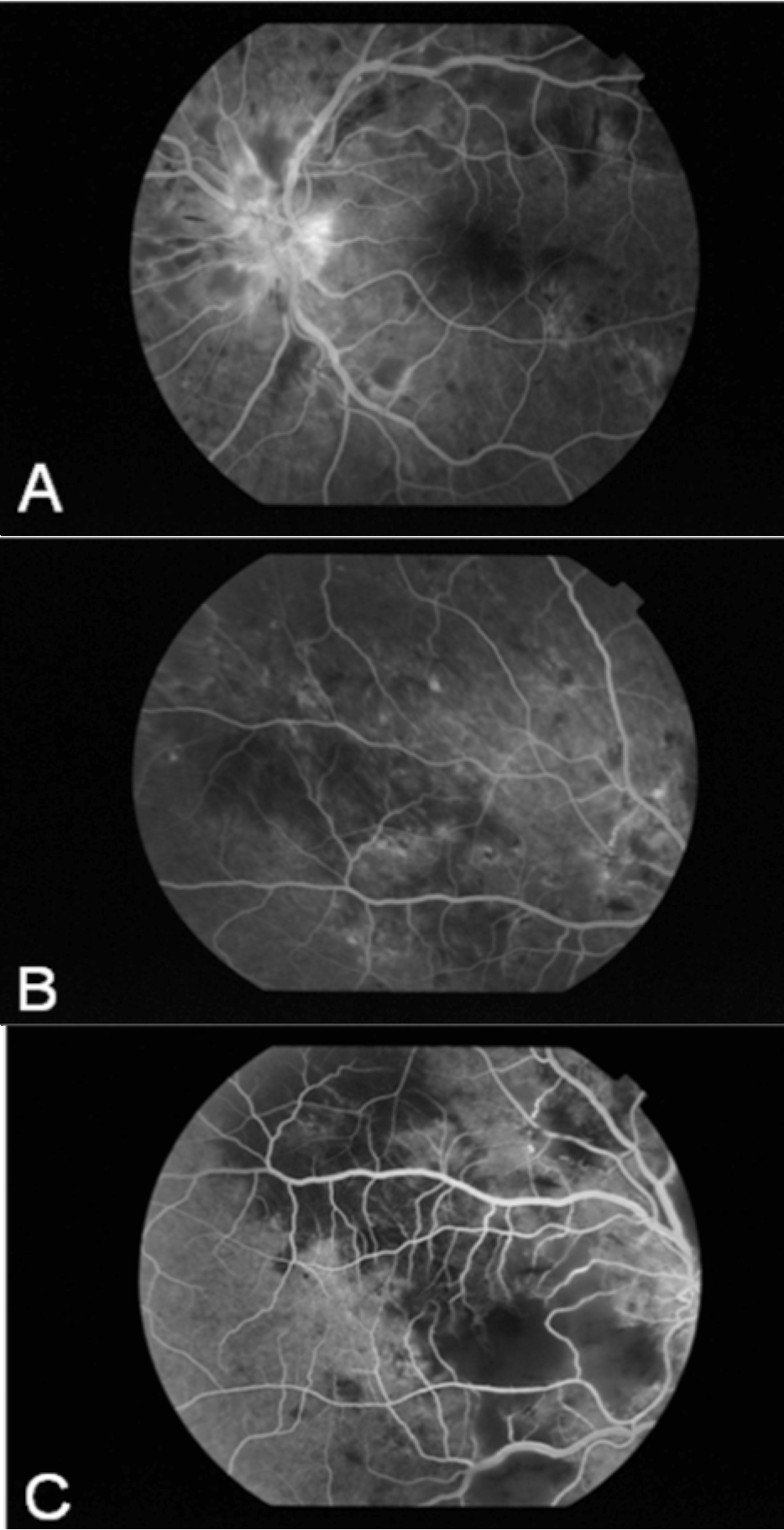
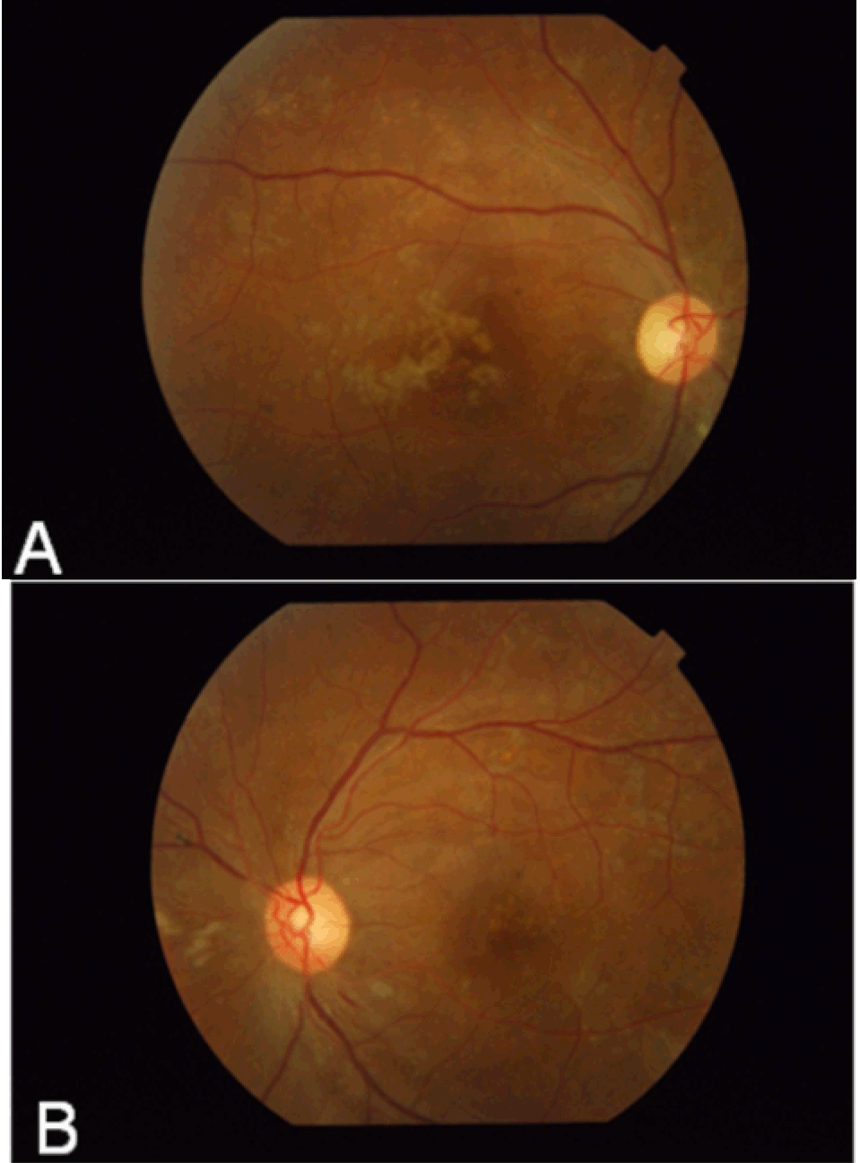
A 27-yr-old male attended the retina clinic with complaints of sudden loss of vision in both eyes for four days. Loss of vision was associated with severe occipital headache, vomiting and irritability. His visual acuity in right eye was perception of hand movement and in left eye was counting fingers at one meter distance. Anterior segment examination was normal. Intraocular pressure was within normal range. Funduscopy revealed multiple yellow-white patches lying deep in the retina at the level of pigment epithelium and choroid. There were numerous areas of localized serous retinal detachments which involved the macula and became confluent and bullous towards the periphery of the both fundi (figures 1 A, B). Blood pressure was noted to be 220/130 mm Hg. The patient was referred to the department of medicine for the cause of hypertension and further management. Antihypertensives such as ß-blockers (metoprolol: 75 mg, twice in a day), calcium channel blockers (nifedipine retard: 20 mg thrice in a day), lasix (frusemide: one ampule containing 40 mg twice in a day for five days) and mannitol (20%, 100 ml intravenously twice in a day for five days) were started to control the blood pressure in the intensive care unit. Fundus fluorescein angiography was performed on the sixth day of presentation after stabilization of blood pressure. In the earliest phase of the angiogram, multiple hypofluorescent areas of delayed choroidal filling were seen. Later, numerous foci of dye leakage appeared within these areas and fluorescein was seen to pool within the subretinal space in regions of retinal detachment (figure 2 A, B). Macular phase, showed irregularly enlarged foveal avascular zone suggesting ischemia (figure 2C). CT scan of brain showed edema involving both occipital lobes. Doppler excluded renal and carotid vessels abnormalities. Ultrasound of abdomen showed bilateral hydroureteronephrosis with high residual urine (>200 ml). Cystoscopy revealed a high bladder neck with bladder neck stenosis. So, a diagnosis of bladder outlet obstruction with obstructive uropathy was made. Patient underwent endoscopic bladder neck incision to relieve bladder outlet obstruction. Control of hypertension resulted in gradual absorption of subretinal fluid and subsequent retinal reattachment over the next four months. There was considerable improvement of visual acuity, 6/18 in right eye and 6/9 in left eye. Funduscopy revealed small greyish scars with central pigmentation, representing healed infarcts of retinal pigment epithelium (figure 3 A, B). | ||||||
| ||||||
|
| ||||||
| ||||||
|
Discussion
| ||||||
|
Hypertensive choroidopathy is seen in patients suffering from acute hypertension. Typically it occurs in relatively young individuals whose vessels are pliable and respond by constriction when the sympathetic vascular tone is increased. It was observed by Kishi S et al. [2] in their experimental study on monkeys that the acute ischemic phase characterized by constriction of choroidal arterioles lead to focal necrosis of choriocapillaries and retinal pigment epithelium. In the chronic occlusive phase, occlusive changes extend to the level of arteries and in the chronic reparative phase recanalization takes place. The early ocular manifestation includes disturbances of the retinal pigment epithelium and choroid with accompanying retinal vascular manifestations (hemorrhages and cotton wool exudates). In the early phase of choroidopathy, the fundus exhibits pale white or yellow patches (acute Elschnig's spots) corresponding to area of hyperperfusion of underlying choriocapillaries resulting from fibrinoid necrosis. [3] [4] There may be a subsequent breakdown of outer blood retinal barrier and focal posterior pole serous detachment. However, there are a few isolated case reports in literature where exudative retinal detachment is a presenting feature. Malhotra et al. [5] described similar presenting features in a young female with bilateral renal artery stenosis. Pierro L et al., [6] and de Venecia G et al. [7] reported a case of exudative retinal detachment in previously diagnosed case of renovascular hypertension. Chronic obstructive uropathy cause chronic interstitial nephropathy which leads to chronic kidney damage as kidneys are exposed to repeated infection and high intra renal hydrodynamic pressure. They may be silent in most cases except for frequency of urine and polyuria. It may produce severe hypertension which is renin dependent. [7] However, obstructive uropathy usually presents with flank pain, urinary tract infection, fever, difficulty while urination, nausea or vomiting. But in our case the patient presented with bilateral exudative retinal detachment which led to the diagnosis of obstructive uropathy with secondary hypertension within four days duration. In this scenario we are emphasizing here the role of the ophthalmologist in first detecting a case of secondary hypertension and quick management of the patient's complaints. It is also clearly demonstrated that ophthalmic manifestations do not need any specific treatment other than controlling blood pressure. Good comprehensive examination of the patient can lead to a systemic diagnosis and control of systemic parameters will help in improving associated ophthalmic features. | ||||||
|
Conclusion
| ||||||
|
In conclusion, our case indicates that accelerated hypertension may precipitate massive spontaneous bilateral exudative retinal detachment in patient with obstructive uropathy in young adults within four days. Obstructive uropathy in young adults should also be included in the list of systematic risk factors for spontaneous exudative retinal detachment of short duration. It is confirmed that bilateral exudative retinal detachment is a catastrophic event that results in devastating vision loss within very short span of time due to accelerated hypertension with obstructive uropathy. | ||||||
|
References
| ||||||
| ||||||


|
[HTML Abstract]
[PDF Full Text]
|
|
Author Contributions:
G Nageswar Rao - Conception and design, Acquisition of data, Analysis and interpretation of data, Drafting the article, Final approval of the version to be published Suresh Chandra Dash - Acquisition of data, Analysis and interpretation of data, Revising the article critically for important intellectual content, Final approval of the version to be published Gayatri Kanungo - Acquisition of data, Analysis of data, Revising the article critically for important intellectual content, Final approval of the version to be published Arttatrana Pal - Conception and design, Analysis and interpretation of data, Drafting the article, Critical revision of the article, Final approval of the version to be published |
|
Guarantor of submission:
The corresponding author is the guarantor of submission. |
|
Source of support:
None |
|
Conflict of interest:
Authors declare no conflict of interest. |
|
Copyright:
© G Nageswar Rao et al. 2011; This article is distributed under the terms of Creative Commons Attribution License which permits unrestricted use, distribution and reproduction in any means provided the original authors and original publisher are properly credited. (Please see www.ijcasereportsandimages.com /copyright-policy.php for more information.) |
|
|