| Table of Contents |  |
|
Case Report
|
| Non-operative management of pneumobilia following blunt abdominal trauma |
| Jorge Zequeira1, Pablo Rodriguez2 |
|
1Resident, Department of Surgery, University of Puerto Rico, San Juan, PR, USA.
2Section Chief, Section of Trauma & Critical Care, Department of Surgery, University of Puerto Rico, San Juan, PR, USA. |
|
doi:10.5348/ijcri-2011-12-72-CR-2
|
|
Address correspondence to: Jorge J. Zequeira Department of Surgery University of Puerto Rico School of Medicine PO Box 365067 San Juan, PR 00936-5067 USA Phone: 787 403-9370 Email: jorge.zequeira@upr.edu |
|
[HTML Abstract]
[PDF Full Text]
|
| How to cite this article: |
| Zequeira J, Rodriguez P. Non-operative management of pneumobilia following blunt abdominal trauma. International Journal of Case Reports and Images 2011;2(12):6-10. |
|
Abstract
|
|
Introduction:
Pneumobilia is an uncommon finding in imaging studies that may have a myriad of clinical connotations. Taking into consideration that only six cases of pneumobilia following blunt abdominal trauma have been reported, we will add an additional case and discuss the ones that have been documented in the world literature.
Case Report: We will report the case of an elderly male who sustained a motor-vehicle accident with diagnoses of a duodenal contusion and a femur fracture. These diagnoses were associated with the presence of pneumobilia on abdominal imaging. The patient was placed on bowel-rest, nasogastric suction, followed with serial physical exams, and discharged home on the ninth day after admission. Conclusion: Pneumobilia has been associated to diseases that mandate a surgical intervention. Six cases of pneumobilia after a blunt traumatic injury have been reported. Two of these patients were managed operatively while the other four were managed expectantly. None of the patients who were explored had findings suggestive of a surgically-correctable cause of pneumobilia. Those who were managed expectantly were discharged home without complications. The proposed mechanism for this event to occur is retrograde flow of air through the sphincter of Oddi as a consequence of sudden application of extrinsic pressure to the abdomen. Pneumobilia is a rare finding after blunt abdominal trauma that may be treated expectantly although more evidence is needed in order to generalize a management strategy. | |
|
Key Words:
Pneumobilia, Trauma, Blunt abdominal trauma
| |
|
Introduction
| ||||||
|
Pneumobilia is an uncommon finding in imaging studies that denotes the presence of air in the biliary tree. Pneumobilia has a myriad of clinical connotations depending on the setting in which it is found. Its significance may range from a simple self-limiting finding to a more serious, life-threatening disorder. Pneumobilia may occur as an expected finding after a biliary-enteric anastomosis, endoscopic biliary manipulation, as a sign of biliary tract infection, biliary-enteric fistula, emphysematous cholecystitis, high gastrointestinal obstruction, [1] ampullary tumors, or an incompetent sphincter of Oddi. [2] [3] [4] [5] We will report the case of a male patient with pneumobilia after a blunt abdominal injury that was managed with bowel rest, nasogastric suction and close surveillance with serial physical examinations. | ||||||
|
Case Report
| ||||||
|
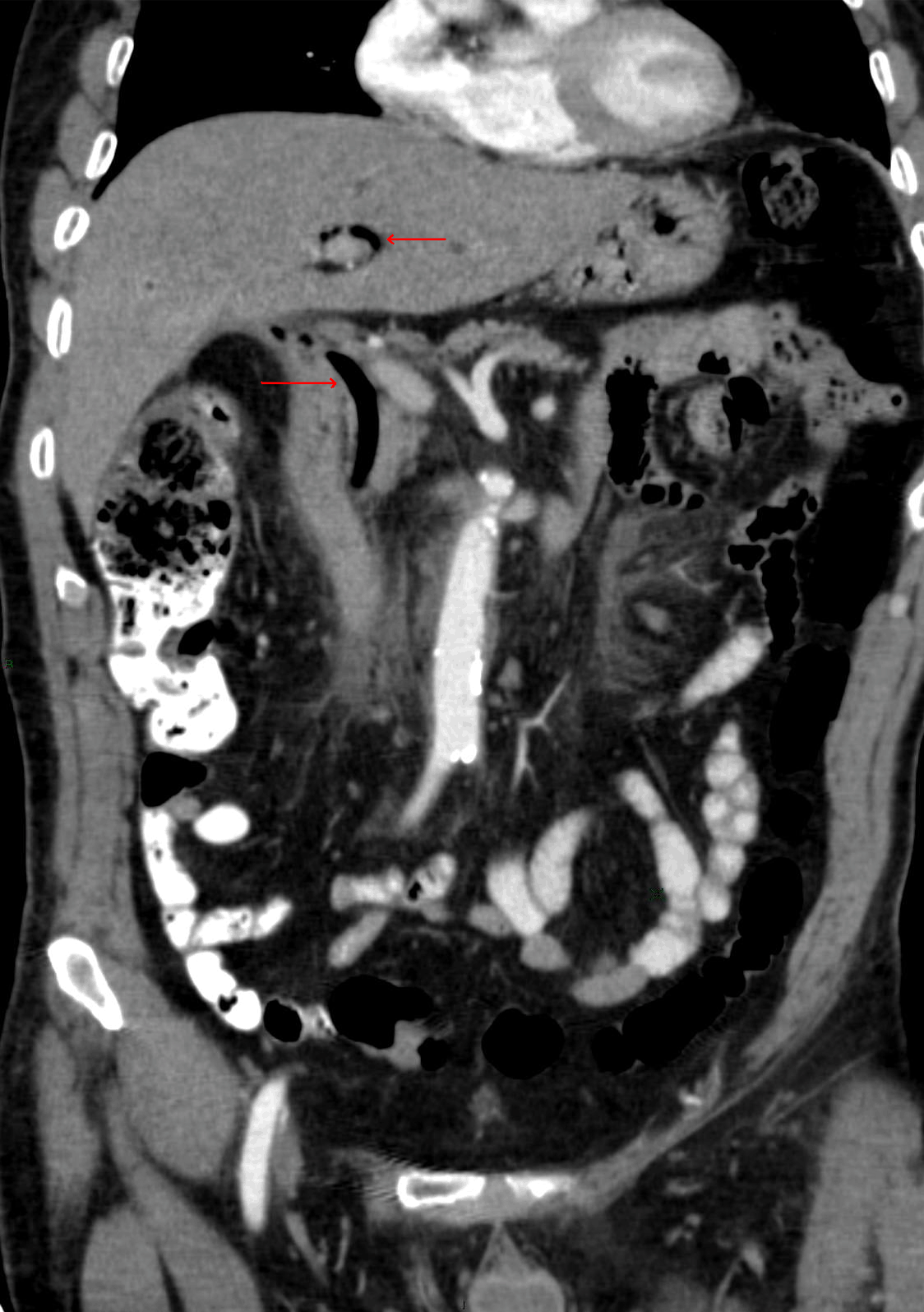
We evaluated a seventy-one year old male patient who sustained a motor vehicle accident as a restrained driver. He was taken to a rural hospital where a primary survey was performed along with its adjuncts. Once his airway and breathing were cleared, peripheral IV lines were inserted along with a foley catheter and subsequently transferred to our institution. The patient arrived at our center two hours after the accident took place. The patient was speaking coherently and grimacing in pain. He was normopneic, ventilating both lung fields, and all his distal pulses were present. He remained normocardic with a heart rate of 72 beats/min, blood pressure of 138/71 mmHg, and normothermic. The patient arrived with peripheral intravenous catheters and a foley catheter in place. Glasgow Coma Scale was reported as 14/15 (E3, V5, M6). On physical examination, pertinent findings included a frontal cephalohematoma, abdominal tenderness in upper quadrants, no rebound tenderness, no involuntary guarding, mild tenderness of lumbar spine, and an obvious deformity at the distal aspect of the left thigh without signs of compartment syndrome. Distal pulses in the extremity were preserved. Computed tomography images revealed a frontal cephalohematoma without evidence of intracranial bleeding or contusions, pneumobilia (figures 1, 2), a duodenal contusion of the 1st - 3rd portions of the duodenum (figure 1), minimal free fluid surrounding the hepatic flexure, and a left transverse process fracture of the 2nd and 3rd lumbar vertebrae. X-rays revealed a left femoral shaft fracture. The laboratory reported hemoglobin of 11.3 g/dL with no electrolyte, glucose, amylase, lipase, coagulation, urine, or bilirubin abnormalities. The patient was admitted, placed on bowel rest, intermittent nasogastric suction, and serial physical exams. An abdominal CT scan with oral and intravenous contrast was repeated the next day and revealed no new findings. Nasogastric suction was used for 48 hours until the patient resumed adequate peristaltic sounds, and abdominal tenderness disappeared. Once the nasogastric tube was removed, the patient was started on a clear liquids diet. On the 4th day post-injury, the patient received two units of packed red blood cells for optimization of hemoglobin of 8 g/dl and was taken to the operating room for an intramedullary nailing of his femoral shaft fracture. The shift in hemoglobin could be explained by a falsely increased value in a hypovolemic patient who has lost a significant amount of blood from his femur fracture. Once the patient was successfully resuscitated the real hemoglobin value of 8 g/dl shows up in the labwork. After surgery, the patient's diet was progressed uneventfully and he was discharged home during the ninth day of his hospital stay. The patient was seen at the trauma surgery outpatient clinics one, three, six, and twelve months after the injury took place. The patient stated that he has not had any episodes of abdominal pain, is tolerating a regular diet, and passing stools of adequate color and consistency. | ||||||
| ||||||
| ||||||
| ||||||
|
Discussion
| ||||||
|
There are different possible causes of pneumobilia. These may include both pathologic and non-pathologic etiologies. Simple causes such as the presence of a biliary-enteric anastomosis, recent biliary instrumentation, an incompetent sphincter of Oddi or trauma may explain the presence of this possibly asymptomatic sign. Other more ominous reasons might be cholangitis or a biliary-enteric fistula as seen with a gallstone ileus. It has been described that the highest pressure in a normal biliary tree may be as high as 30 cmH2O when the gallbladder contracts. This is comparable to the 35 cmH2O baseline pressure in the duodenal lumen. Since the sphincter of Oddi is capable of exerting a resistance of up to 60 cmH2O, the transfer of an extrinsic pressure greater than 60 cmH2O to the duodenal lumen may cause retrograde air flow into the biliary tree. [2] After a thorough review of the existing literature, six cases of pneumobilia in the setting of blunt abdominal injury have been reported (table 1). Gering et al. reported the case of an eighty-nine year-old female who sustained several injuries after a motor vehicle accident. The patient suffered a right midshaft humerus fracture, a left tibial plateau fracture, multiple right-sided rib fractures with an associated hemothorax, mild intracranial bleeding, a duodenal contusion with massive pneumobilia, small pneumoperitoneum, and a small amount of intraperitoneal fluid. In view of the pneumobilia, duodenal thickening, acidosis, scant pneumoperitoneum and episodes of transient hypotension, the patient's abdomen was explored. There were no findings suggestive of hepato-biliary, gastric, enteric, pancreatic, or retroperitoneal injuries. The patient ultimately died due to the development of respiratory failure that subsequently led to ARDS and Multiple Organ Dysfunction Syndrome. [6] Barnes et al. reported three additional cases of traumatic pneumobilia; each managed using a different strategy. The first of these patients was a 47-year-old male who suffered a high-speed motor vehicle accident. The patient sustained an open left olecranon fracture, a left hemo-pneumothorax with associated rib fractures, pneumobilia, and a minor splenic laceration with minimal hemoperitoneum. In view of the pneumobilia, the patient's abdomen was explored. The only finding at laparotomy was the splenic injury. A splenoraphy was performed and the patient was discharged home without further abdominal complications. [7] The second of these patients was a thirty-four year-old male who was also involved in a motor vehicle collision. This patient suffered a left corneal abrasion, a facial laceration, mild closed head injury, a lumbar vertebral (L1) burst fracture, and no abdominal pain. On the second day of his hospital stay the patient developed abdominal distention. An abdominal-pelvic CT scan was performed and revealed a minor hepatic laceration, minimal intraperitoneal fluid, pneumobilia, and a pattern of intestinal dilation suggestive of ileus. An upper gastrointestinal endoscopy was performed and no injury was found. The patient was discharged home on the fourth day of his hospital stay after bowel function returned to normal. [7] The third case presented in the Barnes et al. series involved a 45-year-old female who was involved in a motor vehicle collision. The patient sustained a subarachnoid hemorrhage, thoracic aortic rupture, multiple bilateral rib fractures, a right pneumothorax, sacral fracture, left superior and inferior pubic rami fractures, a left acetabular fracture and pneumobilia. After emergent aortic repair, the patient had a prolonged stay at the ICU, was then transferred to a rehabilitating facility and subsequently discharged home. [7] Thompson et al. reported the case of a 19-year-old female who was involved in a motor vehicle accident. She sustained a depressed parietal skull fracture with an associated epidural hematoma, multiple bilateral rib fractures, a left pneumothorax, a right hemothorax, pneumobilia, a sacral fracture, and bilateral pubic rami fractures. The surgeons evacuated the epidural hematoma and placed bilateral closed thoracostomy drainage tubes. Her pneumobilia was managed expectantly without further complications. [8] Huang et al. reported the case of a 65-year-old female who sustained blunt abdominal trauma as a pedestrian. She was diagnosed with pneumobilia using bedside sonography and confirmed wih a CT scan and MRCP. She was observed for 24 hrs in an ICU setting and discharged home after three days of careful monitoring. [9] Pneumobilia has been historically associated to diseases that mandate a surgical intervention. None of the six cases reported in the literature have had pneumobilia with an etiology that could have mandated surgery. The authors that reported these cases attribute the pneumobilia to retrograde passage of air through the sphincter of Oddi into the biliary tract. This thought was originally entertained when Bautista el al. reported the case of a patient with a proximal small bowel obstruction and incidental pneumobilia. When a nasogastric tube was placed, 5 L of pressurized fluid was drained. This finding suggested that the high pressure being imparted by the fluid inside the upper gastrointestinal tract caused retrograde flow of air through the Sphincter of Oddi into the biliary system. [1] Ladurner et al. reported another case that supports the concept of pneumobilia induced by a sudden increase in intrabdominal pressure. They reported the case of a patient who developed pneumobilia after cardiopulmonary resuscitation. They attributed this finding to the transmission of increased intrathoracic pressure to the abdominal cavity. [10] There have been other non-trauma related cases in which the incidental pneumobilia was attributed to sphincter incompetence. [2] [3] [4] [5] | ||||||
|
Conclusion
| ||||||
|
With this report and review we add another case of pneumobilia after blunt abdominal trauma to the literature. We suggest that the etiology for incidental pneumobilia in these patients is retrograde flow of air from the duodenum into the biliary system as a consequence of sudden application of an extrinsic pressure greater than 60 cm H2O to the abdomen. Taking into consideration the lack of surgical findings in those patients whose abdomen has been explored we preliminarily propose that patients with radiographic evidence of pneumobilia and no other clinical or radiographic indications for laparotomy should be managed expectantly with bowel rest, nasogastric suction, and serial physical examinations. However, we still believe that pneumobilia is a rare finding after blunt abdominal trauma and more evidence is needed in order to generalize a management strategy for these patients. | ||||||
|
References
| ||||||
| ||||||


|
[HTML Abstract]
[PDF Full Text]
|
|
Author Contributions:
Jorge Zequeira - Substantial contributions to conception and design, Acquisition of data, Analysis and interpretation of data, Drafting the article, Revising it critically for important intellectual content, Final approval of the version to be published Pablo Rodriguez - Substantial contributions to conception and design, Acquisition of data, Analysis and interpretation of data, Drafting the article, Revising it critically for important intellectual content, Final approval of the version to be published |
|
Guarantor of submission:
The corresponding author is the guarantor of submission. |
|
Source of support:
None |
|
Conflict of interest:
Authors declare no conflict of interest. |
|
Copyright:
© Jorge Zequeira et al. 2011; This article is distributed under the terms of Creative Commons Attribution License which permits unrestricted use, distribution and reproduction in any means provided the original authors and original publisher are properly credited. (Please see www.ijcasereportsandimages.com /copyright-policy.php for more information.) |
|
|