| Table of Contents |  |
|
Case Report
|
| Spontaneous resolution of a trauma induced direct carotid cavernous fistula |
| Zain A Sobani1, Arshad Ali2 |
|
1Section of Neurosurgery, Dept. of Surgery, Aga Khan University, Stadium Road, Karachi, Pakistan; Affiliation where work was carried out: Elective Student, Department of Neurosurgery, Jinnah Post Graduate Medical Centre, Karachi, Pakistan.
2Pakar Neurosurgeri, Neurosurgeri Klinik, Hospital Sultanah Aminah, Jalan Abu Bakar, Johor Bahru-80100, Johor-Malaysia; Affiliation where work was carried out: Assistant Professor, Department of Neurosurgery, Jinnah Post Graduate Medical Centre, Karachi, Pakistan. |
|
doi:10.5348/ijcri-2011-11-67-CR-5
|
|
Address correspondence to: Arshad Ali Pakar Neurosurgeri, Neurosurgeri Klinik, Hospital Sultanah Aminah Jalan Abu Bakar Johor Bahru-80100 Malaysia. Email: doc4brains@yahoo.com |
|
[HTML Abstract]
[PDF Full Text]
|
| How to cite this article: |
| Sobani ZA, Ali A. Spontaneous resolution of a trauma induced direct carotid cavernous fistula. International Journal of Case Reports and Images 2011;2(11):18-20. |
|
Abstract
|
|
Introduction:
Direct carotid cavernous fistulas (CCF) are abnormal communications between the internal carotid artery and the cavernous sinus usually resulting from high energy trauma to the face and base of skull. The management of direct CCF relies on endovascular procedures repairing the malformation as healing cannot take place in the high flow system.
Case Report: A 16-year-old male presented to our clinic with complaints of a progressively bulging left eye. He had suffered a severe head injury two months back resulting in an extradural hematoma in the left temporoparietal region which was surgically evacuated. Cerebral imaging revealed a fistula between the internal carotid artery (ICA) and cavernous sinus. While awaiting definitive treatment, the patient was advised to compress his left carotid artery manually about 15-20 times a day as per tolerance. After two weeks the patient was prepared for a definitive endovascular procedure; however the CCF had spontaneously thrombosed. Conclusion: Although rare, spontaneous resolution of direct CCF may occur and in such cases there may be a role of conservative management by decreasing the flow through the fistula by carotid massage and temporary occlusion. | |
|
Key Words:
Carotid cavernous fistulas; Conservative management.
| |
|
Introduction
| ||||||
|
Direct carotid cavernous fistulas (CCF) are abnormal communications between the arterial and venous systems of the internal carotid artery and the cavernous sinus respectively usually resulting from high energy trauma to the face and base of skull. Direct CCF results in a transmission of the high pressure carotid blood to the low pressure system of the cavernous sinus; leading to a mass effect on the contents of the cavernous sinus namely the III, IV and VI cranial nerves and an increase in pressure of the ophthalmic veins draining into the cavernous sinus. The systemic manifestations are mainly ocular; however fatal epistaxis has also been reported. [1] The management of direct CCF relies on endovascular procedures repairing the malformation, as healing cannot take place in the high flow system. Here we would like to report a case of a 16-year-old male with angiography proven direct CCF who was managed conservatively with carotid massage while awaiting endovascular repair; however at the time of repair the CCF was found to have spontaneously healed with minimal tissue reaction at the previously recorded site. | ||||||
|
Case Report
| ||||||
|
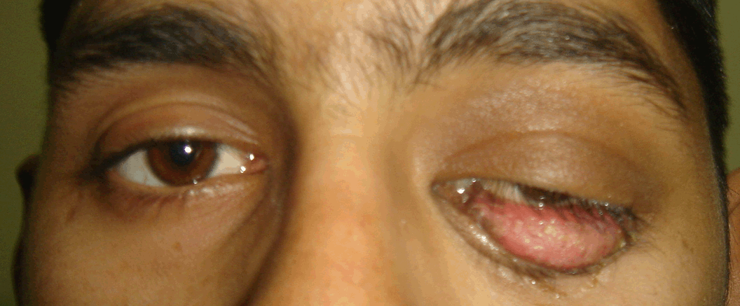
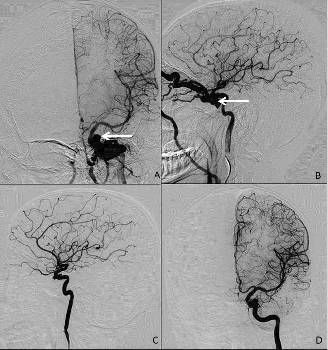
A 16-year-old male presented to our clinic with complaints of a progressively bulging left eye associated with mild pain. It was not associated with any visual disturbances. He gave a history of severe head injury two months ago and as per record, he suffered from a extradural hematoma in the left temporoparietal region. The hematoma was surgically evacuated and he recovered without any neurological deficits. On examination there was severe conjunctival chemosis of left eye with inability to completely close the eye due to significant proptosis (figure 1). Ophthalmological examination revealed a normal sized left pupil with impaired direct light reflex. His visual acuity was normal, but eye movements were restricted along all planes in the left eye. A bruit was also noted on the left eye during palpation which was confirmed by auscultation. The right eye and rest of clinical examination was within normal limits. Contrast enhanced computed tomography (CT) scan of the brain with contrast revealed a prominent superior ophthalmic vein on the left side. Contrast enhancement and prominence were also observed in the left paracavernous area (figure 2). Based on clinical features and CT findings a provisional diagnosis of left carotid-cavernous fistula was considered. Selective digital subtraction angiography of left internal carotid artery (ICA) confirmed a direct fistulous opening of medial wall of ICA into the cavernous sinus. The fistula was draining through the left superior ophthalmic vein into the angular and jugular veins. Dilatation and increased tortuosity of the left superior ophthalmic was also noted with no retrograde venous drainage into the cerebral and petrosal venous systems (figure 3). The right ICA and vertebro-basilar circulation showed normal vasculature. While awaiting definitive treatment, the patient was advised to sit on armless chair and compress his left carotid artery with his hand about 15-20 times a day with duration of compression as per tolerance. After two weeks, the patient was prepared for a definitive endovascular procedure; however during the procedure the pre-embolization injection of left ICA showed that carotid-cavernous fistula had spontaneously thrombosed with subtle contrast spillage at the site of previous fistula (figure 3). The procedure was abandoned after completing the protocol of diagnostic angiography, which was also found to be normal. In retrospect, the patient had shown some signs of improvement before the procedure. There was a decrease in proptosis and improved extra-ocular movements; but the conjunctival chemosis was still prominent. At six week follow-up, his proptosis has regressed and there was a slight residual weakness of the medial rectus (figure 4). | ||||||
| ||||||
| ||||||
|
| ||||||
| ||||||
|
Discussion
| ||||||
|
Direct CCF usually results from trauma to the base of skull and face resulting in high-flow shunts between the arterial and venous systems in the cavernous sinus. Due to the high flow rate through the fistula, healing is not possible and currently mainstay for management is endovascular repair. Various modalities including embolization by detachable balloon or platinum micro-coils and covered-stents have been shown to be safe, effective and reliable. [2] The different modalities may have advantages over each other, however few cases of spontaneous repair or successful conservative management have been reported in literature. Since the first reported case in 1972 by Grunert et al. nine cases or series of cases, have been documented in literature to the best of our knowledge. The mechanism of the event is not well understood however keeping previously described cases in mind we find the healing usually takes place after a diagnostic angiography, Considering the common denominator in these cases we may hypothesize that an event in the diagnostic angiogram may incite the healing process. Whether it is the catheter or contrast medium that irritates the artery inducing thrombosis and healing cannot be determined, but Nishijima et al. in their review of six cases also concluded that the angiography procedure may have a role in the resolution of CCF. [3] A decrease in blood flow through the fistula due to anesthesia has also been considered as a possible mechanism leading to the resolution by allowing the natural healing process to take place. [3] Whang et al also considered that small CCFs may be susceptible to carotid artery compression. [4] Keeping this in mind we decided to conservatively manage our patient by prescribing carotid massage while he was awaiting endovascular management. Even if this may not lead to healing the decreased flow through the fistula will prevent development of further complications and decrease the pressure building in the cavernous sinus. | ||||||
|
Conclusion
| ||||||
|
We conclude that although rare spontaneous recovery of direct traumatic CCF may occur and that there may be a role of conservative management by decreasing the flow through the fistula by carotid massage and temporary occlusion. | ||||||
|
References
| ||||||
| ||||||


|
[HTML Abstract]
[PDF Full Text]
|
|
Author Contributions:
Zain A Sobani - Substantial contributions to conception and design, Acquisition of data, Analysis and interpretation of data, Drafting the article, Revising it critically for important intellectual content, Final approval of the version to be published Arshad Ali - Substantial contributions to conception and design, Acquisition of data, Analysis and interpretation of data, Drafting the article, Revising it critically for important intellectual content, Final approval of the version to be published |
|
Guarantor of submission:
The corresponding author is the guarantor of submission. |
|
Source of support:
None |
|
Conflict of interest:
Authors declare no conflict of interest. |
|
Copyright:
© Arshad Ali et. al. 2011; This article is distributed the terms of Creative Commons Attribution License which permits unrestricted use, distribution and reproduction in any means provided the original authors and original publisher are properly credited. (Please see Copyright Policy for more information.) |
|
|