| Table of Contents |  |
|
Case Report
|
| Management of impacted common bile duct stones in late stage pregnancy and immediately post-partum after failed endoscopic stone extraction |
| Omar K Danner1, Omosalewa Akinyemi2, Kenneth L Wilson1, Moses Owoso3, L Ray Matthews1 |
|
1Assistant Professor of Surgery, Morehouse School of Medicine, 720 Westview Dr. S.W. Atlanta, GA 30310, USA.
2PGY-II, Morehouse School of Medicine, 720 Westview Dr. S.W. Atlanta, GA 30310, USA. 3Assistant Professor of Surgery, Meharry School of Medicine, 720 Westview Dr. S.W. Atlanta, GA 30310, USA |
|
doi:10.5348/ijcri-2011-11-66-CR-4
|
|
Address correspondence to: L. Ray Matthews Morehouse School of Medicine Department of Surgery 720 Westview Dr. S.W. Atlanta GA 30310 USA. Phone: (404)616-1415 Fax: (404)616-1417 Email: odanner@msm.edu |
|
[HTML Abstract]
[PDF Full Text]
|
| How to cite this article: |
| Danner OK, Akinyemi O, Wilson KL, Owoso M, Matthews LR. Management of impacted common bile duct stones in late stage pregnancy and immediately post-partum after failed endoscopic stone extraction. International Journal of Case Reports and Images 2011;2(11):14-17. |
|
Abstract
|
|
Introduction:
Symptomatic gallstone disease is the second most common abdominal emergency in pregnant women after acute appendicitis. Extrahepatic biliary obstruction by gallstones during pregnancy remains one of the most challenging management problems in the field of surgery. Although there have been many advancements in the surgical and endoscopic management of gallstone disease, the risks of these interventions and anesthesia to the developing fetus still prevents their routine application during the gestational period. Traditionally, these types of disorders during pregnancy are managed conservatively or via appropriately timed cholecystectomy. However, when the late-term pregnant patient presents acutely with evidence of biliary tract obstruction and sepsis, it represents a medical or surgical emergency.
Case Report: We present the case management of a 25-year-old late term pregnant female with an impacted common bile duct stone and impending biliary sepsis as a result of failed postpartum endoscopic stone extraction. Conclusion: Due to the risk of potential harm to the fetus, this patient subset must be managed in clinically sound and technically proficient manner, which does not allow time for conservative management. Consequently, appropriate management algorithms for conservative therapy versus endoscopic or surgical interventions need to be clearly outlined and defined. | |
|
Key Words:
Management, Post-Partum, Stones
| |
|
Introduction
| ||||||
|
Symptomatic gallstone disease is the second most common abdominal emergency in pregnant women after acute appendicitis. The physiologic changes in the biliary system in the presence of obstructing gallstones during the later stages of pregnancy increase the risk of septic complications, such as, cholecystitis, cholangitis and gallstone pancreatitis. If these disease processes are left untreated, they can lead to overwhelming sepsis and potentially death. The management of the late term pregnant female with gallstone disease or evidence of biliary tract obstruction as a consequence of an impacting common bile duct stone can be a clinically challenging and stressful situation. When conservative or endoscopic therapy fails, surgical treatment options should be evaluated and applied. Extrahepatic biliary obstruction by gallstones during pregnancy remains one of the most challenging management problems in the field of surgery. [1] Although there have been many advancements in the surgical and endoscopic management of gallstone disease, the risks to the developing fetus still prevents their routine application during the gestational period. [2] Traditionally, these types of disorders during pregnancy have been managed conservatively or via cholecystectomy, depending on the nature and severity of the biliary tract disease. [3] However, in one review of six studies comparing conservative therapy with surgical management of cholecystitis during pregnancy, no significant difference in incidence of preterm delivery was shown (3.5% versus 6.0%, p= 0.33), or fetal mortality (2.2% versus 1.2%, p = 0.57). [4] In another report of 12 studies evaluating the management of gallstone pancreatitis, fetal mortality was 8.0% in conservatively treated patients versus 2.6% in the surgical treatment group (p = 0.28). This supports the need for earlier, prudent surgical intervention in order to prevent potentially catastrophic complications. Therefore, appropriate management algorithms for conservative therapy versus endoscopic or surgical interventions need to be clearly elucidated. | ||||||
|
Case Report
| ||||||
|
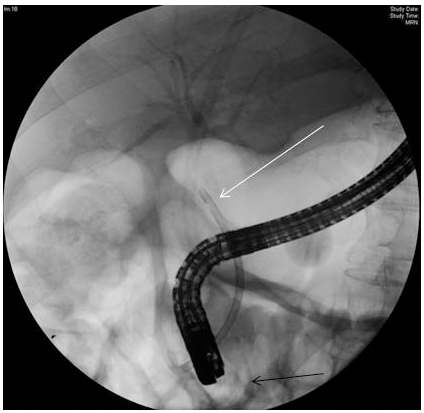
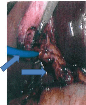
A 25-year-old African American female G5, P4-0-0-4 at 35 weeks gestation presented with a two day history of constant, sharp epigastric and right upper quadrant abdominal pain rated 7/10. She had associated loss of appetite, nausea and vomiting of bilious, non bloody fluid. Patient was somnolent on arrival to the emergency department. She admitted to cocaine use prior to admission. Patient denied contractions or vaginal bleeding. On examination, patient was afebrile with elevated blood pressures with systolics in the 140s and diastolics in the 90s. She was found to have a soft, gravid abdomen, with right upper quadrant tenderness, no palpable contractions, and normal bowel sounds. Her WBC count was 5.2x10³/mm3 hematocrit was 32%, and liver enzymes were noted to be elevated with ALP of 403IU/L, Total bilirubin was 2.4 mg/dl and direct bilirubin of 1.8 mg/dl. A right upper quadrant abdominal ultrasound demonstrated cholelithiasis with gallbladder wall thickening at 5 mm, dilated intra and extrahepatic biliary ducts. The dilated common bile duct measured up to 1.5 cm with a non-shadowing echogenic focus within the distal common duct in the region of the pancreatic head, consistent with a large, impacted gallstone. Patient was admitted to the obstetrics service for further evaluation and PIH labs were ordered to rule out preeclampsia. Results were within normal limits. Two days after presentation, the patient had a spontaneous preterm rupture of membranes at 35-1/7 weeks and was transferred to labor and delivery, where she had a spontaneous vaginal delivery of a healthy, female infant. Postpartum, the patient continued to have right upper quadrant abdominal pain and thus, an ERCP with a sphinterotomy was performed, which revealed impacted, large common bile duct stones that were not able to be removed. Although the patient delivered prior to biliary decompression via gallstone extraction and biliary stent placement, this intervention is often utilized as a temporizing maneuver until definite therapy can be instituted. Consequently, a biliary stent was placed for decompression to prevent cholangitis. The patient was taken to the operating room where she underwent laparoscopic cholecystectomy with intraoperative cholagiogram and laparoscopic common bile duct exploration and stone extraction. Post operatively, the patient recovered uneventfully with her liver enzymes trending downwards and relief of her right upper quadrant abdominal pain. Patient was discharged on post operative day two in good condition. | ||||||
| ||||||
|
| ||||||
| ||||||
|
Discussion
| ||||||
|
Over the past two decades, endoscopic retrograde cholangiopancreatography and sphincterotomy have been performed safely in pregnancy with minimal radiation exposure. [1] Now with advances in fiber-optic technology over the past 10 to 20 years, modern endoscopic techniques have allowed endoscopy to be used as a safe, effective and definitive treatment alternative for pancreatobiliary disease in pregnancy. [3] Typically, endoscopic management should be considered in women presenting with acute cholangitis, impacted common bile duct stones, and/or gallstone pancreatitis during pregnancy. Although the first line of management of bile duct stones usually includes endoscopic retrograde cholangiopancreatography (ERCP) with or without sphincteromy and biliary stent placement, complications such as, pancreatitis, bleeding, and visceral or ductal perforation are serious potential concerns. Prior to cholecystectomy, ERCP is indicated in the presence of cholangitis, persistent jaundice, and choledocholithiasis on ultrasound, or the combination of dilated ducts and abnormal liver function tests (LFT's). [5] [6] When endoscopic stone removal fails, especially during pregnancy, biliary decompression still has to be and usually can be achieved via endoscopic biliary stent placement by skilled gastroenterogist. Satisfactory drainage was achieved through the biliary stent. When available, balloon lithotripsy and debulking of the common bile duct stone is another option in the treatment armamentarium. However, this modality of treatment may not be readily available at all medical facilities or institution, which limits its widespread and/or universal application. Although it has been shown to be an effective treatment option, lithotripsy was unavailable at the institution where this patient received her care. [7] [8] [9] Thus, other minimally invasive surgical treatment options for CBD stone extraction such as laparoscopic stone removal using a fogarty cathether or dormia basket technique should be considered. In this particular case, we were successfully able to remove the impacted stone and clear the CBD utilizing a combination of balloon extraction and a forward flushing technique via the cholangiocatheter via the cystic duct stump. After the stone was removed the post-procedure cholangiogram revealed adequate clearing of the biliary tract in our patient. Thus, there was no need for the intra-operative placement of a T-Tube, as the endoscopically placed internal biliary stent remained in the CBD. These procedures are followed by laparoscopic cholecystectomy during the same operative setting, which is usually performed in the immediate post-partum period after successful delivery. [10] Even though open cholecystectomy with common bile duct exploration is a viable option, additional consideration should be given to the possibility of compromising the mother's ability to care for her newborn infant during the postoperative convalescence period. Therefore, we recommend gallbladder removal and stone extraction utilizing minimally invasive techniques whenever possible to minimize postoperative morbidity after delivery and to expedite recovery. [11] In our case study, we present the management of a 25-year-old, late term pregnant female with cholecystitis, choledocholithiasis and dilatation of her biliary anatomy as a result of an impacted common bile duct stone. After successful delivery of a healthy baby, a postpartum endoscopic stone extraction with sphincterotomy failed to dislodge the impacted stone. A guide-wire was able to be maneuvered past the obstructing stone and an internal biliary stent was placed and used as a bridge to prevent biliary sepsis until definitive surgical intervention could be performed. Even when endoscopic stone extraction is unsuccessful, these patients can be temporized before definitive therapy such as laparoscopic common bile duct exploration, intraoperative cholangiography and laparoscopic gallstone extraction can be performed, thus avoiding the need for a conventional open surgical procedure. The approach of bridging with an endoscopic biliary stent followed by a laparoscopic common bile duct management combined with cholecystectomy is a safe and effective alternative for pregnant patients with impacting common bile duct stones. However, this management strategy is best accomplished via a multidisciplinary team approach and requires close collaboration between the obstetrician, primary physician, gastroenterologist and surgeon. Surgeons should also be appropriately trained to utilize these advanced laparoscopic surgical techniques. | ||||||
|
Conclusion
| ||||||
|
Treatment of late stage pregnant patients with obstructive biliary tract disease remains a significant challenge in the modern medicine. Although open cholecystectomy and common bile duct exploration are a viable options, additional consideration should be given to minimally invasive techniques, such as, laparoscopic bile duct exploration with stone extraction to minimize postoperative morbidity of the peri- or postpartum patient. In addition, consideration should be given to the possibility of compromising the mother's ability to care for her newborn infant during her postoperative convalescence. Consequently, gallbladder removal and stone extraction utilizing laparoscopic and/or endoscopic techniques is recommended whenever possible, in order to minimize postoperative recovery and postpartum convalescence. | ||||||
|
References
| ||||||
| ||||||

|
[HTML Abstract]
[PDF Full Text]
|
|
Author Contributions:
Omar K Danner - Substantial contributions to conception and design, Acquisition of data, Analysis and interpretation of data, Drafting the article, Final approval of the version to be published Omosalewa Akinyemi - Acquisition of data, Drafting the article, Final approval of the version to be published Kenneth L Wilson - Substantial contributions to conception and design, Revising it critically for important intellectual content, Final approval of the version to be published Moses Owoso - Analysis and interpretation of data, Revising it critically for important intellectual content, Final approval of the version to be published L Ray Matthews - Analysis and interpretation of data, Revising it critically for important intellectual content, Final approval of the version to be published |
|
Guarantor of submission:
The corresponding author is the guarantor of submission. |
|
Source of support:
None |
|
Conflict of interest:
Authors declare no conflict of interest. |
|
Copyright:
© Danner et al. 2011; This article is distributed the terms of Creative Commons Attribution License which permits unrestricted use, distribution and reproduction in any means provided the original authors and original publisher are properly credited. (Please see Copyright Policy for more information.) |
|
|