
| Table of Contents |  |
|
Case Report
|
| Massive chondrosarcoma of sternum: A surgical challenge in resection and reconstruction of a rare tumor |
| R.S. Mohil1, Anurag Pandey2, Sujata Sarabahi3, V.K. Tiwari4 |
|
1Consultant & Associate Professor, Department of Surgery Vardhman Mahavir Medical College & Safdarjung Hospital, New Delhi, India.
2Senior Resident, Department of Burns, Plastic and Reconstructive Surgery Vardhman Mahavir Medical College & Safdarjung Hospital, New Delhi, India. 3Senior Plastic Surgeon, Department of Burns, Plastic and Reconstructive Surgery Vardhman Mahavir Medical College & Safdarjung Hospital, New Delhi, India. 4Professor & Consultant, Department of Burns, Plastic and Reconstructive Surgery Vardhman Mahavir Medical College & Safdarjung Hospital, New Delhi, India. |
|
doi:10.5348/ijcri-2011-07-42-CR-2
|
|
Address correspondence to: R S Mohil DII / 114 West Kidwai Nagar New Delhi 110023 India Phone: 9868952676,91-11-26882272 Email: rsmohil@yahoo.com, rsmohil@gmail.com |
|
[HTML Abstract]
[PDF Full Text]
|
| How to cite this article: |
| Mohil RS, Pandey A, Sarabahi S, Tiwari VK. Massive chondrosarcoma of sternum: A surgical challenge in resection and reconstruction of a rare tumor. International Journal of Case Reports and Images 2011;2(7):4-7. |
|
Abstract
|
|
Introduction:
Reconstruction of chest wall defects remains a very challenging issue and often requires prosthetic repair to prevent flail chest and paradoxical breathing.
Case Report: We report a case of chest wall reconstruction following resection of a long standing, massive, well differentiated, primary chondrosarcoma of the sternum which was involving the entire body of sternum and adjoining costal cartilages. Excision of entire sternum (except manubrium) and 3rd to 8th ribs left a large defect (20x15 cm) in the chest wall which was reconstructed with a simple and new three layer technique using Prolene-Vicryl mesh, long split rib grafts and lattissimus dorsi muscle (LD) flap. Patient was weaned off the ventilator on seventh post operative day and discharged after four weeks. Conclusion: This is a simple chest wall reconstruction technique for wide chest wall defects and is effective, safe, low cost and easily reproducible. | |
|
Key Words:
Sternal chondrosarcoma, Chest wall repair
| |
|
Introduction
| ||||||
|
Sternum is an unusual site for malignant bone tumors; chondrosarcoma being the commonest with an annual incidence of less than 0.5 per million per year. [1] The management of chondrosarcoma with respect to both radical excision and reconstruction of subsequent wide chest wall defect has always been a challenging problem because of anticipated postoperative flailing of chest and alteration in the pulmonary physiology. Optimal reconstructive procedure still remains controversial. Various types of materials and techniques have been used in the past but the primary aim remains to give a rigid thoracic cage support with soft tissue cover in order to give chest wall stability, good lung function as well as protection to the endothoracic organs. [2] [3] [4] We report a successful wide excision of a massive chondrosarcoma of sternum (50x40 cm) and reconstruction with Prolene-Vicryl mesh, autogenous split rib graft and a lattismus dorsi (LD) muscle flap. The simple technique used for reconstruction proved to be effective in preventing flail chest and paradoxical breathing, restoring chest wall stability, obliterating underlying dead space, providing coverage to intrathoracic organs and also restoring aesthetic appearance. | ||||||
|
Case Report
| ||||||
|
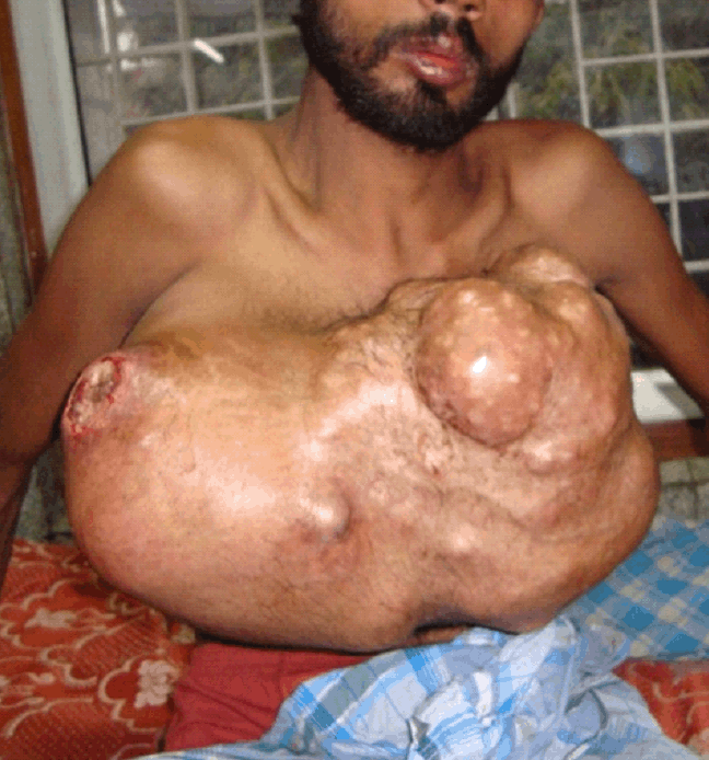
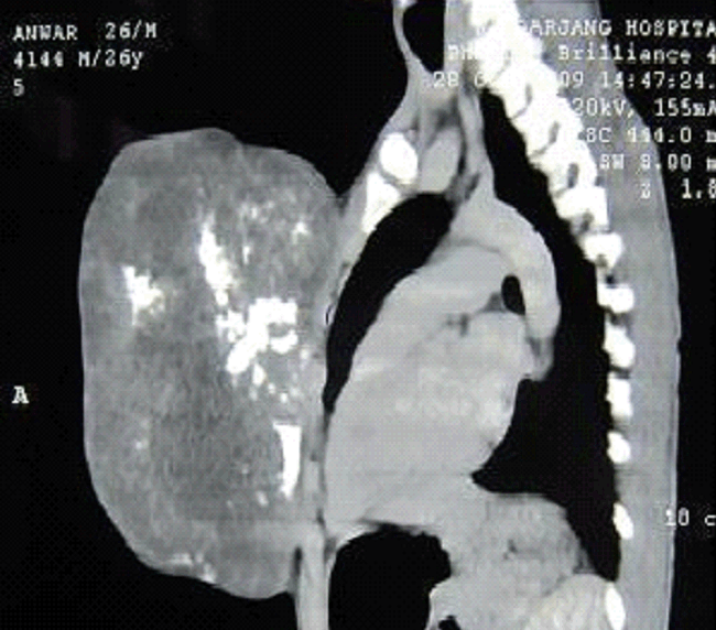
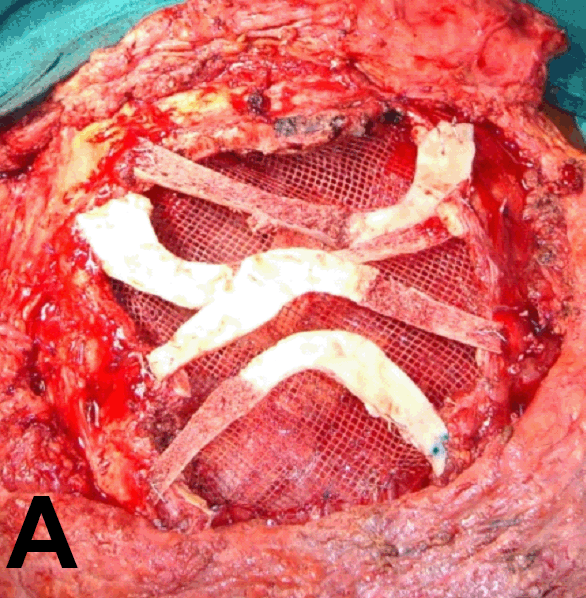
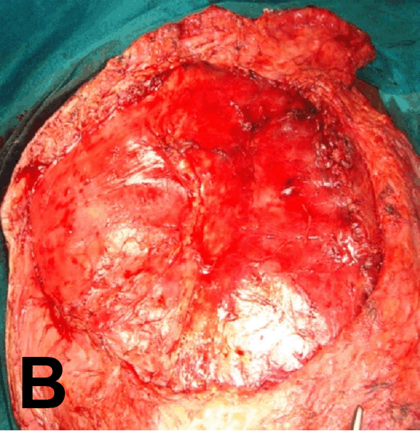
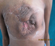
A 35-year-old male, was admitted to our hospital with a large 50x40 cm swelling over anterior chest wall for 20 years which in last six months rapidly increased in size, ulcerated and became painful (Figure 1). Computed tomography (CT) scan revealed a large tumor arising from the whole of sternum, sparing the manubrium with no intrathoracic extension, pleural or pericardial involvement and no evidence of lung or visceral metastasis (Figure 2). Patient was unable to perform any pulmonary function test due the size of the tumor. An incisional biopsy was confirmative of a well differentiated chondrosarcoma. Excision involved subtotal sternectomy (leaving only manubrium sterni with sternoclavicular joints), bilateral 3rd to 8th rib till 4 cm beyond the visible margin of tumor, partial pectoralis major muscles and part of right pleura which were adherent to the tumor. Both internal mammary arteries were also sacrificed. Large 20x15 cm defect over anterior chest wall was created after resection of the tumor which weighed eight kilograms. Reconstruction of the defect was done in layers - innermost being Prolene-Vicryl combination mesh. Over this four split rib grafts harvested from seventh rib each side and vertically split into two providing four rib grafts were placed with their medullary part facing ventrally. They were fixed with resected margins of ribs with steel wires. LD muscle flap was harvested on its thoracodorsal vascular pedicle and placed over the rib grafts and fixed to adjacent intercostal muscles thus sealing the defect (Figure 3 A, B). The expanded skin overlying the tumor was sufficient to cover the entire defect (Figure 4). Intercostal drains were placed bilaterally before commencing reconstruction. Postoperatively patient was kept on ventilatory support which was removed on seventh postoperative day. There was marginal skin flap necrosis which was excised and closed primarily. A seroma developed at LD muscle donor site requiring drainage. Histopathology showed a well differentiated chondrosarcoma with more than three cm clear resected margin. | ||||||
| ||||||
| ||||||
|
| ||||||
|
| ||||||
| ||||||
|
Discussion
| ||||||
|
Adequate excision of primary chondrosarcoma with clear margins is the cornerstone for disease eradication. [1] Full thickness chest wall resections leave a segment of the chest wall vulnerable to paradoxical movement during respiration. Reconstruction of the defect is important for providing chest wall stability, reducing the duration of ventilatory support, adequate pulmonary function, protection of intrathoracic organs from trauma and infection and improved cosmesis. [2] [3] [4] Though the importance of reconstruction has been well described, there are still controversies as to which lesions should be reconstructed. [2] [3] [4] [5] Defects which are smaller than 5 cm in any location and those up to 10 cm in size posteriorly need no reconstruction. Larger defects, most anterior defects or defects in proximity to the tip of scapula must be reconstructed. [2] [5] Different types of materials and techniques like polytetrafluroethylene (PTFE) patch, stainless steel mesh, resin plate or metal plate have been used to stabilize the chest wall depending upon site and size of defect, surgeon's preference and availability of the material, its adaptability and durability. Rigid reconstruction with methylmethacrylate spread between layers of Prolene mesh is usually used after total sternal resection as it restores chest wall stability and geometry of the thoracic cage. However, its use is associated with more wound complications, fracture, tilting or extrusion of implants. [3] [4] [5] [6] [7] On other hand non-rigid prosthesis using Prolene, Marlex or PTFE is indicated for small or subtotal sternectomy where a small part of the manubrium (with sternoclavicular joint) or lower part of sternum are conserved. [2] [3] [4] [5] [8] [9] There is no significant difference in outcomes or complications based on the type of material used. [6] In the present case body of sternum with a substantial part of anterior rib cage was removed creating a very large defect measuring 15x20 cm, exposing right lung, part of left pleura and pericardium. Since we were able to preserve manubrium and both the sternoclavicular joints it was decided to use a combination of Prolene-Vicryl mesh and rib grafts for thoracic cage replacement over the more expensive rigid prosthesis like titanium plates for its easy handling, availability and reported safety. Soft tissue cover was then provided by latissimus dorsi muscle flap to obliterate the dead space, cover the exposed mesh with overlying ribs and control infection. LD Flap was the only choice for cover as the defect was in middle and lower chest and both internal mammary arteries had been sacrificed which ruled out the use of pectoralis major and rectus abdominis muscles respectively. The length and bulk of the robust LD muscle provided extensive vascular cover over the mesh and rib grafts and additional stability to the chest wall. The ribs were split because the graft survival is better when their cancellous portion is kept approximated to the vascularised LD flap. Patient's respiratory functions improved gradually and he could be weaned off the ventilator by seventh postoperative day. Using incentive spirometry patient could achieve normal lung function before discharge by the end of four weeks. Patient is free of the disease at one year follow up and has not developed any major respiratory complications. | ||||||
|
Conclusion
| ||||||
|
Although a longer follow up is essential, the good, early, aesthetic and respiratory physiological result proves beyond doubt that this is a simple and effective chest wall reconstructive technique which is safe, low cost and easily reproducible for wide chest wall defects such as in this case. | ||||||
|
References
| ||||||
| ||||||

|
[HTML Abstract]
[PDF Full Text]
|
|
Author Contributions:
R. S. Mohil - Substantial contributions to conception and design, Drafting the article, Revising it critically for important intellectual content, Final approval of the version to be published Anurag Pandey - Substantial contributions to conception and design, Drafting the article, Revising it critically for important intellectual content, Final approval of the version to be published Sujata Sarabahi - Substantial contributions to conception and design, Drafting the article, Revising it critically for important intellectual content, Final approval of the version to be published V. K. Tiwari - Substantial contributions to conception and design, Drafting the article, Revising it critically for important intellectual content, Final approval of the version to be published |
|
Guarantor of submission:
The corresponding author is the guarantor of submission. |
|
Source of support:
None |
|
Conflict of interest:
Authors declare no conflict of interest. |
|
Copyright:
© R.S. Mohil et. al. 2011; This article is distributed the terms of Creative Commons Attribution License which permits unrestricted use, distribution and reproduction in any means provided the original authors and original publisher are properly credited. (Please see Copyright Policy for more information.) |
|
|