| Table of Contents |  |
|
CLINICAL IMAGE
|
| Large calcified patent ductus arteriosus |
| János Tomcsányi1, Miklós Somlói2, Béla Bózsik2, Zsófia Farbaky3 |
|
1Head of Cardiology Department, St. John of God Hospital, Budapest, Hungary.
2Assistant Professor of Cardiology Department, St. John of God Hospital, Budapest, Hungary. 3Head of Radiology, St. John of God Hospital, Budapest, Hungary |
|
doi:10.5348/ijcri-2011-06-40-CI-5
|
|
Address correspondence to: János Tomcsányi 1023 Árpád fejedelem u.7 Hungary Phone/Fax: 003614388560 Email: tomcsanyi.janos@t-online.hu |
|
[HTML Abstract]
[PDF Full Text]
|
| How to cite this article: |
| Tomcsányi J, Somlói M, Bózsik B, Farbaky Z. Large calcified patent ductus arteriosus. International Journal of Case Reports and Images 2011;2(6):20-22. |
|
Clinical Image
| ||||||
|
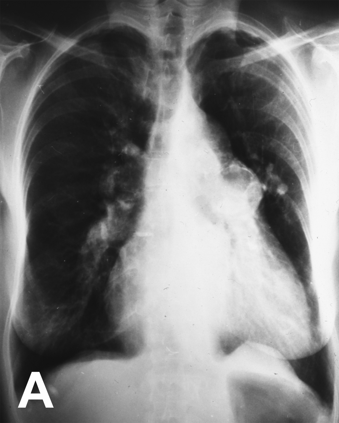
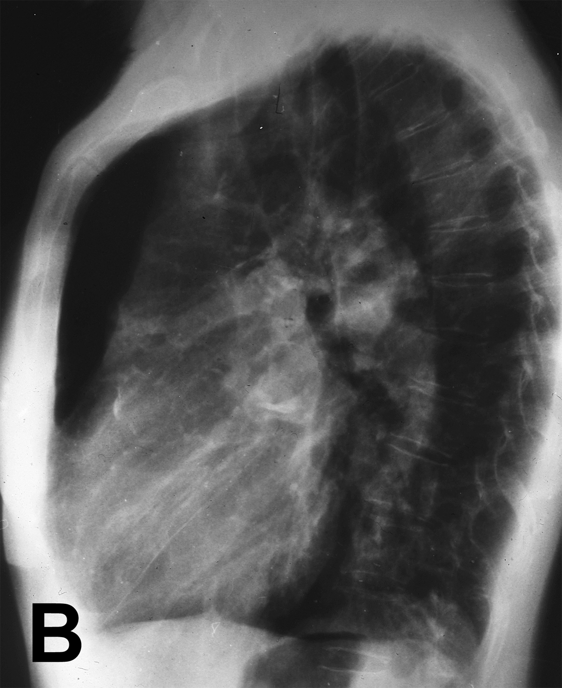
Aortopulmonary window is a rare defect caused by failure of fusion of the two opposing conotruncal ridges which are responsible for separating the truncus arteriosus into the aorta and pulmonary tract. A case of long survival with aortopulmonary window causing Eisenmenger syndrome is presented. A 65-year-old female patient was admitted with cardiorespiratory failure. She had had effort dyspnoea for 30 years but had refused investigations. Physical examination revealed generalised cyanosis, marked anasarca and a holosystolic murmur superimposed on an accentuated S2. Oxygen saturation was 95% but the arterial blood gas showed significant hypoxia (pO2 65 mmHg). Blood pressure was 150/80 mmHg. The ECG on admission showed pulmonary P-waves and signs of right ventricular hypertrophy. Echocardiography revealed a grossly dilated right ventricle with severe tricuspid regurgitation and mild pulmonary insufficiency. The estimated pulmonary pressure of 140/70 mmHg was nearly identical with the systemic blood pressure. The dimensions of the left atrium and left ventricle were within normal limits with good left ventricular systolic function. Chest X-ray showed significantly dilated pulmonary vessels and a round calcified mass in the left pulmonary hilum (Figures 1A, 1B). The patient died suddenly on the following day. Autopsy revealed a large (28 mm) communication between the pulmonary trunk and the aorta (Figure 2).
| ||||||
| ||||||
|
| ||||||
| ||||||
|
| ||||||
| ||||||
|
Discussion
| ||||||
|
The diagnosis of aortopulmonary window or patent ductus arteriosus in the presence of pulmonary hypertension can be challenging due to the fact that physical signs such as machinery heart murmur may be absent. [1] [2] Calcification of the duct is common in adults, which may provide a diagnostic clue. [3]
| ||||||
|
Conclusion
| ||||||
|
The presented case demonstrates that the development of Eisenmenger syndrome with equalised systemic and pulmonary vascular resistance permits a relatively long survival in some cases.
| ||||||
|
References
| ||||||
| ||||||
| [HTML Abstract] [PDF Full Text] |
|
Author Contributions:
János Tomcsányi - Conception and design, Acquisition of data, Analysis and interpretation of data, Drafting the article, Critical revision of the article, Final approval of the version to be published Miklós Somlói - Conception and design, Acquisition of data, Analysis and interpretation of data, Drafting the article, Critical revision of the article, Final approval of the version to be published Béla Bózsik - Conception and design, Acquisition of data, Analysis and interpretation of data, Drafting the article, Critical revision of the article, Final approval of the version to be published Zsófia Farbaky - Conception and design, Acquisition of data, Analysis and interpretation of data, Drafting the article, Critical revision of the article, Final approval of the version to be published |
|
Guarantor of submission:
The corresponding author is the guarantor of submission. |
|
Source of support:
None |
|
Conflict of interest:
The author(s) declare no conflict of interests |
|
Copyright:
© János Tomcsányi et al. 2010; This article is distributed under the terms of Creative Commons Attribution License which permits unrestricted use, distribution and reproduction in any means provided the original authors and original publisher are properly credited. (Please see copyright policy for more information.) |
|
|


