| Table of Contents |  |
|
CASE REPORT
|
| An unusual presentation of thyroglossal duct cyst associated with papillary thyroid carcinoma |
| A. Chuathong1, S. Ouawathanamongkol1, T. Maipang1, N. Chongchitnant2 |
|
1Surgical Oncology Unit, Bangkok Hospital Group, 2, Soi. Soonvijai 7, New-Petchburi Road, Bangkok, Thailand.
2Pathology Unit, Bangkok Hospital Group, 2, Soi. Soonvijai 7, New-Petchburi Road, Bangkok, Thailand. |
|
doi:10.5348/ijcri-2011-05-33-CR-3
|
|
Address correspondence to: Dr. Chuathong A Surgical Oncology Unit Bangkok Hospital Group 2, Soi. Soonvijai 7 New-Petchburi Road Bangkok Thailand Phone: +662-3103000 Fax: +662-3181546 Email: attawut9@msn.com |
|
[HTML Abstract]
[PDF Full Text]
|
| How to cite this article: |
| Chuathong A, Ouawathanamongkol S, Maipang T, Chongchitnant N. An unusual presentation of thyroglossal duct cyst associated with papillary thyroid carcinoma. International Journal of Case Reports and Images 2011;2(5):12-15. |
|
Abstract
|
|
Introduction:
Thyroglossal duct cyst is the most common form of congenital malformations in the neck. Thyroglossal duct cyst presents characteristically as a midline cervical mass at the level of thyrohyoid membrane. Approximately 25% are located within 2 cm. of midline. Carcinoma arising in thyroglossal duct cyst constitutes a very uncommon clinicopathological entity (about 1% of thyroglossal duct cyst)
Case Report: This is a report of an unusual location of thyroglossal duct cyst (supraclavicular region) associated with papillary carcinoma. Conclusion: Papillary thyroid carcinoma is the most common cancer of the thyroid gland (about 90% of cases) and represents also the most frequently encountered carcinoma in thyroglossal duct cyst. | |
|
Key Words:
Papillary carcinoma, Thyroglossal duct cyst, Thyroid carcinoma
| |
|
Introduction
| ||||||
|
The thyroglossal duct cyst (TDC) is the most common congenital neck mass, comprising 70% of all congenital neck anomalies. [1] Normal thyroid gland descends along thyroglossal duct , embryonic midline duct originated from foramen cecum through the floor of mouth, and passes anteriorly to inferior part of the neck where thyroid gland is found in adult. Abnormal involution of the duct during gestation may give rise to TDC. [1] [2] TDC is usually located in the midline or, less frequently, in the antero-lateral part of the neck (25%). [1] Relatively normal thyroid tissue has been found in the cyst walls in as many as 62% of cases. [3] [4] [5] TDC is most frequently seen between the thyroid gland and the hyoid bone (61%), followed in order of frequency by the suprahyoid (24%), suprasternal (13%) and intralingual (2%) regions. [3] Generally, TDC is benign, but about 1% change into carcinomas. [1] The histologic findings of thyroglossal duct carcinoma is most commonly papillary carcinomas (75-80%), but other thyroid tumors such as follicular, Hurthle cell, and mixed papillary and follicular carcinomas have been reported. [4] Malignancy may arise from thyroid tissue presented in the wall of TDC, or metastasize from thyroid carcinoma. We report an unusual location of TDC associated with papillary carcinoma with a papillary carcinoma in the thyroid gland.
| ||||||
|
Case Report
| ||||||
|
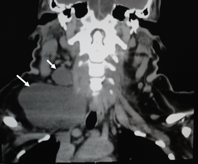
A 35-year-old woman visited the Surgical Oncology Unit, Wattanosoth-Bangkok hospital group in July 2008, with the complaint of an 8 cm, cystic lump on her right supraclavicular region for two months. No changes in voice, dysphagia or pain at the lump were noticed. Only two years history of type II diabetes mellitus which was well controlled by oral hypoglycemic drugs was recorded. No history of head and neck irradiation or surgery was confirmed. Physical examination revealed an 8.0 x 6.0 cm, cystic mass at the right supraclavicular region. It was mobile with movement when swallowing. The thyroid gland was of normal size with no palpable nodules. Cervical lymph nodes were not palpable. Hematological and biochemical tests revealed no abnormalities. Computed tomography (CT) of the neck showed two non-calcified cystic masses measured 8.6 x 5.2 cm and 3.0 x 2.7 cm. Both were located at the right mid-jugular and right supraclavicular regions, respectively (Figure 1). Lower border of this cystic mass is seen in figure 2.
| ||||||
| ||||||
|
| ||||||
| ||||||
|
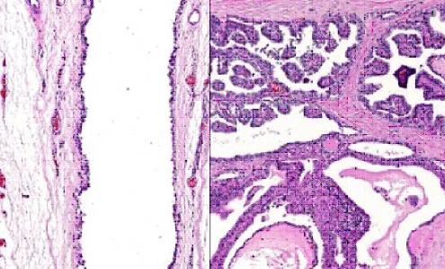
The patient underwent neck exploration for removal of the mass. At the time of operation two cystic masses were found at the right mid jugular region (3.0 x 2.7 cm in diameter) and right supraclavicular region (8.6 x 5.0 cm in diameter) with no adjacent organ invasion and no association to hyoid bone. The patient developed chyle leakage on the first post- operative day, but resolved after five days of conservative treatment. Histopathological examination showed cystic spaces lined with simple columnar epithelium cells and foci of thyroid tissue at the outer aspect of the cyst wall. A small papillary carcinoma was present in this area which showed finger-like branching papillary projections (arrows) lined by neoplastic cells (Figure 3).
| ||||||
|
| ||||||
|
However, ultrasonographic evaluation on the patient's thyroid gland was performed and revealed one cm diameter nodule at the superior pole of the right lobe (Figure 4). Unsatisfactory result performed by ultrasound-guided core needle biopsy lead the surgeon to perform right lobectomy of the thyroid gland. Papillary carcinoma was found by intraoperative frozen section, thus the total thyroid was removed without postoperative complication. Ablative radioactive iodine was scheduled at four week after operation but the patient didn't come back for follow up.
| ||||||
| ||||||
|
Discussion
| ||||||
|
Although some authors consider the thyroglossal duct as a natural way of spread of an occult thyroid cancer, [5] relatively normal thyroid tissue has been found in the cyst walls in as many as 62% of cases. [3] [9] [10] [11] Its presence is most probably due to the origin of duct in relation to thyroid tissue. Thyroglossal duct carcinoma is most commonly papillary carcinoma (75-80%), but other thyroid tumors such as follicular, Hurthle cell, and mixed papillary and follicular carcinomas have been reported. Malignancy may arise from thyroid tissue presente in the wall of TDC or metastasize from thyroid carcinoma, although it may be difficult to determine which is the primary tumor and which is the metastasis. [5] [10] Primary carcinoma of the TDC should conform to criteria such as localization of the carcinoma to a clearly demonstrable TDC or tract, and absence of carcinoma after histopathologic examination of the thyroid gland. [7] In our case, metastasized papillary carcinoma of thyroid gland was most likely the provisional diagnosis. This is also the first case to report the unique location of TDC. TDC is usually located in the mid-line or slightly off-midline. Of those in a para-median location , ninety five percent occur on the left for unclear reasons and 5% are on the right. [12] Seventy-three percent occur below the hyoid bone, 24% are suprahyoid, and 2% occur intralingually. [8] In addition, thyroglossal ducts rarely occur in a lateral/cranial/caudal location or even localize to the mediastinum or larynx. [13] We believe this is an atypical location of TDC, confirmed by presence of simple columnar epithelium. Moreover, the coincidence with metastasized papillary carcinoma of thyroid gland may be found up to 14% of TDC cases. [6] Papillary carcinoma within thyroglossal duct cyst usually presents as an asymptomatic mass. Diagnostic imaging modalities (ultrasonography, CT scan, MRI and PET/CT scan) are usually unable to diagnose pre-operative malignant condition. [14] The surgical treatment of papillary carcinoma of TDC is controversial. From the study of Patel et al. [15] which analyzed 62 cases of these carcinomas, Sistrunk procedure is adequate for most patients and has low rates of recurrence (1.8%). [16] All patients should receive suppressive doses of thyroid hormone. If co-incident papillary thyroid carcinoma was detected, removal of thyroid should be done.
| ||||||
|
Conclusion
| ||||||
|
The incidence of carcinomas arising from TDC is very small. Most of the cases are papillary carcinoma in nature. Malignancy may arise from thyroid tissue presented in the wall of TDC or metastasize from thyroid carcinoma although it may be difficult to determine which is the primary tumor, and which is the metastasis. We report here a case of papillary carcinoma arising in a TDC localized at an unusual location together with a papillary carcinoma in the thyroid gland. Careful evaluation of thyroid gland is essential for complete removal of possible residual malignancy.
| ||||||
|
References
| ||||||
| ||||||


| [HTML Abstract] [PDF Full Text] |
|
Author Contributions:
Chuathong A - Substantial contributions to conception and design, Acquisition of data, Analysis and interpretation of data, Drafting the article, Revising it critically for important intellectual content, Final approval of the version to be published Ouawathanamongkol S - Substantial contributions to conception and design, Acquisition of data, Analysis and interpretation of data, Drafting the article, Revising it critically for important intellectual content, Final approval of the version to be published Maipang T - Substantial contributions to conception and design, Acquisition of data, Analysis and interpretation of data, Drafting the article, Revising it critically for important intellectual content, Final approval of the version to be published Chongchitnant N - Substantial contributions to conception and design, Acquisition of data, Analysis and interpretation of data, Drafting the article, Revising it critically for important intellectual content, Final approval of the version to be published |
|
Guarantor of submission:
The corresponding author is the guarantor of submission. |
|
Source of support:
None |
|
Conflict of interest:
The author(s) declare no conflict of interests |
|
Copyright:
© Chuathong A et. al. 2011; This article is distributed under the terms of Creative Commons Attribution License which permits unrestricted use, distribution and reproduction in any means provided the original authors and original publisher are properly credited. (Please see Copyright Policy for more information.) |
|
|