
| Table of Contents |  |
|
CASE REPORT
|
| Iatrogenic tracheobronchial rupture: A case report and review of the literature |
| Alexander Zlotnik1, Shaun E. Gruenbaum2, Benjamin F. Gruenbaum1, Michael Dubilet1, Evgenia Cherniavsky3 |
|
1Department of Anesthesiology and Critical Care, Soroka Medical Center, Ben Gurion University, Beer Sheva, Israel
2 Department of Anesthesiology, Yale University School of Medicine, New Haven, CT, USA. 3 Department of Radiology, Soroka Medical Center, Ben Gurion University, Beer Sheva, Israel. |
|
doi:10.5348/ijcri-2011-03-24-CR-4
|
|
Address correspondence to: Alexander Zlotnik Department of Anesthesiology Soroka Medical Center POB 151, Beer-Sheva, 84105 Israel Phone: 972-8-6400262 Fax: 972-8-6403795 Email: zlotnika@bgu.ac.il |
|
[HTML Abstract]
[PDF Full Text]
|
| How to cite this article: |
| Zlotnik A, Gruenbaum SE, Gruenbaum BF, Dubilet M, Cherniavsky E. Iatrogenic tracheobronchial rupture: A case report and review of the literature. International Journal of Case Reports and Images 2011;2(3):12-16. |
|
Abstract
|
|
Introduction:
We present a case of a 37-year-old female who suffered an iatrogenic tear of the trachea following a non-traumatic intubation for an uncomplicated, elective cesarean section under general anesthesia.
Case Report: Fifty minutes after extubation, the patient developed hemodynamic instability and respiratory distress, which required re-intubation, and the trachea was surgically repaired. Tracheal rupture is a rare but potentially life-threatening lesion, and a delay in the diagnosis and treatment is associated with a poor outcome. Conclusion: The diagnosis and treatment of tracheal rupture is discussed in this report, especially in the context of pregnant patients. The authors recommend early surgical repair of the trachea in patients who suffer post-intubation tracheal injury complicated by respiratory failure or hemodynamic instability. | |
|
Key Words:
Cesarean section, Iatrogenic Tracheobronchial rupture, Intubation
| |
|
Introduction
| ||||||
|
Tracheal rupture is a rare but potentially life-threatening lesion. Tracheal rupture is usually iatrogenic or traumatic in origin and can sometimes be spontaneous. Here, we present a case of iatrogenic tracheobronchial rupture following an uneventful intubation for elective cesarean section, that was diagnosed postoperatively. We discuss the diagnosis and treatment options available for this life-threatening problem.
| ||||||
|
Case Report
| ||||||
|
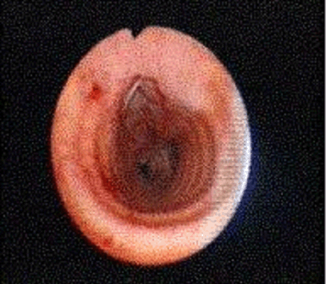
A 37-year-old-female was scheduled for cesarean section (CS) under general anesthesia (G5 P4 A39+6W). The CS was performed due to two prior CS. A review of her medical history and physical examination was significant for arterial hypertension, long-term heavy smoking, and short stature. Her weight measured 83 kg and her height was 155 cm (BMI of 35 kg/m2). Her previous experiences under general anesthesia were without any complications. The anesthesia with preoxygenation and rapid sequence induction with Sellic's maneuver was initiated with sodium thiopenthal 400 mg and succinylcholine 100 mg intravenously. The direct laryngoscopy was performed with a Mackintosh with blade number 3 and revealed Laryngoscopy Lehane's Class II with visualization of posterior part of the vocal chords. The trachea was intubated by an experienced anesthesiologist without the use of stylet, with a cuffed endotracheal tube (ETT) of internal diameter 7.0. The cuff was inflated with 3-4 ml of air. The intubation was performed without any difficulties. The patient was positioned in the Trendelenburg position and was mechanically ventilated with positive pressure ventilation at the following setting parameters: tidal volume of 450 ml, respiratory rate of 10 per minute, and peak inspiratory pressure of 25 cm H2O. The anesthesia was maintained using 1:2 oxygen to nitrous oxide. A one percent inspired concentration of isoflurane and 0.01 mg of fentanyl was used for analgesia. Muscle relaxation was achieved with 25 mg of atracurium besylate. The patient was stable throughout the duration of the surgery. The surgery was uneventful and lasted 50 minutes. At the end of surgery, the patient was fully awake and was extubated and transported to the postanesthesia care unit. Fifty minutes after extubation the patient developed a cough and massive, palpable, upper thoracic and cervical subcutaneous emphysema developed. Physical examination revealed a smooth bulging of the skin of the neck and chest, and there were decreased breath sounds over the lungs bilaterally with crepitus on auscultation. The patient remained stable at this time without any respiratory insufficiency or hypoxemia. Chest radiography demonstrated extensive subcutaneous emphysema and pneumomediastinum (Figure 1). A thoracic helical CT scan (Figure 2) showed a pneumomediastinum, small left pneumothorax, massive subcutaneous emphysema, and focal defect in the posterior membranous part of the mid trachea. The patient was transferred to the ICU while breathing spontaneously. The diagnosis of tracheal tear was confirmed by bronchoscopy. A 2-3 cm vertical tear was visualized in the posterior membranous wall of the distal intrathoracic trachea (Figure 3). Soon after bronchoscopy, the patient became hemodynamically unstable and developed respiratory distress. She was again intubated with a regular ETT without any difficulties.
| ||||||
| ||||||
|
| ||||||
|
| ||||||
| ||||||
|
The decision was made to immediately repair the injured trachea. The ETT was replaced with a left sided, double- lumen ETT 35F in the operating room. The trachea was approached through a right thoracotomy. Revision discovered a tracheal tear 1.5 cm long, located 2 cm above the carina in the posterior membranous part. No esophageal injury, inflammatory tissues or pus was noted. The injury was closed using interrupted buttressed sutures, and a mediastinal pleural flap was raised to cover the suture line. The total surgery time was fifty minutes. Prophylactic antibiotic therapy with amoxicillin-clavulanic acid (Augmentin®) and gentamicin was applied before the surgery to prevent mediastenitis. Two large bore pleural chest drains were inserted. Following completion of the surgery, the double-lumen ETT was replaced with a regular 7 mm internal diameter ETT. Extubation was performed five hours after finishing the surgery, and thoracal drains were removed 36 hours after finishing the surgery. The subcutaneous emphysema and pneumomediastinum resolved over the next four days. There was no respiratory infection or mediastenitis, and the patient's recovery was uneventful. She was discharged home on the 9th postoperative day. At one month follow up, the patient was doing well with no evidence of respiratory compromise or complications.
| ||||||
|
Discussion
| ||||||
|
We presented a case of tracheal rupture which occurred following general anesthesia involving an endotracheal intubation. The cause of the rupture can be considered iatrogenic in nature. Iatrogenic tracheal injury is a rare (less than 0.01%) but severe complication after airway access techniques such as endotracheal intubation and tracheostomy. [1] [2] In decreasing frequency, the causes of tracheal rupture include trauma, complications from intubation, complications from percutaneus tracheostomy, and spontaneous rupture. [1] [3] Endotracheal intubation is an important cause of tracheal rupture. Many iatrogenic tracheal lesions are the consequence of emergency intubations in stressful situations. [1] [3] Cases of post-intubation tracheal rupture described in literature have generally been attributed to the potentially traumatic process of endotracheal intubation. [4] The pointed tip of the intubation stylet of an ETT can injure the trachea, although the stylet is usually pointed anteriorly, away from the fragile membranosa. Prolonged endotracheal intubation can cause pressure necrosis or erosion of the lower cervical trachea. Hyperinflation of the cuff causes overstretching of the membranosa, which can separate longitudinally. [5] This complication is less frequent due to the routine use of highly compliant, low-pressure sealing cuffs. [5] Several authors have suggested that female gender and short height are important risk factors of tracheobronchial injury. [3] [4] Excessive coughing against an inflated cuff can cause tracheal laceration, especially in elderly patients or patients under corticosteroid therapy with frail tissues. [1] [3] [4] Other important risk factors for tracheal rupture include advanced age, poor medical condition, short stature, chronic obstructive pulmonary disease, and tracheomalacia. [3] Postintubation tracheal rupture typically occurs in female patients with a median age of 45 years. In women, anatomical and technical factors may be responsible for causing tracheal tears, including the downward placement of the tube too far into a short trachea [1], the use of an oversized tube or overinflation of the cuff in a narrow trachea, and the vulnerability and weakness of the trachea in women. In the present case, the patient was female, had a short stature, and was obese. The complication developed after coughing against an ETT with an inflated cuff. In our opinion, one possible explanation for why tracheal tears occur more frequently in pregnant women is due to the presence of chronic chemical tracheitis during pregnancy. This condition is the result of chronic exposure of the tracheal mucous membranes to the extremely acidic gastric content that results from regurgitation. [6] Increased progesterone concentrations lead to both a decreased gastric pH and decreased tonus of the gastroesophageal sphincter, which in turn predisposes pregnant women to develop tracheal mucosal injury. [7] This mucosal injury then increases the risk of its laceration during traumatic intubation. [6] The time elapsed before the diagnosis of tracheobroncheal injury is made may be as short as few minutes or as long as 72 hours after intubation. [1] A delay in diagnosis can be explained by preservation of the integrity of paratracheal connective tissue sheath or by temporary occlusion of a small tear by the endotracheal balloon. [9] A delayed diagnosis and treatment is associated with a poor outcome. [8] [10] Suspicion of tracheal rupture is primarily based on clinical signs. The most frequent sign (present in about 80% of patients) is the sudden onset of subcutaneous emphysema located in the upper thorax and neck. [10] Hemoptysis is a less common sign. Respiratory failure may develop because of the air leak, resulting in dyspnea and cyanosis. Few patients remain asymptomatic. [10] A pneumothorax can occur if the tracheal air leak communicates with the pleural space due to a tear of the mediastinal pleura. This can be confirmed with chest X-ray. A CT scan with the appropriated window settings allows for direct visualization of the tracheal injury in 70% of cases. [5] [11] Bronchoscopy is regarded as the most effective method for confirming the diagnosis and for determining the exact location and extent of the tear. [5] [12] Post-intubational iatrogenic tracheal injuries are usually linear, involving the membranous portion of the intrathoracic segment of the trachea. [13] [14] Most often, localization of the tracheal injuries is in the lower third of the trachea (60-80%). Less often, they may be localized in the middle trachea. [1] The management of iatrogenic TBR is controversial. Localization of the rupture, its nature and extent, the mechanism of the injury, the patient's age, and the patient's pre-existing risk factors are taken into consideration to define the optimal therapeutic option. [1] Early surgical repair has traditionally been considered as the cornerstone of the management of tracheal ruptures. [1] Conservative treatment is recommended for small lesions of the trachea that do not have any clinical or radiological signs of mediastinal involvement. However, close follow-up with brochoscopy and CT-scan is required. [15] There is growing evidence to suggest that iatrogenic TBR can be treated conservatively in select cases. [3] Selection criteria for non-operative management are still currently being debated. Most authors agree that there should be no evidence of respiratory or hemodynamic instability, esophageal injury, mediastinitis, or progressive pneumomediastinum. [3] [15] The size and the depth of the defect is also important, and surgery should be advocated whenever full-thickness or lengthy lacerations (more then one cm) are present. [15] Critically ill patients, especially in whom TBR results from emergency intubation, may fulfill all the criteria for surgical repair. In a relatively large series of patients, early surgical treatment was associated with a significantly lower mortality rate and a better outcome. [10]
| ||||||
|
Conclusion
| ||||||
|
In the present case, the patient presented with a pneumomediastinum and with signs of developing respiratory failure. She underwent early surgical repair and was discharged with a good clinical outcome. Our case represents an excellent example of the success of early surgical treatment of iatrogenic tracheal rupture, which was followed by the patient's full recovery. In our opinion, early surgical intervention for repair of the tracheal injury remains the safest choice in the management of patients suffering from postintubational tracheal injury.
| ||||||
|
References
| ||||||
| ||||||



| [HTML Abstract] [PDF Full Text] |
|
Author Contributions:
Alexander Zlotnik - Conception and design, Drafting the article, Critical revision of the article, Final approval of the version to be published Shaun E. Gruenbaum - Conception and design, Drafting the article, Critical revision of the article, Final approval of the version to be published Michael Dubilet - Conception and design, Drafting the article, Critical revision of the article, Final approval of the version to be published Evgenia Cherniavsky - Conception and design, Drafting the article, Critical revision of the article, Final approval of the version to be published Benjamin F. Gruenbaum - Conception and design, Drafting the article, Critical revision of the article, Final approval of the version to be published |
|
Guarantor of submission:
The corresponding author is the guarantor of submission. |
|
Source of support:
None |
|
Conflict of interest:
The author(s) declare no conflict of interests |
|
Copyright:
© Alexander Zlotnik et. al. 2010; This article is distributed the terms of Creative Commons Attribution License which permits unrestricted use, distribution and reproduction in any means provided the original authors and original publisher are properly credited. (Please see Copyright Policy for more information.) |
|
|