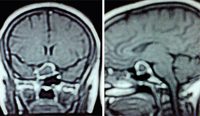
Left: MRI T1W Saggital cuts with contrast showing 20x13
milimeters hypointense mass in the sellar area. Right: MRI T1W Coronal cuts with contast showing 15x13
milimeters heterogeneously enhancing well encapsulated mass in the sellar area with right paracavernous extension. |