| |
|
|
|
Case Report
| ||||||
| Synchronous dual malignancy of papillary carcinoma thyroid and squamous cell carcinoma tongue: A case report | ||||||
| Soni Tej Prakash1, Goel Sajal2, Sharma Lalit Mohan3, Gupta Anil Kumar4, Sharma Shantanu5, Gothwal Ravindra6 | ||||||
|
1MD, Consultant, Department of Radiation Oncology, Bhagwan Mahaveer Cancer Hospital & Research Centre, Jaipur, Rajasthan, India.
2DNB, Junior Consultant, Department of Radiation Oncology, Patel Hospital, Jalandhar, Punjab, India. 3MD, Sr. Consultant, Department of Medical Oncology, Bhagwan Mahaveer Cancer Hospital & Research Centre, Jaipur, Rajasthan, India. 4MS, Sr. Consultant, Department of Surgical Oncology, Bhagwan Mahaveer Cancer Hospital & Research Centre, Jaipur, Rajasthan, India. 5MD, Associate Professor, Department of Radiotherapy, S.M.S. Medical College & Hospital, Jaipur, Rajasthan, India. 6MD, (Associate Professor, Department of Radiotherapy, S.M.S. Medical College & Hospital, Jaipur, Rajasthan, India. | ||||||
| ||||||
|
[HTML Abstract]
[PDF Full Text]
[Print This Article]
[Similar article in Pumed] [Similar article in Google Scholar] 
|
| How to cite this article |
| Soni TP, Goel S, Sharma LM, Gupta AK, Sharma S, Gothwal R. Synchronous dual malignancy of papillary carcinoma thyroid and squamous cell carcinoma tongue: A case report. Int J Case Rep Images 2017;8(3):209–212. |
|
Abstract
|
|
Introduction:
Patients with thyroid carcinoma have increased risk for development of second malignancy either synchronous or metachronous. We are reporting a rare case of synchronous squamous cell carcinoma oral tongue and papillary thyroid carcinoma.
Case Report: A 60-year-old smoker male was presented with the complaints of non-healing ulcer (2x2 cm) at right anterolateral tongue since six months. Excision biopsy of tongue ulcer was reported as pT 0.8x0.8 cm, moderately differentiated squamous cell carcinoma. Wide local excision of tongue lesion with right modified neck dissection was done. Histopathology was reported as no residual primary tongue tumor seen, 1 out of 28 lymph nodes showed metastatic deposits of squamous cell carcinoma. Unexpectedly another one lymph node was positive of metastatic thyroid papillary carcinoma. Total thyroidectomy with central compartment neck dissection and left modified neck dissection was done. Histopathology was reported as pT 2x1 cm, papillary carcinoma right lobe of thyroid, 7 out of 32 lymph nodes showed metatstatic paillary carcinoma thyroid. Although exact cause of second malignancy is unknown but field cancerization, genetic predisposition, history of prior radiation or chemotherapy, improved survival can be attributed for increased incidence of second cancer. Conclusion: Double malignancies require multidisciplinary management approach. | |
|
Keywords:
Carcinoma tongue, Papillary thyroid carcinoma, Synchronous
| |
|
Introduction
| ||||||
|
Double malignancy now comprises the sixth most common cancers and it makes 16% of all incident cancers [1]. Head and neck is a common site for synchronous malignancy owing to field cancerization with use of tobacco but the co-existence of tongue and thyroid primaries are rare [1]. Incidental cervical lymph node metastasis from papillary thyroid carcinoma, detected in lymph node dissection done for separate head neck primary cancer is extremely rare. A case of synchronous papillary thyroid carcinoma and squamous cell carcinoma tongue is being reported with review of literature. | ||||||
|
Case Report
| ||||||
|
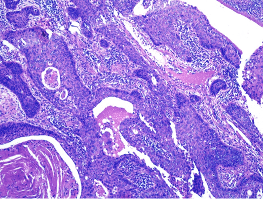
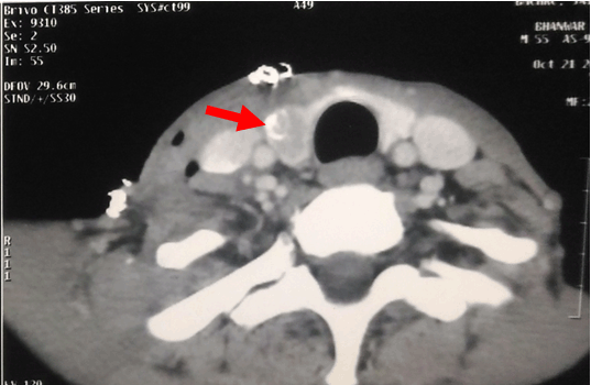
A 60-year-old smoker male was presented with the complaints of non-healing ulcer over right side tongue since six months. On examination there was an ulcer (1x1 cm) at right anterolateral tongue and palpable ipsilateral submandibular lymph node size 2x2 cm. Excision biopsy of tongue ulcer was reported as pT 0.8x0.8 cm, moderately differentiated squamous cell carcinoma, depth 0.5 mm, no lymphovascular or perineural invasion, cut margins free of tumor. Fine needle aspiration cytology (FNAC) from right cervical lymph node was reported as metastatic squamous cell carcinoma. Metastatic work-up was done. All routine blood investigations, X-ray chest, ultrasound-whole abdomen were within normal limits. Wide local excision of tongue lesion with right modified neck dissection was done. Histopathology was reported as no residual primary tongue tumor seen, 1 out of 28 lymph nodes showed metastatic deposits of squamous cell carcinoma with no perinodal spread (Figure 1), 3 nodes showed epithelioid granulomas reaction, 1 lymph node showed a focus of follicles filled with colloid and lined by ovoid cells with optically clear nuclei suggestive of metastatic thyroid papillary carcinoma (Figure 2). Computed tomography scan neck showed an enhancing nodular lesion in right lobe of thyroid with large ring calcification measuring 2.9x1.7x1.8 cm in size with surgical changes in overlying cutaneous and subcutaneous tissues of neck (Figure 3). Total thyroidectomy with central compartment neck dissection and left modified neck dissection was done. Histopathology was reported as pT 2x1 cm, papillary carcinoma right lobe of thyroid, 7 out of 32 lymph nodes showed metastatic papillary carcinoma thyroid, maximum nodal size 2 cm, no extracapsular extension or lymphovascular invasion. Finally, he was diagnosed as a case of synchronous carcinoma anterior tongue pT1pN1M0 (stage III) and papillary carcinoma thyroid pT2pN1bM0. Expert opinion was sought from physician to rule out active tuberculosis. He received adjuvant external beam radiation to face/neck region to dose of 60Gy in 30 fractions over six weeks duration. He tolerated the treatment well. Whole body I-131 scan showed residual functioning thyroid tissue in thyroid bed. He received single dose of 150 mCi of I-131 orally. He was started on tablet levothyroxine 100 mg once a day and tablet calcium 500 mg twice a day. | ||||||
| ||||||
| ||||||
| ||||||
|
Discussion
| ||||||
|
Thyroid cancer is the fifth most common cancer in females and accounts for 1% of all cancers. Incidence of carcinoma thyroid has increased significantly in last two decades. Surveillance epidemiology and end results database also reveals a 2.4-fold rise in thyroid cancer incidence from 3.6 per 100,000 in 1973 to 8.7 per 100,000 in 2002 consisting primarily of papillary thyroid carcinoma [2]. Wide use and availability of ultrasound and other imaging techniques contributes at least partly of this rise in incidence of carcinoma thyroid. Papillary thyroid cancer is the most common type of thyroid cancer and represents about 75% of all thyroid malignancies. Cervical lymph node metastasis is common in papillary thyroid cancer (ranging from 15–65% cases) and it is associated with a significant probability for loco-regional recurrence [3]. Lymphatic metastasis from papillary thyroid cancer occurs to neck lateral nodal groups of levels II, III, IV and central (level VI) lymph nodes. Thyroid cancer patients have an increased risk of developing a second cancer as either synchronous or metachronous [4]. In a pooled analysis of primary carcinoma thyroid patients from 13 registries, Sandeep et al. [4] found a 30% increase in risk of second cancers including salivary gland, kidney, prostate, skin, breast, brain, myeloma, leukemia, and non-Hodgkin lymphoma. On another side primary head neck cancer patients also have increased incidence (2–3% per year) of development of second primary cancer [5]. Barnes [6] found 0.7% of patients with squamous of carcinoma cell carcinoma of the head and neck cancers had lymphatic metastasis of papillary thyroid carcinoma. Singh J et al. [7] also reported a similar case of synchronous double malignancy of thyroid carcinoma and tongue carcinoma. The exact mechanism of double malignancy is unknown. The predisposing risk factors for dual malignancy include field cancerization, tobacco and alcohol, genetic predisposition (Li–Fraumeni syndrome and Beckwith–Weidemann syndrome, Cowden syndrome), history of prior external radiation, radioiodine ablation or chemotherapy, environmental risk factors and improved survival [8]. Field cancerization also promotes transformation of an existing precancerous lesion into a malignancy and causes multifocal and second cancer in individual sites. Carcinogen exposure creates multiple genetic abnormalities in the whole tissue region and explores development of many foci of malignant transformation. Thyroid cancer is associated with genetic mutations and abnormalities in tumor suppressor genes and cell cycle proteins which activates development of second cancers. Mutation of CHEK2 protein and RET genes are associated with an increased risk of thyroid, breast, prostate, and colon cancers [9]. | ||||||
|
Conclusion
| ||||||
|
In conclusion, incidence of second cancer has increased substantially. For early detection of second primary malignancy in head neck region complete and thorough examination and investigations including contrast enhanced computed tomography scan and pan-endoscopy are warranted. Squamous cell carcinoma of oral cavity along with synchronous papillary thyroid carcinoma is a rare situation. In such cases of double malignancy, multidisciplinary management and team work is required with the consensus of surgical oncology, radiation oncology, pathology, nuclear medicine and radiology experts. Oral cancers require aggressive treatment on priority as thyroid carcinoma has excellent prognosis. Metastatic workup followed by wide excision of primary oral cancer, total thyroidectomy and neck node dissection is required for adequate staging, proper diagnosis and management. | ||||||
|
References
| ||||||
| ||||||
|
[HTML Abstract]
[PDF Full Text]
|
|
Author Contributions
Tej Prakash Soni – Substantial contributions to conception and design, Drafting the article, Final approval of the version to be published Sajal Goel – Acquisition of data, Drafting the article, Final approval of the version to be published Lalit Mohan Sharma – Substantial contributions to conception and design, Drafting the article, Final approval of the version to be published Anil Kumar Gupta – Acquisition of data, Revising it critically for important intellectual content, Final approval of the version to be published Shantanu Sharma – Acquisition of data, Revising it critically for important intellectual content, Final approval of the version to be published Ravindra Gothwal – Acquisition of data, Revising it critically for important intellectual content, Final approval of the version to be published |
|
Guarantor of submission
The corresponding author is the guarantor of submission. |
|
Source of support
None |
|
Conflict of interest
Authors declare no conflict of interest. |
|
Copyright
© 2017 Tej Prakash Soni et al. This article is distributed under the terms of Creative Commons Attribution License which permits unrestricted use, distribution and reproduction in any medium provided the original author(s) and original publisher are properly credited. Please see the copyright policy on the journal website for more information. |
|
|