
  |

|
 |
|
Clinical Image
| ||||||
| 18Fluorine fluorodeoxyglucose positron emission tomography diagnosis of an aortic thoracic prosthesis infection by slow-growing bacteria | ||||||
| Geraldine Celine Bera1, Patrick Farahmand2, Françoise Cavailloles3, Charlotte Lepoutre-lussey4 | ||||||
|
1MD, University Medical Assistante, Department of Nuclear Medicine, AP-HP Hospital Pitié-Salpêtriére, 47-83 Bd de l'hôpital 75651 Paris Cedex 13 France and Departement of Biophysic UPMC Paris VI, 91 Bd de l'hôpital 75651 Paris Cedex 13, France.
2MD, Practitioner Attached, Department of Cardiovascular and Thoracic Surgery, Institute of Cardiology, AP-HP Hospital Pitié-Salpêtriére, 52 Bd Vincent Auriol 75013 Paris Cedex 13, France. 3MD, Department of Nuclear Medicine, Private Hospital of Antony, 1 rue Velpeau 92166 Antony Cedex, France. 3MD, Practitioner Attached, Department of Biochemistry INSERM 0970, AP-HP European Hospital Georges-Pompidou, and Department of Nuclear Medicine AP-HP Hospital Pitié-Salpêtriére, 56 rue Leblanc 75015 Paris, France. | ||||||
| ||||||
|
[HTML Abstract]
[PDF Full Text]
[Print This Article]
[Similar article in Pumed] [Similar article in Google Scholar] 
|


| How to cite this article |
| Bera GC, Farahmand P, Cavailloles F, Lepoutre-lussey C. 18Fluorine fluorodeoxyglucose positron emission tomography diagnosis of an aortic thoracic prosthesis infection by slow-growing bacteria. Int J Case Rep Images 2015;6(6):381–383. |
|
Case Report
|
|
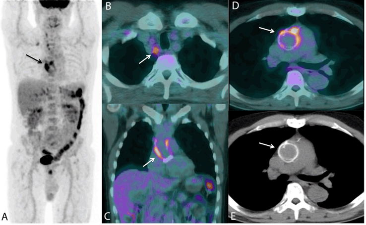
A 66-years-old male with a history of aortic root aneurysm and bicuspid aortic valve underwent aortic root replacement with a Bentall procedure in 2009. Four years later, he presented to the emergency department with a fever and general malaise. Serial blood cultures were negative and a transthoracic echocardiogram showed no evidence of vegetations or an aortic root abscess. Although there was no evidence to support a diagnosis of infective endocarditis (IE) empirical antibiotic therapy, comprising vancomycin, gentamicin and rifampicin, was commenced. Two weeks later, a18fluorine-fluorodeoxyglucose positron emission tomography (18[F]-FDG PET) was performed under a diet (low-carbohydrate and high-fat during 24-hours) designed to suppress myocardial activity. One hour after the injection of 295MBq of 18F-FDG radioisotope, 3 foci were highlighted around the aortic prosthesis (SUVmax = 6, graft-to-mediastinum ratio SUVmax = 3.2) (Figure 1A, 1D, 1E) with additional uptake within mediastinal lymph adenopathies (SUmax = 2.6) (Figure 1B). There was no pathological uptake noticed around the adjacent aortic valve prosthesis (Figure 1C). This result was highly suggestive of an infection of the aortic tubular prosthesis and so surgery was undertaken to replace the aortic tubular prosthesis with a mechanical valve. During surgery, macroscopic examination confirmed the presence of pus inside the envelope surrounding the prosthesis. Cultures were taken during the surgery and analysis via polymerase chain reaction revealed the growth of Kingella kingae, the fifth member of the HACEK (Haemophilus, Aggregation bacter, Cardiobacterium hominis, Eikenella corrodens and Kingella species) bacteria group. |
|
|
|
Discussion
|
|
The HACEK microorganisms, commensal residents of the oropharynx, are responsible for ~3% of cases of IE. They have an incidence of ~2–3 cases per 100,000 of population per annum and their mortality can reach up to 14%. These gram-negative bacilli are slow-growing, likely explaining the frequently negative blood cultures [1]. Diagnosis of cardiovascular prosthesis infections can be challenging with 30% having normal or inconclusive echocardiographic findings [2]. This often leads to a serious delay in instigating medical and, more importantly, surgical treatment. Saby et al. [2] have demonstrated that if used as a major criterion 18[F]-FDG PET increased the sensitivity of the modified Duke classification from 80–97%, without compromising its specificity. Thoracic aortic prosthetic graft infection is particularly rare and associated with very high morbidity and mortality [3]. Its diagnosis by conventional imaging is difficult due to non-specific nature of the findings and the sensitivity of magnetic resonance imaging remains unclear [2][3][4]. This report shows a high added value of 18F-FDG PET in the diagnosis of a sub-acute endocarditis even after fifteen days of a broad-spectrum antibiotic therapy. Focal 18F-FDG uptake around the cardiovascular prosthesis has a sensitivity of 93%, specificity of 91%, positive predictive value of 88% and negative predictive value of 96% for the diagnosis of prosthetic vascular graft infection [4]. A non-homogeneous uptake pattern around the cardiovascular prosthesis is described as a poor diagnostic marker [4]. While SUVmean and graft-to-mediastinum ratio represent the overall metabolic activity in the whole graft more accurately than SUVmax, [4] a SUVmax > 8 in the surrounding graft area has been described as a potential cut-off value for distinguishing infected from non-infected grafts with sensitivity and specificity of 100% and 80%, respectively [3]. |
|
Conclusion
|
|
This report illustrates that 18F-FDG PET with a characteristic uptake pattern (focal uptake), a SUVmax ≥ 6 and a graft-to-mediastinum ratio SUVmax ≥ 3.2, allowed the detection of a cardiovascular prosthesis infection due to a slow-growing bacteria even following 2 weeks of antibiotic treatment. |
|
Acknowledgments
|
|
Professor Aurelie KAS MD.PhD., Department of Biophysic UPMC Paris VI, LIB, INSERM UMR 678, 91 Bd de l'hôpital 75651 Paris Cedex 13 France and Department of Nuclear Medicine, AP-HP Hospital Pitié-Salpêtrière, 47–83 Bd de l'hôpital 75651 Paris Cedex 13 France. Nathanaëlle YENI MD., Contractual Hospital Practitioner, Department of Nuclear Medicine, AP-HP Hospital Pitié-Salpêtrière, 47-83 Bd de l'hôpital 75651 Paris Cedex 13 France and Department of Biophysic UPMC Paris VI, LIB, INSERM UMR 678, 91 Bd de l'hôpital 75651 Paris Cedex 13 France. Françoise CAVAILLOLES MD., Department of Nuclear Medicine, Private Hospital of Antony, 1 rue Velpeau 92166 Antony Cedex, France. |
|
References
|
|

|
[HTML Abstract]
[PDF Full Text]
|
|
Author Contributions
Geraldine Celine Bera – Conception and design, Analysis and interpretation of data, Drafting the article, Final approval of the version to be published Patrick Farahmand – Acquisition of data, Analysis and interpretation of data, Critical revision of the article, Final approval of the version to be published Françoise Cavailloles – Acquisition of data, Analysis and interpretation of data, Critical revision of the article, Final approval of the version to be published Charlotte Lepoutre-lussey – Conception and design, Critical revision of the article, Final approval of the version to be published |
|
Guarantor of submission
The corresponding author is the guarantor of submission. |
|
Source of support
None |
|
Conflict of interest
Authors declare no conflict of interest. |
|
Copyright
© 2015 Geraldine Celine Bera et al. This article is distributed under the terms of Creative Commons Attribution License which permits unrestricted use, distribution and reproduction in any medium provided the original author(s) and original publisher are properly credited. Please see the copyright policy on the journal website for more information. |
|
|